
Revision sheet
Partial nephrectomy
6 steps, each one the authored title and caption from the interactive animation, with the frame captured from that step. Read it before the case, or print it and work through the steps in order.
Teaching only, not for diagnosis or treatment decisions. The text on this sheet is the animation's own step copy, reproduced verbatim.
UrOops3D · UroRef · © Nity G
- 01
Read the model
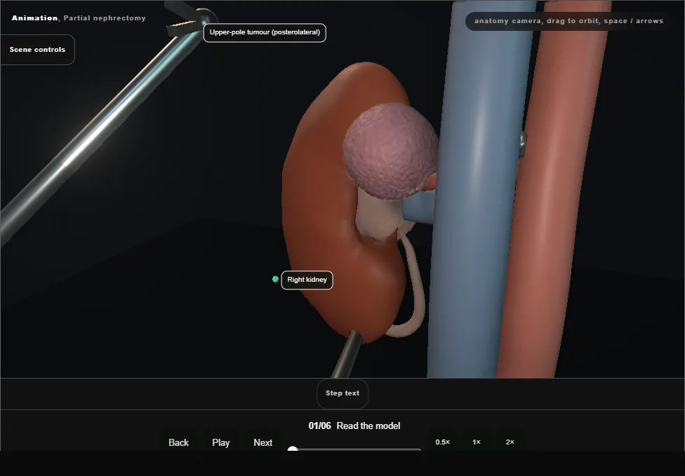
- Right kidney: an exophytic upper-pole, posterolateral tumour: depth into the parenchyma, the arterial branch map and the collecting system decide the operation before the clamp goes on.
- 02
Hilum exposed + artery slung
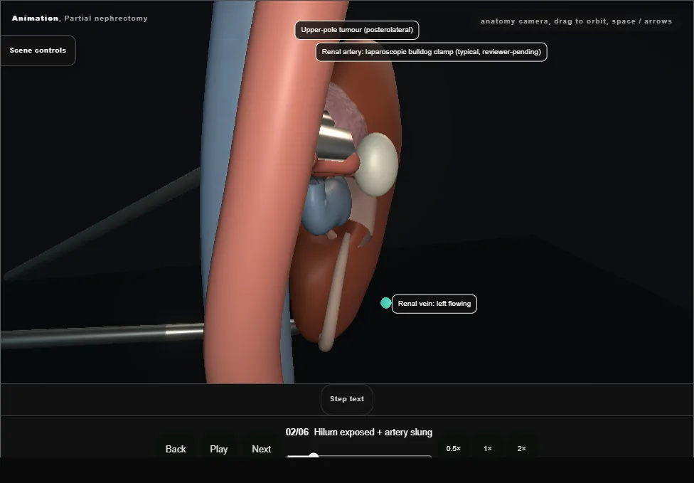
- Colon reflected, renal artery skeletonised enough for a clean bulldog.
- The vein sits anterior to the artery at the hilum and is exposed but left flowing; check the pre-op map for accessory arteries; two clamps beat one leaky one.
- 03
Clamp + excision
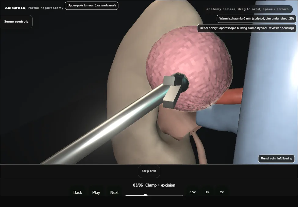
- The bulldog closes, the kidney pales, the clock starts.
- Excision takes the tumour with a thin visible rim: the pale ring you can see around the base is the plane, and it is decided now, not on histology.
- 04
Inner layer (renorrhaphy 1)
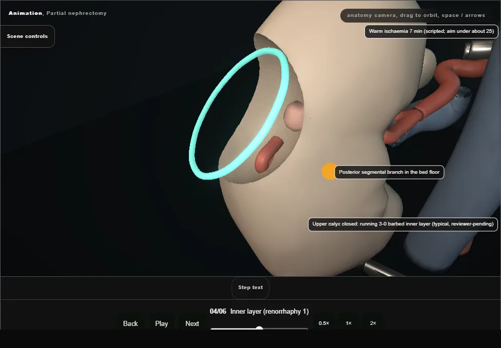
- The bed shows the stack: the opened upper calyx crossing the floor and the cut segmental branch.
- The running inner layer closes the calyx watertight; this layer is what stands between the patient and a urinoma on day three.
- 05
Outer layer (renorrhaphy 2)
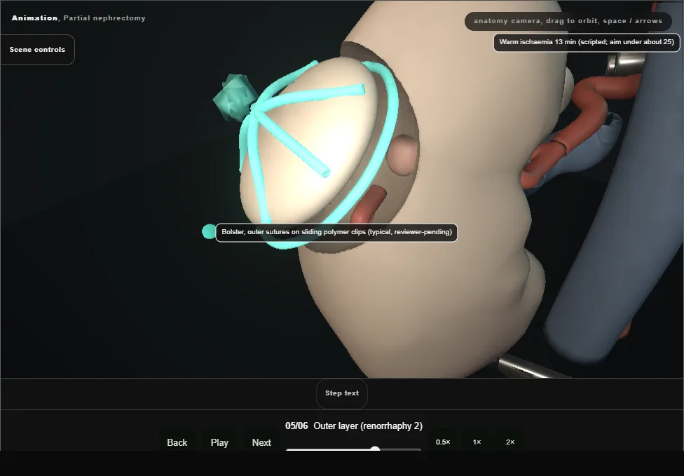
- A bolster caps the defect and sliding clips tension the outer sutures over it: haemostasis by compression, designed to hold on reperfusion.
- 06
Reperfusion + check
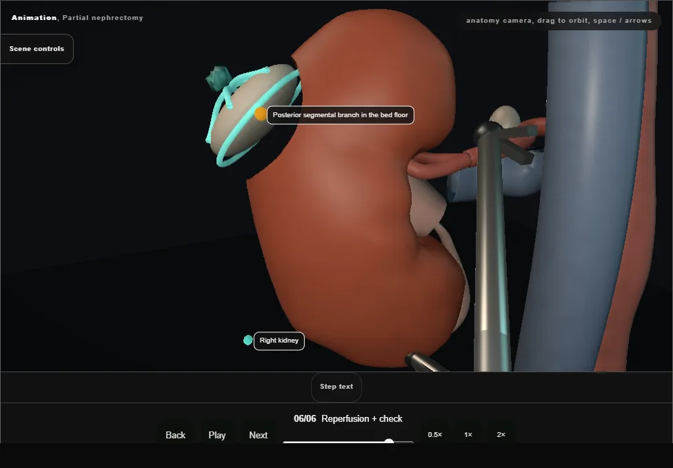
- Clamp off, colour back.
- Watch the bed: ooze settles with patience; anything pulsatile gets a stitch now.
- Delayed gross haematuria days later is a segmental-artery pseudoaneurysm until proven otherwise; the vessel it comes from is the one stitched in this bed.
Derived from PartialNephrectomyV2Scene.tsx at build time: every step title and caption above is the authored
ProcedureDef text, reproduced verbatim, and this page shares its readable-points renderer with the animation
itself. 6 of 6 steps have a captured frame.
Text fingerprint b36410457a3784bb. A caption edit without a re-capture fails the production
check, so the sheet and the animation cannot drift apart silently.