Al-Ghorab open distal shunt for ischaemic priapism
The open corporoglanular shunt for ischaemic priapism that has beaten aspiration, irrigation, phenylephrine and a percutaneous distal shunt.
A tourniquet at the base, a transverse incision in the dorsum of the glans, a segment of tunica excised from the dorsal tip of each corpus, the stagnant blood pressed out until it runs bright, and the glans closed over two windows that stay open.
The lesson is where the excision goes, what counts as success, and what the patient is told afterwards.
Advanced tierFRCS andrology, emergency, sexual medicineInteractive 3D
How to use this module. Play through the eight steps, or use Back and Next. Watch the glans open, a window cut from each dorsal tip, dark clot give way to bright blood, and the drainage path stay open under the closed glans. The triggers show a firm penis the next morning, a ventral excision and a dusky glans.
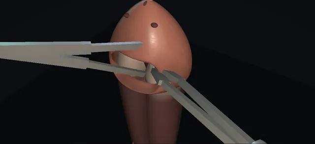
The dorsum of the glans from the transverse incision down into the corporal tips. Schematic, not to scale; relationships only.
Seen from above the glans, the two corporal tips lie side by side just under the dorsal glans tissue, the septum between them and the urethra below and behind everything.
A transverse incision in the dorsum of the glans opens on to both tips at once; a round window cut from the dorsal tunica of each tip opens the ischaemic compartment into the glans spongy tissue, which drains separately.
Closing the glans over the open windows leaves that path in place: corpus, window, glans.
›Corpora cavernosa - the compartment: dark, rigid and tender; their dorsal tips under the glans are where the windows are cut.
›Tunica albuginea of the tips - the white layer the Allis grasps and the scissors excise; its thickness is what the rim of the window shows.
›Glans penis - soft and uninvolved; incised transversely on its dorsum and closed over the open windows.
›Glans spongy tissue - the drainage bed: each window opens into it, and it drains through the separate glanular and spongiosal venous system.
›Intercavernous septum - between the two windows and left intact; each corpus gets its own window.
›Corpus spongiosum and urethra - ventral to the whole field, marked by the optional catheter; the structure a ventral excision would injure.
›Glans arterial supply - the dorsal arteries run on to the glans; the transverse incision is the moment they can be compromised.
›Base of the penis - where the tourniquet sits during the excision and comes off after closure.
Refractory ischaemia: the indicationA man in his thirties with ischaemic priapism, confirmed on corporal blood gas, after repeated aspiration and irrigation, phenylephrine under monitoring, and a percutaneous distal shunt that did not hold. The corpora are dark and rigid; the glans and spongiosum are soft because they drain separately. The two healed puncture marks lateral to the meatus are the history of the failed percutaneous attempt. The alternatives are named before the knife: another percutaneous distal shunt with tunnelling, the Burnett modification with retrograde dilators through the windows, and an early prosthesis conversation when the episode is delayed or refractory. where it bites: the indication is failure of the ladder, not a first choice; the European guideline ranks no distal shunt technique above another.
Tourniquet and set-upSupine, prepared and draped, antibiotics given. A tourniquet goes round the base of the penis so the excision is done in a still field; it stays on until the glans is closed. A urethral catheter is optional: it marks the urethra, which lies ventral to everything that follows, and makes a ventral wander obvious at once. where it bites: the urethra is ventral; everything in this operation happens on the dorsum.
Transverse dorsal glans incisionA transverse incision in the dorsum of the glans, distal to the corona, taken down sharply through the dartos and Buck's fascia to the tunica albuginea. The lips part and the white dorsal tips of both corpora cavernosa lie in the floor of the wound, with the glans spongy tissue round them. where it bites: a transverse glans incision can compromise the glans arterial supply; it stays transverse, dorsal and no longer than it needs to be.
Excise the first segment: the patient's left tipAllis forceps lift the dorsal tunica of the first corporal tip. A segment of tunica is excised with scissors, leaving a round window with a cut rim; dark stagnant blood follows at once. The excised segment leaves the field. The window is on the dorsal tip, away from the septum and well away from the ventral spongiosum. where it bites: excise from the dorsal tip; a segment taken ventrally is an excision toward the urethra.
Excise the second segment: the patient's right tipThe same on the other corpus, grasped the same way and excised to the same depth. Two windows now sit side by side, one in each tip, with the intercavernous septum intact between them. The excision is bilateral, as both the operative text and the guideline describe it: one window per corpus. where it bites: symmetry and the same depth on both sides; the septum between the windows is left alone.
Evacuate: compression from base to tipExternal pressure on the corpora, milked from the base toward the tip, pushes the stagnant dark blood and clot out through both windows. The colour at the windows is the endpoint: dark, then dusky, then bright red as arterial inflow returns, and the shaft softens with it. If clot will not come, a dilator passed through each window into the corpus clears the channel (the Burnett modification). where it bites: bright blood at the windows is the sign of restored inflow; softening alone can be emptying without reperfusion.
Close the glans; tourniquet offThe glans incision is closed with interrupted absorbable sutures over the open windows; a running closure is also described. The windows are not closed: the shunt is the open window under the closed glans, a path from each corpus through its window into the glans spongy tissue, which drains freely. The tourniquet comes off and haemostasis is checked with the shunt working. where it bites: the windows stay open; closing the tunica closes the shunt.
Reassess and counselA loose dressing, the penis supported against the abdomen to limit swelling. A firm penis afterwards may be oedema or reactive hyperaemia rather than persistent ischaemia, so the decision to go further rests on a repeat corporal blood gas or direct observation, not on feel or colour Doppler alone. Before discharge the conversation is honest: duration drives smooth muscle death and fibrosis, erectile function may not recover, and in a delayed or refractory episode an early prosthesis is discussed rather than waiting for dense fibrosis. where it bites: judge success by gas or direct observation; a prosthesis after a shunt is delayed, and in the delayed or refractory case it is raised early.
The spatial relationships the animation is built to make automatic. Before you read each one, ask yourself: at this point in the operation, what is about to go wrong, and what is my first move? The save for each sits in the Complications tab.
RiskThe dorsal tip, not the ventral - the excision belongs on the dorsal tunica of each tip; carried ventrally it heads for the spongiosum and the urethra.
RiskThe glans arterial supply - a transverse glans incision can compromise it; glans ischaemia and necrosis are the late price of a careless incision.
RiskThe endpoint at the windows - bright blood is restored inflow; a soft penis with dark windows has been emptied, not reperfused.
RiskThe firm penis afterwards - oedema and hyperaemia feel firm too; only a repeat corporal blood gas or direct observation justifies another shunt.
Trigger each of these live in the Animation tab, then read the save: the first move, the escalation trigger, and the line that goes in the op note.
Persistent ischaemia after the shunt
the shaft stays firm after the windows are cut and the glans is closed; firmness can be oedema or hyperaemia, so the question is whether the compartment is still ischaemic.
The save
Repeat the corporal blood gas before acting; firmness alone is not persistent ischaemia.first move
Ischaemia confirmed: re-open the pathway, tunnel through each window into the corpus with a dilator, or add another distal option.
Persistent ischaemia despite distal shunting with tunnelling needs an andrology surgeon now, not a repeat of the same operation.escalation trigger
In a delayed or refractory episode, raise the early prosthesis conversation rather than waiting for fibrosis.
Op note: duration of the episode, each blood gas with its time, every step of the ladder and this operation, and the counselling given.document
Urethral injury
the excision carried too deep or too ventral, toward the spongiosum and the urethra, which lie beneath and behind the dorsal field.
The save
Stop. Inspect over the catheter, or pass one now: a breach shows as blood at the meatus or the catheter in the wound.first move
Close the breach in the spongiosum primarily and leave the catheter in.
A breach that cannot be defined or closed, or one with urethral tissue loss, needs a reconstructive opinion at the time.escalation trigger
Finish the shunt on the dorsal tip where it belongs; the injury does not change the need to drain the corpora.
Op note: the site and size of the injury, the repair, the catheter and its planned duration, and the imaging before it comes out.document
Glans ischaemia or necrosis
a dusky glans in the days after the operation; a transverse glans incision can compromise the glans arterial supply.
The save
Assess perfusion of the glans early and often: colour, capillary return, temperature, and a low threshold for imaging.first move
Stop anything that adds pressure: a tight dressing, a tourniquet that stayed on too long.
A glans that is losing colour goes to an andrology or reconstructive surgeon the same day.escalation trigger
Necrosis declares itself over days; debridement waits for demarcation and is a specialist decision.
Op note: the incision as made, tourniquet time, the state of the glans at closure and at each review.document
When the case turns: the problem, then the next move.
ActionClot will not come through the windows - pass a dilator through each window into the corpus to break up the clot and open a channel, then milk from the base again (the Burnett modification); do not enlarge the windows ventrally.
ActionDark blood keeps coming and never brightens - keep milking and give it time; if it stays dark, the compartment is still ischaemic and tunnelling is next, with the duration of the episode framing the prosthesis conversation.
ActionThe corporal tips are hard to find in the wound - the incision is dorsal and distal to the corona; deepen in the midline to the white tunica and work laterally to each tip rather than extending the skin incision.
ActionBleeding from the glans after the tourniquet comes off - pressure first; the windows must stay patent, so a brief pressure dressing, not a tight prolonged one.
ActionWhich distal shunt to choose - the European guideline ranks no distal technique above another: percutaneous options (Winter, Ebbehoj, T-shunt with tunnelling) and the open Al-Ghorab are listed side by side; the open shunt is the operation for the patient in whom the percutaneous ones have failed.
The examiner stems this scene prepares you for. Try to answer out loud first, then reveal, and learn the discriminator, the detail that turns a pass into a strong pass.
QDescribe the Al-Ghorab shunt and where it sits in the management of ischaemic priapism.reveal answer
The Al-Ghorab shunt is an open corporoglanular shunt.
Through a transverse incision in the dorsum of the glans, distal to the corona, both distal corpora cavernosa are exposed;
the dorsal tunica of each is grasped and a segment excised bilaterally, the stagnant blood and clot are pressed out until arterial inflow returns, and the glans is closed over the open windows.
A tourniquet at the base keeps the field still and comes off after closure.
It sits at the end of the ladder: corporal aspiration and irrigation, intracavernosal sympathomimetic under monitoring, then distal shunting.
The European guideline puts distal shunts before proximal, ranks no distal technique above another, and describes the Al-Ghorab as bilateral excision of distal tunical segments through the glans with closure.
I would use it when the percutaneous distal shunts have failed, with tunnelling if ischaemia persists afterwards, and I would judge success by repeat corporal blood gas or direct observation rather than by firmness.
Strong pass
Say both
Stating that no distal technique is ranked above another
Naming the endpoint as blood gas or direct observation rather than feel
Examiner note
Stating that no distal technique is ranked above another, and naming the endpoint as blood gas or direct observation rather than feel.
QTalk me through the operation step by step.reveal answer
Supine, prepared, antibiotics, and a tourniquet round the base.
A catheter is optional and marks the urethra ventrally.
I make a transverse incision in the dorsum of the glans distal to the corona and take it down through dartos and Buck's fascia to the tunica of both distal corpora.
I grasp the dorsal tunica of the first tip with an Allis and excise a segment with scissors, leaving a window; dark blood comes.
The same on the second tip to the same depth, keeping the septum between them.
I then milk the corpora from base to tip until clot and dark blood give way to bright red blood, which tells me inflow is back; if clot will not clear I pass a dilator through each window.
Once detumescence is adequate I close the glans incision with interrupted absorbable sutures over the open windows, take the tourniquet off and check haemostasis.
A loose dressing, the penis supported against the abdomen, and an honest conversation before discharge.
Strong pass
Say all three
Keeping the excision on the dorsal tip on both sides
Bright blood as the endpoint
The windows left open under the closed glans
Examiner note
Keeping the excision on the dorsal tip on both sides, bright blood as the endpoint, and the windows left open under the closed glans.
QThe penis is still firm the next morning. What do you do?reveal answer
I do not assume failure.
After a shunt the penis is often firm from oedema and reactive hyperaemia, and the guideline is explicit that colour Doppler during that hyperaemic phase misleads.
So the first thing is a repeat corporal blood gas, or direct inspection of what comes from the windows.
If the gas is normal I leave it alone and review.
If it shows persistent ischaemia I go back: tunnel through the existing windows into each corpus with a dilator, or add another distal option, and if that fails the case goes to an andrology surgeon.
Proximal shunts are expert options, not the routine next rung.
Throughout, the clock frames everything: the longer the episode, the more smooth muscle has died, and in a delayed or refractory case the guideline supports an early prosthesis conversation rather than waiting for fibrosis to mature.
All of it is documented with times.
Strong pass
Say all three
Repeating the blood gas before reoperating
Naming hyperaemia as the trap
Raising the early prosthesis conversation in the delayed or refractory case
Examiner note
Repeating the blood gas before reoperating, naming hyperaemia as the trap, and raising the early prosthesis conversation in the delayed or refractory case.
QWhat are the specific complications of this operation and how do you avoid them?reveal answer
Bleeding, infection, persistent priapism and erectile dysfunction are the ones the operative text lists.
The anatomical ones are urethral injury and glans ischaemia.
Urethral injury comes from an excision carried ventrally or too deep toward the spongiosum; I avoid it by excising from the dorsal tip of each corpus, and a catheter in the urethra makes a ventral wander obvious.
Glans ischaemia or necrosis follows a transverse glans incision that compromises the glans arterial supply;
I keep the incision transverse, dorsal and no longer than needed, take the tourniquet off promptly after closure, and avoid a tight dressing.
Persistent priapism is addressed by judging success properly, by gas, and by tunnelling or escalating when ischaemia truly persists.
Erectile dysfunction is driven mostly by the duration of the episode rather than by the shunt, and I say so to the patient before he goes home.
Strong pass
Say both
Separating the anatomical complications (urethra, glans supply) from the outcome ones
Tying erectile dysfunction to duration rather than to the shunt
Examiner note
Separating the anatomical complications (urethra, glans supply) from the outcome ones, and tying erectile dysfunction to duration rather than to the shunt.
Curriculum. Supports learning relevant to the ISCP Urology Curriculum syllabus topic Priapism. Not a workplace-based assessment and not evidence of competence.
Two minutes
Close the lap: recall it before you leave.
Say it aloud, then reveal. Reading is not remembering.
01The relationshipSay the critical spatial relationship out loud, then reveal.reveal
Memory frame
Seen from above the glans, the two corporal tips lie side by side just under the dorsal glans tissue, the septum between them and the urethra below and behind everything.
Why?
A transverse incision in the dorsum of the glans opens on to both tips at once; a round window cut from the dorsal tunica of each tip opens the ischaemic compartment into the glans spongy tissue, which drains separately. Closing the glans over the open windows leaves that path in place: corpus, window, glans.
02The first movePersistent ischaemia after the shunt. Your first move?reveal
First move
Repeat the corporal blood gas before acting; firmness alone is not persistent ischaemia.
03The escalation triggerPersistent ischaemia after the shunt. What makes you call for help?reveal
Escalate when
Persistent ischaemia despite distal shunting with tunnelling needs an andrology surgeon now, not a repeat of the same operation.
04The discriminatorWhat turns a pass into a strong pass?reveal
Strong pass
Say both
Stating that no distal technique is ranked above another
Naming the endpoint as blood gas or direct observation rather than feel
Examiner note
Stating that no distal technique is ranked above another, and naming the endpoint as blood gas or direct observation rather than feel.
05The pitfallWhere does this operation bite first?reveal
Pitfall
The dorsal tip, not the ventral
Why?
the excision belongs on the dorsal tunica of each tip; carried ventrally it heads for the spongiosum and the urethra.
Revision practice with self-marked answers, not an assessment of clinical competence. Kept in
this browser only; it does not sync between devices.