
UrOops3D, danger-zone atlas
Operative animation, IVC tumour thrombectomy

Radical nephrectomy with caval tumour thrombectomy

Renal cancer grows along the renal vein into the cava as a tongue of tumour that can reach the liver, the diaphragm and the right atrium, and it is still curable if it comes out whole.

The operation controls the cava above and below the thrombus, opens it, delivers the thrombus in one piece with the kidney, and closes the vein.

This page is the staging by level, the team that each level needs, the exposure, the clamps in order, the cavotomy, the closure, and the embolus and the bleeding that end lives.

Advanced tier FRCS uro-oncology, locally advanced renal cancer Interactive 3D

Operative visualisation

Schematic scene preview: Renal vein and caval tumour thrombectomy
Preparing the 3D anatomy…

UrOops3D · UroRef · © Nity G

Click me next Section 2 of 8 Anatomy The structures this operation works between, and the one it must not cross.

The structures in the scene

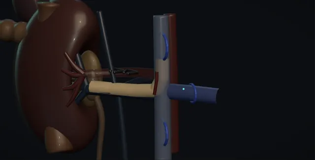
IVC tumour thrombectomy: how the structures relateOpen chevron, right colon and duodenum swept medially, the cava exposed. Inferior vena cava: cranial clamp above the top before handling Tumour thrombus, staged by its top (danger). Inferior vena cava: enters opposite; clamped second, not too long Contralateral renal vein (danger). Inferior vena cava: level III reaches them; liver mobilised Hepatic veins and the liver (danger). Inferior vena cava: enter from behind; every one ligated Lumbar veins (danger). Inferior vena cava: ligated first, before vein or thrombus Renal artery. Inferior vena cava: level IV reaches it; opened on bypass Right atrium.Inferior vena cavaTumour thrombus,staged by its top!Contralateral renalvein!Hepatic veins andthe liver!Lumbar veinsRenal arteryRight atriumcranial clamp abovethe top beforehandlingenters opposite;clamped second, nottoo longlevel III reachesthem; livermobilisedenter from behind;every one ligatedligated first,before vein orthrombuslevel IV reachesit; opened onbypassTargetDangerLandmarkContext
Open chevron, right colon and duodenum swept medially, the cava exposed. Schematic, not to scale; relationships only.
  • The abdomen is open through a chevron with the right colon and duodenum swept medially, and the cava runs up the right of the aorta from the iliac confluence to disappear behind the liver.
  • The right renal vein is short and enters the cava directly with the thrombus continuing up the lumen as a filling defect that expands the vein, its top somewhere between the renal vein and the hepatic veins.
  • The left renal vein crosses in front of the aorta to enter the cava opposite, and the lumbar veins enter the cava from behind;
  • the clamps sit below the renal veins, on the left renal vein, and above the top of the thrombus.
  • ›The inferior vena cava - exposed from the iliac confluence to the liver with the duodenum kocherised; clamped at three points, opened along its anterior wall.
  • ›The tumour thrombus - a tongue of tumour on a stalk from the renal vein, staged by its top: renal vein, less than two centimetres, below the hepatic veins, retrohepatic, above the diaphragm.
  • ›The contralateral renal vein - clamped second so the other kidney does not bleed into the cavotomy and is not lost.
  • ›The hepatic veins and the liver - a level III thrombus reaches them; the liver is mobilised and the porta clamped so the cava can be controlled above.
  • ›The lumbar veins - enter the posterior cava in the clamped segment; every one is ligated or they bleed back into the open vein.
  • ›The renal artery - ligated first, behind the cava on the right, at the aorta on the left, before the vein or the thrombus is handled.
  • ›The right atrium - a level IV thrombus reaches it; opened by the cardiac surgeon on bypass and the thrombus pushed down.
Click me next Section 3 of 8 Steps The operation in order, with the landmark that guards each move.

Operative steps

  1. Who: the tumour that grows up the vein Renal cell carcinoma extends into the renal vein in about a tenth of cases and into the cava in fewer, more often from the right because the right renal vein is short. The thrombus is tumour, not clot, growing along the lumen on a stalk from the primary, sometimes with bland thrombus on top of it and sometimes invading the caval wall. The patient may have leg oedema, a varicocele that does not empty lying down, dilated abdominal wall veins or a pulmonary embolus, or nothing at all. Without metastases complete removal offers a real chance of cure, with five year survival of about half where the thrombus does not invade the wall, and the operation is offered after staging and an MDT.
  2. Staging the thrombus: the levels The cranial extent decides everything and is mapped by MRI or a fine-cut contrast CT within a fortnight of surgery, because thrombus grows. The Mayo classification: level 0 confined to the renal vein; level I in the cava less than two centimetres above the renal vein; level II above that but below the hepatic veins; level III retrohepatic, reaching the hepatic veins but below the diaphragm; level IV above the diaphragm or into the right atrium. TNM makes it T3a for the renal vein, T3b for the cava below the diaphragm and T3c for the cava above the diaphragm or wall invasion. Wall invasion is suspected when the cava is expanded and the thrombus enhances against it; complete occlusion with collaterals means the cava may have to go.
    where it bites: a thrombus staged a month ago is staged wrong; the imaging is repeated close to the date because a level II can become a level III.
  3. The team and the plan Level 0 and I are the urologist's operation with vascular control below the hepatic veins. Level II needs the cava mobilised and the contralateral renal vein and lumbar veins controlled. Level III needs the liver mobilised off the cava and the hepatic veins controlled with a Pringle manoeuvre, so a hepatobiliary or transplant surgeon is in the room. Level IV needs the cardiothoracic team, with cardiopulmonary bypass and often deep hypothermic circulatory arrest, or a veno-venous bypass, and a transoesophageal echo probe to watch the thrombus. Anaesthesia has large-bore access above and below the diaphragm, cross-matched blood and a cell saver, and a plan for the clamp. The consent names death at about five to ten per cent for the high levels.
  4. Exposure: the kidney, the vein and the cava A chevron or midline incision, extended to a sternotomy for level IV. On the right the colon is reflected and the duodenum kocherised widely to expose the cava from the iliac confluence to the liver. The renal artery is found first, on the right behind the cava between it and the aorta, on the left at the aorta, and ligated and divided before the vein is touched, because an artery in continuity keeps the thrombus swollen and the collaterals full. The kidney is mobilised outside Gerota, leaving it attached only by the renal vein and its thrombus. The cava is exposed circumferentially above and below the renal vein and every lumbar vein entering that segment is ligated, because they bleed back into the opened cava.
    where it bites: the artery before the vein; a thrombus handled with the artery intact is a swollen vein that bleeds and a thrombus that breaks off.
  5. Control above and below For a level I or II thrombus the cava is controlled with vessel loops or a Satinsky clamp at three points, applied in an order that keeps the thrombus in place and the other kidney safe: the infrarenal cava first, then the contralateral renal vein, then the cava above the thrombus, its top confirmed by palpation and intraoperative ultrasound, not by the scan alone. Heparin is given before the clamps. For level III the liver is mobilised, the short hepatic veins divided, the porta clamped with a Pringle, and the cava clamped above the hepatic veins below the diaphragm; some thrombi can be milked down below the hepatic veins with the anaesthetist watching. Venous return falls when the cava is clamped; the anaesthetist is warned before each clamp.
    where it bites: a clamp placed below the top of the thrombus, or a thrombus that has broken off as the cava is handled, is an embolus to the heart; the top is found by ultrasound and the echo watches.
  6. The cavotomy and the thrombus With the three clamps on and the field still, the cava is opened longitudinally from the renal vein orifice cranially along the anterior wall as far as the thrombus reaches, so the whole tongue is seen. The thrombus is delivered downward in one piece with the kidney attached, freed from the caval wall with a gentle finger, a swab on a stick or an endarterectomy spatula where it is adherent; a thrombus that will not come away is invading the wall, which is excised with it. The lumen is inspected and irrigated with heparinised saline and any bland thrombus above or below is removed. In a level IV case on bypass the atrium is opened by the cardiac surgeon and the thrombus pushed down from above into the abdominal field.
    where it bites: a thrombus that fragments is a cava full of tumour and a lung full of embolus; it comes out downward, whole, with the kidney, not piecemeal.
  7. Closing the cava The cavotomy is closed with a running 4-0 or 5-0 polypropylene from each end, with the clamps released in reverse order: the cranial clamp is loosened briefly to flush air and debris out through the suture line before the last throws, then the contralateral renal vein, then the infrarenal clamp. If the wall has been excised the defect is closed primarily where the lumen will stay above half its diameter, otherwise it is patched with bovine pericardium, autologous vein or PTFE, or the cava is replaced with a ring-reinforced graft. Where the cava was already occluded and collaterals have formed, the infrarenal cava can be ligated below the contralateral renal vein without oedema. Protamine may be given and the lumbar vein ties checked.
  8. The nephrectomy With the cava closed and the thrombus delivered, the kidney is completed as a radical nephrectomy: the renal vein stump, now oversewn at its orifice as part of the caval closure, the ureter divided and ligated, the remaining posterior attachments taken, and the adrenal removed with the specimen when the tumour is upper pole or the adrenal looked abnormal. The specimen is sent intact with the thrombus attached and orientated for the pathologist, who reports the level, the wall invasion and the margin at the caval surface. Regional nodes are taken where enlarged. Haemostasis is checked with the pressure normal, the liver is inspected after a Pringle, and a drain left beside the cava. The abdomen is closed as a mass closure.
  9. After Critical care with an arterial line and a low threshold for blood: the risks in the first two days are bleeding from the cava or a lumbar vein, acute kidney injury in the remaining kidney from the clamp time and the blood loss, a pulmonary embolus of tumour or clot, liver injury after a Pringle, and, after bypass, coagulopathy and stroke. Thromboprophylaxis starts as soon as the bleeding risk allows, and a patient with a caval graft is anticoagulated. Leg oedema after caval ligation settles with elevation and stockings. The histology at the MDT decides on adjuvant treatment: pembrolizumab for a year is offered for high-risk resected disease. Imaging is six-monthly for two years and then yearly, the cava included, because recurrence can be in the vein.
Click me next Section 4 of 8 Danger zones Where this operation bites, and why it bites there.
Critical relationship

Danger zones

The spatial relationships the animation is built to make automatic. Before you read each one, ask yourself: at this point in the operation, what is about to go wrong, and what is my first move? The save for each sits in the Complications tab.

  • RiskThe top of the thrombus - a clamp below it, or a thrombus handled before the cranial clamp is on, is a tumour embolus to the right heart and the lungs.
  • RiskThe lumbar veins and the posterior cava - torn during mobilisation or left unligated in the clamped segment; they bleed from behind where the clamp does not reach.
  • RiskThe hepatic veins and the retrohepatic cava - the level III thrombus; liver mobilisation and a Pringle are the price of control, and a torn hepatic vein is the bleed that kills.
  • RiskThe contralateral renal vein - left unclamped it fills the cavotomy; clamped too long it costs the only kidney left.
Practise it Take me into the scene Opens the animation at a danger moment and asks you to predict the mechanism and your first move before it reveals.
Click me next Section 5 of 8 Complications The save for each one: the first move, the escalation trigger, the op-note line.

Complications and immediate saves

Trigger each of these live in the Animation tab, then read the save: the first move, the escalation trigger, and the line that goes in the op note.

Massive haemorrhage

  • from a torn lumbar vein, a hepatic vein during liver mobilisation, the cava itself at a clamp or the suture line, or collaterals in a pelvis and retroperitoneum congested by a blocked cava.
The save
  1. Pack and press, tell the anaesthetist the estimate and that the major haemorrhage protocol is now active, and get a second suction and a second pair of senior hands. first move
  2. Proximal and distal control of the cava with clamps or manual compression on the aorta and cava against the spine; a lumbar vein is oversewn from inside the opened cava if it cannot be reached behind.
  3. A hepatic vein or retrohepatic caval tear: total vascular exclusion of the liver with the Pringle on and the cava clamped above and below, and the hepatobiliary surgeon repairs it.
  4. Bleeding that will not stop with the patient cold, acidotic and coagulopathic: pack, close and return in twenty-four hours; the operation is abandoned before the patient is. escalation trigger
  5. Op note: the source, the control, the clamp times, the blood and products given, and whether the abdomen was left packed. document

Tumour embolus and pulmonary embolism

  • sudden hypotension, a falling end-tidal carbon dioxide and a rising right heart on the echo as the cava or the kidney is handled: thrombus has broken off and gone to the lungs.
The save
  1. Stop handling the cava; tell the anaesthetist; the transoesophageal echo looks at the right atrium and pulmonary artery. first move
  2. Supportive resuscitation with the anaesthetist, vasopressors and fluid for the right heart; the cranial clamp is placed above whatever thrombus remains.
  3. Prevention is the operation: the artery ligated first, the top of the thrombus confirmed by ultrasound, the cranial clamp on before the vein is touched, and the thrombus delivered downward whole.
  4. A large embolus with collapse: cardiothoracic help for bypass and pulmonary embolectomy where it can be reached; otherwise the patient is lost on the table. escalation trigger
  5. Op note: the moment of the event, the echo findings, the resuscitation, and the outcome; the family told plainly. document

Caval thrombosis, stenosis and lower body oedema

  • a narrowed or ligated cava, a graft that clots, or a contralateral renal vein compromised by the closure: bilateral leg oedema, a swollen abdominal wall, or a rising creatinine from the remaining kidney.
The save
  1. Duplex ultrasound of the cava, the iliac veins and the remaining renal vein at the first sign of oedema or a creatinine that rises after day two. first move
  2. A caval graft is anticoagulated; a primary closure that has narrowed is anticoagulated and watched; elevation and compression for the legs.
  3. The closure is planned to keep the lumen above half its diameter, or a patch is used; a ligated cava is acceptable only where collaterals were already formed.
  4. A thrombosed cava with a compromised remaining renal vein, or a rising creatinine with oliguria: CT venography, catheter thrombolysis or thrombectomy with vascular surgery, and nephrology for the kidney. escalation trigger
  5. Op note: the closure method, the patch or graft, the estimated residual lumen, and the anticoagulation plan. document

Acute kidney injury in the remaining kidney

  • oliguria and a rising creatinine after the operation: the contralateral renal vein clamped, hypotension and blood loss, contrast, and a kidney that was already borderline.
The save
  1. Keep the contralateral renal vein clamp time as short as possible and the pressure up during it; the anaesthetist is told the clamp is on and when it comes off. first move
  2. Hourly urine output and a creatinine at six hours and daily; fluids and pressure targeted with critical care, nephrotoxins stopped.
  3. Duplex of the remaining kidney if oliguria persists, because a compromised renal vein is treatable and tubular necrosis is not.
  4. Anuria, hyperkalaemia or fluid overload: renal replacement therapy with nephrology, expected to be temporary. escalation trigger
  5. Op note: the clamp times for each clamp, the lowest pressure, and the blood loss, so the nephrologist can read the injury. document

Positive caval margin and tumour spill

  • the pathologist reports tumour at the caval surface, or the thrombus fragmented at delivery and pieces were left in the lumen or the field: the operation that was meant to cure has not.
The save
  1. Recognise wall invasion in the room: a thrombus that will not peel off is taken with the wall, and the lumen is inspected and irrigated before closure. first move
  2. Fragments in the lumen are retrieved before the clamps come off; the field is irrigated, and the specimen is sent orientated with the caval margin inked.
  3. The MDT reviews the margin with the imaging; the cava is imaged early for residual thrombus.
  4. A positive margin or residual thrombus: adjuvant immunotherapy is discussed, and re-resection of the cava only in the fit patient with no other disease. escalation trigger
  5. Op note: whether the thrombus came out whole, the wall findings, what was excised, and the orientation of the specimen for the pathologist. document
Click me next Section 6 of 8 Troubleshooting When the case turns: the problem, then the next move.

Intra-operative troubleshooting

When the case turns: the problem, then the next move.

  • ActionThe top of the thrombus is higher than the scan showed - do not clamp; intraoperative ultrasound to find the true top, mobilise the liver if it is now retrohepatic, and call the hepatobiliary or cardiac team before going further.
  • ActionThe right renal artery cannot be reached behind the cava - approach it between the aorta and the cava from the left side of the cava with the cava retracted laterally, or from below the left renal vein; it is ligated there before the vein is handled.
  • ActionVenous return collapses when the cava is clamped - release the cranial clamp for a moment, fill the patient, and reapply; if the patient cannot tolerate it, a veno-venous bypass or a shorter clamp with the thrombus milked down.
  • ActionThe thrombus will not come away from the wall - it is invading; do not tear it out. Excise the wall with it and plan a patch or a graft, with vascular help if the defect is large.
  • ActionBleeding from inside the opened cava with all clamps on - a lumbar vein or an unclamped tributary: find the orifice from inside and oversew it with 5-0 polypropylene, or pack it with a finger and clamp it from behind.
  • ActionAir or debris in the cava at closure - flush by loosening the cranial clamp briefly before the last throws, aspirate through the suture line, and only then release the other clamps.
Click me next Section 7 of 8 FRCS viva Say it out loud, then mark yourself against the model answer.

FRCS viva questions

The examiner stems this scene prepares you for. Try to answer out loud first, then reveal, and learn the discriminator, the detail that turns a pass into a strong pass.

QHow is caval tumour thrombus classified and why does the level matter?reveal answer
  • The Mayo classification describes the cranial extent of the thrombus: level 0 is confined to the renal vein;
  • level I extends into the cava less than two centimetres above the renal vein;
  • level II extends further but stays below the hepatic veins;
  • level III is retrohepatic, reaching the hepatic veins but remaining below the diaphragm;
  • level IV extends above the diaphragm into the intrathoracic cava or the right atrium.
  • In TNM, the renal vein is T3a, the cava below the diaphragm T3b, and above the diaphragm or wall invasion T3c.
  • The level matters because it dictates the exposure, the vascular control and the team.
  • Level 0 and I need only local caval control below the liver.
  • Level II requires full mobilisation of the cava with the contralateral renal vein and lumbar veins controlled.
  • Level III requires the liver mobilised off the cava, the short hepatic veins divided and a Pringle manoeuvre so the cava can be clamped above the hepatic veins, and a hepatobiliary or transplant surgeon.
  • Level IV requires a cardiothoracic team with cardiopulmonary bypass and often deep hypothermic circulatory arrest.
  • The level also predicts blood loss and mortality, about five to ten per cent at the higher levels.
  • The imaging must be repeated within a fortnight of surgery because the level changes.
Strong pass

Say both

  • The five levels with their anatomical boundaries and the team each one summons
  • The imaging repeated close to the date because thrombus grows
Examiner note
The five levels with their anatomical boundaries and the team each one summons, and the imaging repeated close to the date because thrombus grows.
QDescribe the sequence of vascular control for a level II thrombus and explain the order.reveal answer
  • I would first ligate the renal artery, on the right by approaching it between the aorta and the cava, before the vein or the thrombus is touched, because an artery in continuity keeps the thrombus turgid and the collaterals full.
  • I would mobilise the kidney so it hangs on the renal vein alone, expose the cava from the iliac confluence to the liver with the duodenum kocherised, and ligate every lumbar vein entering the segment to be clamped, because they bleed back into an opened cava.
  • I would confirm the top of the thrombus by palpation and intraoperative ultrasound.
  • After heparin, I would apply the clamps in this order: the infrarenal cava first, so that nothing from below can push the thrombus up;
  • then the contralateral renal vein, so that the other kidney does not bleed into the cavotomy;
  • then the cava above the thrombus, placed with a margin above the top confirmed by ultrasound, warning the anaesthetist that venous return will fall.
  • I would then open the cava along its anterior wall from the renal vein orifice cranially, deliver the thrombus downward whole with the kidney, inspect and irrigate the lumen, close with running polypropylene, flush air out by briefly releasing the cranial clamp before the last throws, and release the clamps in reverse order.
Strong pass

Say all three

  • The artery before anything else with the reason
  • The cranial clamp placed on an ultrasound-confirmed top
  • The clamps released in reverse with the flush before the last throws
Examiner note
The artery before anything else with the reason, the cranial clamp placed on an ultrasound-confirmed top, and the clamps released in reverse with the flush before the last throws.
QThe blood pressure falls suddenly while you are mobilising the cava. What has happened and what do you do?reveal answer
  • The diagnosis until proved otherwise is tumour embolus: a fragment of thrombus has broken off and travelled to the right heart and pulmonary arteries, and the other possibilities are a major venous bleed I have not yet seen, or a fall in venous return from caval compression.
  • I would stop handling the cava, look at the field for bleeding, and ask the anaesthetist what the end-tidal carbon dioxide, the airway pressures and the filling look like, and ask for the transoesophageal echo to be looked at, which shows thrombus in the atrium or pulmonary artery and a dilated, failing right ventricle.
  • If it is an embolus, the anaesthetist resuscitates with vasopressors and fluid for the right heart, and if the patient stabilises I would place the cranial caval clamp above whatever thrombus remains before doing anything else, so that no more can go, and continue.
  • If the patient does not stabilise, the only definitive treatment is cardiothoracic: bypass and pulmonary embolectomy, and without that team the patient will probably die on the table.
  • This is why prevention is written into the operation: the artery is ligated first, the top of the thrombus is confirmed by ultrasound, the cranial clamp goes on before the vein is handled, and the echo probe is in for anything above level I.
Strong pass

Say both

  • The end-tidal carbon dioxide and the echo as the way to confirm it
  • The cranial clamp placed above the remaining thrombus as the first surgical move once the patient is stable
Examiner note
The end-tidal carbon dioxide and the echo as the way to confirm it, and the cranial clamp placed above the remaining thrombus as the first surgical move once the patient is stable.
QWhat are the postoperative complications of caval thrombectomy and how are they followed?reveal answer
  • The early complications are bleeding, from the caval suture line, a lumbar vein or the retroperitoneal collaterals, managed with a low threshold for re-exploration;
  • acute kidney injury in the remaining kidney from the renal vein clamp, the hypotension and the blood loss, managed with critical care support and renal replacement if needed;
  • pulmonary embolism of tumour or bland thrombus, which can occur postoperatively as well as on the table;
  • caval thrombosis or stenosis after a narrowed closure, a patch or a graft, presenting with leg oedema or a rising creatinine if the remaining renal vein is involved, managed with duplex, anticoagulation and vascular intervention;
  • liver dysfunction after a Pringle;
  • and, after bypass, coagulopathy, stroke and a prolonged critical care stay.
  • Mortality is about five to ten per cent for the higher levels.
  • Thromboprophylaxis starts as soon as it is safe and a caval graft is anticoagulated.
  • Late problems are chronic lower body oedema after ligation and recurrence in the cava, the retroperitoneum or at distant sites.
  • Follow-up is by the histology: patients with T3 disease are at intermediate to high risk and are offered adjuvant pembrolizumab for a year, with CT of the chest, abdomen and pelvis at six months for two years and then yearly, the cava reviewed on each scan.
Strong pass

Say both

  • Naming the remaining kidney's vein as the structure at risk in caval thrombosis
  • The adjuvant immunotherapy discussion as part of the follow-up for T3 disease
Examiner note
Naming the remaining kidney's vein as the structure at risk in caval thrombosis, and the adjuvant immunotherapy discussion as part of the follow-up for T3 disease.
Click me next Section 8 of 8 Evidence The guidance each of these answers is built on.

Evidence and guidance

Two minutes

Close the lap: recall it before you leave.

Say it aloud, then reveal. Reading is not remembering.

  1. 01 The relationship Say the critical spatial relationship out loud, then reveal. reveal
    Memory frame

    The abdomen is open through a chevron with the right colon and duodenum swept medially, and the cava runs up the right of the aorta from the iliac confluence to disappear behind the liver.

    Why?
    The right renal vein is short and enters the cava directly with the thrombus continuing up the lumen as a filling defect that expands the vein, its top somewhere between the renal vein and the hepatic veins. The left renal vein crosses in front of the aorta to enter the cava opposite, and the lumbar veins enter the cava from behind; the clamps sit below the renal veins, on the left renal vein, and above the top of the thrombus.
  2. 02 The first move Massive haemorrhage. Your first move? reveal
    First move

    Pack and press, tell the anaesthetist the estimate and that the major haemorrhage protocol is now active, and get a second suction and a second pair of senior hands.

  3. 03 The escalation trigger Massive haemorrhage. What makes you call for help? reveal
    Escalate when

    Bleeding that will not stop with the patient cold, acidotic and coagulopathic

    More detail
    Bleeding that will not stop with the patient cold, acidotic and coagulopathic: pack, close and return in twenty-four hours; the operation is abandoned before the patient is.
  4. 04 The discriminator What turns a pass into a strong pass? reveal
    Strong pass

    Say both

    • The five levels with their anatomical boundaries and the team each one summons
    • The imaging repeated close to the date because thrombus grows
    Examiner note
    The five levels with their anatomical boundaries and the team each one summons, and the imaging repeated close to the date because thrombus grows.
  5. 05 The pitfall Where does this operation bite first? reveal
    Pitfall

    The top of the thrombus

    Why?
    a clamp below it, or a thrombus handled before the cranial clamp is on, is a tumour embolus to the right heart and the lungs.

Revision practice with self-marked answers, not an assessment of clinical competence. Kept in this browser only; it does not sync between devices.