
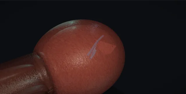
UrOops3D, danger-zone atlas
Operative animation, Retrograde and JJ stent

Retrograde pyelogram and JJ stent insertion

The night-time operation of urology: a kidney obstructed by a stone, a ureter injured by someone else, or a system that has to be kept open, drained from below with a wire and a stent through an orifice the size of a grain of rice.

This page is the decision, the orifice, the retrograde done gently, the wire that must curl, the stent that must be the right length, and the register that stops it being forgotten.

Core tier FRCS core urology, stones and upper-tract drainage Interactive 3D

Operative visualisation

Schematic scene preview: Retrograde pyelogram and JJ stent
Preparing the 3D anatomy…

UrOops3D · UroRef · © Nity G

Click me next Section 2 of 8 Anatomy The structures this operation works between, and the one it must not cross.

The structures in the scene

Retrograde and JJ stent: how the structures relateFrom the cystoscope and the screening monitor, orifice to renal pelvis. Trigone and the ureteric orifice: thick band into the orifice, golden pusher tip seen Stent and pusher. Trigone and the ureteric orifice: pushed against resistance, the wire leaves Intramural ureter (danger). Intramural ureter: ureter runs up to cross the vessels Crossing of the iliac vessels. Crossing of the iliac vessels: past the kink where a wire buckles Renal pelvis. Renal pelvis: curls here to prove it is up The wire, soft tip first.BladderKidneyTrigone and the uretericorificeStent and pusher!Intramural ureterCrossing of the iliacvesselsRenal pelvisThe wire, softtip firstthick band into theorifice, goldenpusher tip seenpushed against resistance, thewire leavesureter runs up to cross thevesselspast the kink where a wirebucklescurls here to proveit is upTargetDangerLandmarkContext
From the cystoscope and the screening monitor, orifice to renal pelvis. Schematic, not to scale; relationships only.
  • From the cystoscope the orifice is a slit at the far corner of the trigone, and everything beyond it is on the screening monitor: the intramural tunnel, the ureter running up to cross the iliac vessels, and the renal pelvis where the wire must curl.
  • The stent is right when the thick band has gone into the orifice, the golden tip of the pusher has come into view from the bladder neck, and both curls are seen, one in the pelvis on the screen and one in the bladder through the lens.
  • ›Trigone and the ureteric orifice - a slit at the lateral corner that opens with each jet; centred in the view before anything goes in.
  • ›Intramural ureter - the oblique tunnel through the bladder wall: the first place a wire meets resistance and the first place it can leave the lumen.
  • ›The crossing of the iliac vessels - the kink where a wire buckles and a stiff stent catches.
  • ›Renal pelvis - where the wire curls to prove it is up and where the proximal curl of the stent sits.
  • ›The ureteric catheter - 5 French, open-ended: the retrograde, and support for a wire past an obstruction.
  • ›The wire - 0.035 inch, soft tip first; hydrophilic to pass, stiff to work over.
  • ›The stent and pusher - 6 French, 24 or 26 cm; four thin dashes then a thick band mark the distal end, and the golden tip of the pusher says stop. The pusher holds it while the wire comes out.
Click me next Section 3 of 8 Steps The operation in order, with the landmark that guards each move.

Operative steps

  1. Why a stent, and why tonight The indications: an obstructed kidney with a stone, pain that will not settle or a single kidney; a ureteric injury to splint; a ureter to keep open before or after ureteroscopy, and the pre-stenting that makes a later ureteroscopy easier. The obstructed infected kidney is the emergency: fever, a raised white cell count or lactate, and an obstruction on imaging. It needs draining tonight, and the choice between a retrograde stent and a nephrostomy is made with the radiologist and the anaesthetist according to the patient, the stone and who is available, not by one person alone. Antibiotics go in before any instrument.
  2. Consent and the trolley The consent names stent symptoms in most people: frequency, urgency, discomfort in the loin and bladder, blood in the urine; infection; failure to pass the stone or the stricture, in which case a nephrostomy; and the need to remove or change the stent by a date. On the trolley: a rigid cystoscope with a bridge, a 5 French open-ended ureteric catheter, dilute contrast in a syringe, a 0.035 inch guidewire with a soft tip and a hydrophilic one, a 6 French JJ stent of 24 or 26 centimetres with its pusher, and the image intensifier with the screen where the surgeon can see it without turning.
  3. Cystoscopy: the orifice The bladder is surveyed, then the trigone found on the floor with an orifice at each corner. The orifice on the side to be stented is a slit at the lateral corner that opens with each jet of urine, and the scope is turned so that it sits in the centre of the view. In an oedematous or bleeding bladder, or after radiotherapy, the orifice can be hard to find: the jet, the interureteric ridge and a few minutes of patience find it. A screening image of the pelvis with the scope in confirms the level. A lateral orifice needs the scope turned, not pushed: on the right, the 12 o'clock arrow is turned to face 3 o'clock and the catheter or wire then leaves the channel angled towards 9 o'clock, onto the orifice; on the left, the arrow to 9 o'clock and the wire comes out towards 3.
    where it bites: a wire pushed into a fold of the trigone next to the orifice makes a false passage; the orifice is seen and centred before anything goes into it.
  4. The retrograde pyelogram The ureteric catheter goes through the bridge and its tip is brought up to the orifice. If it does not go up at once, a guidewire is passed through the catheter with its soft tip showing just beyond the end, the wire tip is led into the orifice, and the catheter follows over it a centimetre or two; the wire then comes out for the retrograde. Dilute contrast is injected gently under screening and outlines the ureter, the stone, the stricture, the leak or the filling defect, and shows the pelvicalyceal system and where the stent has to reach. A few millilitres, slowly; over-pressure refluxes contrast into the renal parenchyma and the veins, and a rigor follows. In an infected system the retrograde is minimal or omitted and the wire goes up with drainage as the only aim.
  5. The guidewire The 0.035 inch wire goes up the ureteric catheter soft tip first and on under screening. It should pass easily; resistance is a stone, a stricture, a kink at the crossing of the iliac vessels or a false passage, and the answer is not force. When the sensor wire will not pass a stone: nudge the ureteric catheter up to just below the stone, because its support often lets the wire through; if not, inject a little contrast to see whether the stone is blocking; if no contrast passes, a Terumo fully hydrophilic wire will sometimes go past. Once a Terumo is up, follow it with the ureteric catheter and the hydrophilic wire is exchanged for a stiffer one, a sensor wire, because it is hard to stent over a Terumo and it slips out. If nothing passes, call the consultant, who will usually fragment the stone with the semirigid ureteroscope so that a wire can pass. On the screen the wire curls in the renal pelvis: it is up, and the ureteric catheter is withdrawn over the wire. A wire that stops and buckles has met something and is not pushed.
    where it bites: a wire forced past an impacted stone perforates the ureter; the wire curling in the renal pelvis is the only proof that it is up.
  6. The stent over the wire The length comes from the screening image or the patient's height: 24 centimetres for most, 26 for the tall, shorter for the small. A 6 French stent is threaded over the wire and fed in with its pusher under screening. The stent carries four thin black dashes and then a thick black band near its distal end. Watch the dashes pass into the orifice one after another; when the thick band is going into the orifice, withdraw the scope to the bladder neck and push until the golden tip of the pusher comes into view, then stop. Ask for the guidewire to be removed completely: it is withdrawn while the pusher holds the stent still, the proximal curl forms in the renal pelvis and the stent's own distal end curls in the bladder. Pulling the wire without the pusher pulls the stent down with it.
  7. The check The distal curl is seen lying free in the bladder, the stent's own end and not a separate loop, not stuck in the orifice and not in the urethra, and the screen shows a full proximal curl in the renal pelvis. A stent whose whole length has gone up the ureter has no distal curl and is retrieved now, while the wire can be re-passed through it. The bladder is emptied through the sheath. A catheter overnight is left only for infection, bleeding or a patient who will not void; most go home the same day.
  8. The note and the register The note records the side, the length and French, the wire used, the screening findings including the level and nature of the obstruction, the ease or difficulty of passage, and the date the stent must come out or be changed. The patient goes onto the stent register before leaving theatre, with the date, and is told in words and on the discharge letter that there is a stent inside that has to be removed. A stent nobody remembers is the complication that comes back years later, encrusted at both ends.
Click me next Section 4 of 8 Danger zones Where this operation bites, and why it bites there.
Critical relationship

Danger zones

The spatial relationships the animation is built to make automatic. Before you read each one, ask yourself: at this point in the operation, what is about to go wrong, and what is my first move? The save for each sits in the Complications tab.

  • RiskThe intramural ureter and the fold beside the orifice - the wire leaves the lumen here if it is pushed against resistance.
  • RiskThe impacted stone - a wire forced past it perforates; the answer is a nephrostomy, not push.
  • RiskThe infected system - contrast under pressure into it produces a rigor and a falling blood pressure.
  • RiskThe stent nobody records - encrusts and obstructs the kidney it was placed to save.
Practise it Take me into the scene Opens the animation at a danger moment and asks you to predict the mechanism and your first move before it reveals.
Click me next Section 5 of 8 Complications The save for each one: the first move, the escalation trigger, the op-note line.

Complications and immediate saves

Trigger each of these live in the Animation tab, then read the save: the first move, the escalation trigger, and the line that goes in the op note.

Wire perforation and false passage

  • the wire outside the line of the ureter on the screen, will not curl, blood from the orifice.
The save
  1. Stop advancing. Withdraw the wire into the bladder. first move
  2. Re-enter the orifice with a fresh soft-tipped wire through the ureteric catheter under the retrograde image; the true lumen is usually found.
  3. If it is found, the stent goes in and splints the injury for four to six weeks.
  4. If it is not found, or the perforation is large: nephrostomy, and an antegrade attempt later. escalation trigger
  5. Note: the level, the image, whether the lumen was regained, the stent or nephrostomy, and the date of review. document

The wire will not pass

  • a stone the sensor wire will not pass, even with the catheter for support.
The save
  1. Do not force it. Nudge the ureteric catheter up to just below the stone; its support often lets the wire through. first move
  2. Inject a little contrast through the catheter to see whether the stone is blocking the lumen.
  3. If no contrast passes, a Terumo fully hydrophilic wire will sometimes go past. Follow it with the ureteric catheter and exchange to a sensor wire; it is hard to stent over a Terumo.
  4. Still no wire: call the consultant, who will usually fragment the stone with the semirigid ureteroscope so a wire can pass. An obstructed infected kidney that cannot be stented is drained by nephrostomy tonight. escalation trigger
  5. Note: the level reached, each wire tried, the contrast finding, and the drainage decision. document

Stent too short, too long or too high

  • no distal curl in the bladder, or a distal end in the urethra, or the whole stent up the ureter.
The save
  1. Recognise it on the screen and through the lens before the scope comes out: two curls, one in each place. first move
  2. A stent that has gone up: re-pass the wire through it and pull it down with graspers, or exchange it for the right length.
  3. A distal end in the urethra: the stent is too long and is exchanged.
  4. A migrated stent found later needs ureteroscopy to retrieve. escalation trigger
  5. Note: the length placed, both curls seen, and the check image. document

Sepsis after the retrograde

  • contrast under pressure into an infected system: rigors, tachycardia and a falling blood pressure within the hour.
The save
  1. Sepsis six with the anaesthetist: cultures, antibiotics, fluids, oxygen, lactate. first move
  2. Make sure the kidney is draining: the stent in, or a nephrostomy if it is not.
  3. A patient who does not respond goes to critical care. escalation trigger
  4. Prevention: antibiotics before the scope and a few millilitres of contrast, slowly, or none at all in an infected system.
  5. Note: the timing, the contrast volume, the drainage, and the antibiotics. document

The forgotten stent

  • a stent left in for years, encrusted at both ends, obstructing the kidney and seeding infection.
The save
  1. The register, the date on the discharge letter, and the patient told in words are what prevent it. first move
  2. When it is found: imaging of the stone burden at each end, and a staged plan of cystolitholapaxy, ureteroscopy and percutaneous surgery as needed.
  3. An infected obstructed kidney behind a forgotten stent is drained by nephrostomy first. escalation trigger
  4. Note: the date placed, the burden found, the plan, and the register corrected. document
Click me next Section 6 of 8 Troubleshooting When the case turns: the problem, then the next move.

Intra-operative troubleshooting

When the case turns: the problem, then the next move.

  • ActionCannot find the orifice - wait for the jet, follow the interureteric ridge, empty and refill the bladder; intravenous indigo carmine or methylene blue as a last resort.
  • ActionThe wire stops in the intramural ureter - the angle: the ureteric catheter up to the orifice first, the soft wire tip just beyond its end to lead it in, the scope turned so the wire leaves the channel towards the orifice.
  • ActionA lateral orifice the catheter will not line up with - turn the scope, do not push: right side, 12 o'clock arrow to 3 o'clock and the wire angles towards 9; left side, arrow to 9 and the wire angles towards 3.
  • ActionThe wire will not pass the stone - catheter up to just below the stone, a little contrast, then a Terumo; follow it with the catheter and exchange to a sensor wire; then the consultant and the semirigid scope.
  • ActionThe wire stops at the pelvic brim - the iliac crossing: a Terumo hydrophilic wire with the catheter alongside, gently; screen to see whether it buckles.
  • ActionThe wire keeps coming back - a hydrophilic wire slips: exchange it for a stiffer wire through the ureteric catheter before the stent.
  • ActionThe stent will not advance over the wire - a kink or a tight stricture: straighten with a stiffer wire, or place a ureteric catheter for drainage and stent later.
  • ActionThe distal curl went up with the wire - the pusher was not holding: re-pass the wire through the stent and reposition, or retrieve and replace.
Click me next Section 7 of 8 FRCS viva Say it out loud, then mark yourself against the model answer.

FRCS viva questions

The examiner stems this scene prepares you for. Try to answer out loud first, then reveal, and learn the discriminator, the detail that turns a pass into a strong pass.

QA patient presents with a 7 mm upper ureteric stone, fever and a raised lactate. How do you manage them?reveal answer
  • This is an obstructed infected kidney and a urological emergency: the priority is resuscitation and decompression of the kidney, not the stone.
  • I would start sepsis management, with blood and urine cultures, broad-spectrum intravenous antibiotics according to local policy, fluids, oxygen and monitoring, and check renal function and clotting.
  • The kidney needs draining as soon as possible, tonight, by either a retrograde JJ stent or a percutaneous nephrostomy.
  • The choice depends on the patient and the resources: a nephrostomy avoids a general anaesthetic and manipulation of the infected ureter and gives a monitorable drain, and is preferred in the unstable patient or with a large stone;
  • a stent avoids an external tube and can be done in theatre where a nephrostomy service is unavailable.
  • I would make the decision with the radiologist and the anaesthetist.
  • If stenting, I would use antibiotics first, minimal or no contrast, and a wire and stent with drainage as the sole aim.
  • Definitive stone treatment follows once the infection has settled, usually after two weeks.
Strong pass

Say all three

  • Decompression tonight with the choice made with radiology and anaesthetics
  • Minimal contrast in the infected system
  • The stone left for later
Examiner note
Decompression tonight with the choice made with radiology and anaesthetics, minimal contrast in the infected system, and the stone left for later.
QTalk me through inserting a JJ stent.reveal answer
  • With antibiotics given and the image intensifier positioned, I would perform a rigid cystoscopy, survey the bladder and centre the ureteric orifice on the side to be treated.
  • I would pass a 5 French open-ended ureteric catheter a centimetre into the orifice and perform a gentle retrograde pyelogram with dilute contrast to define the anatomy and the obstruction.
  • I would pass a 0.035 inch guidewire soft tip first through the catheter and up the ureter under screening until it curls in the renal pelvis, using a hydrophilic wire with the catheter alongside if there is resistance and exchanging it for a stiffer wire afterwards.
  • I would choose the stent length from the image or the patient's height, usually 24 or 26 centimetres and 6 French, and feed it over the wire with the pusher, watching the four thin dashes and then the thick band pass into the orifice, withdraw the scope to the bladder neck and push until the golden tip of the pusher comes into view.
  • Holding the pusher, I would have the wire removed completely so the proximal curl forms in the renal pelvis and the stent's own distal end curls in the bladder.
  • I would confirm both curls, empty the bladder, document the details and enter the stent on the register with a removal date.
Strong pass

The wire curling in the renal pelvis as the proof, the marks and the golden tip, the pusher holding while the wire comes out, both curls confirmed, and the register.

QWhat are the complications of ureteric stents and how do you counsel a patient about them?reveal answer
  • Almost everyone has stent symptoms: frequency, urgency, dysuria, haematuria and discomfort in the loin or bladder, especially on voiding, from the distal curl irritating the trigone and reflux up the stent.
  • Alpha-blockers and anticholinergics reduce them, and a shorter stent with the distal curl clear of the trigone helps.
  • Infection can occur, and encrustation increases with time, particularly in stone formers and pregnancy, so the stent has a defined lifespan.
  • Migration up or down, fracture and, above all, the forgotten stent are the device-related complications, prevented by the register, a removal date on the discharge letter and telling the patient in words.
  • Procedural complications are false passage and perforation, sepsis from a pressurised retrograde in an infected system, and failure to pass.
  • I would counsel that the stent is temporary, that symptoms are common and treatable, that they must attend for removal on the date given, and that fever means coming back.
Strong pass

Say both

  • Stent symptoms as near-universal with their treatment
  • The forgotten stent named with the register and removal date as the prevention
Examiner note
Stent symptoms as near-universal with their treatment, and the forgotten stent named with the register and removal date as the prevention.
QDuring a retrograde the wire will not pass an impacted stone at the pelvic brim. What do you do?reveal answer
  • I would not force it: a wire forced past an impacted stone perforates the ureter and can push the stone through the wall.
  • I would nudge the open-ended ureteric catheter up to just below the stone, because its support often lets the sensor wire through, inject a little contrast to see whether the stone is blocking, and if nothing passes try a Terumo hydrophilic wire, following it with the catheter and exchanging to a sensor wire because it is hard to stent over a Terumo.
  • If the wire still will not pass I would call the consultant, who would usually fragment the stone with the semirigid ureteroscope so a wire can pass, and the next step depends on whether the kidney is infected.
  • An infected obstructed kidney is drained by a percutaneous nephrostomy that night, with antegrade treatment or a later ureteroscopy once the infection has settled.
  • An uninfected kidney with tolerable pain can have a planned semi-rigid ureteroscopy, where the stone is seen and either the wire is passed under vision or the stone is fragmented in situ;
  • a ureteric catheter can be left for drainage in the meantime if the retrograde suggested obstruction.
  • I would document the level reached and the reason for stopping and would speak to the patient about the change of plan.
Strong pass

Catheter support, contrast, Terumo then exchange, then the consultant and the semirigid scope, with the infected kidney drained by nephrostomy if none of it passes.

Click me next Section 8 of 8 Evidence The guidance each of these answers is built on.

Evidence and guidance

Two minutes

Close the lap: recall it before you leave.

Say it aloud, then reveal. Reading is not remembering.

  1. 01 The relationship Say the critical spatial relationship out loud, then reveal. reveal
    Memory frame

    From the cystoscope the orifice is a slit at the far corner of the trigone, and everything beyond it is on the screening monitor: the intramural tunnel, the ureter running up to cross the iliac vessels, and the renal pelvis where the wire must curl.

    Why?
    The stent is right when the thick band has gone into the orifice, the golden tip of the pusher has come into view from the bladder neck, and both curls are seen, one in the pelvis on the screen and one in the bladder through the lens.
  2. 02 The first move Wire perforation and false passage. Your first move? reveal
    First move

    Stop advancing.

    More detail
    Withdraw the wire into the bladder.
  3. 03 The escalation trigger Wire perforation and false passage. What makes you call for help? reveal
    Escalate when

    If it is not found, or the perforation is large: nephrostomy, and an antegrade attempt later.

  4. 04 The discriminator What turns a pass into a strong pass? reveal
    Strong pass

    Say all three

    • Decompression tonight with the choice made with radiology and anaesthetics
    • Minimal contrast in the infected system
    • The stone left for later
    Examiner note
    Decompression tonight with the choice made with radiology and anaesthetics, minimal contrast in the infected system, and the stone left for later.
  5. 05 The pitfall Where does this operation bite first? reveal
    Pitfall

    The intramural ureter and the fold beside the orifice

    Why?
    the wire leaves the lumen here if it is pushed against resistance.

Revision practice with self-marked answers, not an assessment of clinical competence. Kept in this browser only; it does not sync between devices.