UrOops3D, danger-zone atlas

Schematic model.

Operative animation, Male urethral sling

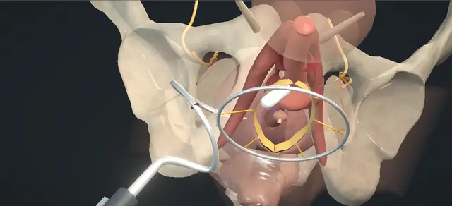
Male urethral sling: a transobturator sling under the bulb

A fixed transobturator sling for mild to moderate stress incontinence after radical prostatectomy. Through a midline perineal incision the bulb is exposed and the central tendon divided.

A helical passer is taken from each groin crease, round the ischiopubic ramus and through the obturator foramen, and each sling arm is drawn back out along its track. The body is fixed on the ventral bulb and the arms are tensioned, which displaces the urethra proximally.

The lesson is the passer's route, where the sling lies, and how far moving the bulb is from obstructing it.

Advanced tier FRCS continence, post-prostatectomy incontinence, functional urology Interactive 3D

Operative visualisation

Schematic scene preview: Male urethral sling
Preparing the 3D anatomy…

UrOops3D · UroRef · © Nity G

How to use this module. Play through the ten steps, or use Back and Next. Watch each passer turn round the ramus and through the foramen, the arms drawn out, the body fixed at the mark and the bulb move proximally as the arms are pulled. The triggers show retention, persistent leakage and erosion.

Click me next Section 2 of 8 Anatomy The structures this operation works between, and the one it must not cross.

The structures in the scene

Male urethral sling: how the structures relateThe bulb of the urethra, what the sling passes round, and what lies beside its track. Bulb of the urethra: carries the sling body: lies between the two rami, below them Ischiopubic ramus: the passer goes round it. Bulb of the urethra: carries the sling body: arms run from its sides up through each foramen Obturator foramen: each arm passes through. Bulb of the urethra: carries the sling body: far from it: the passer crosses low, on the ramus Obturator nerve and vessels, top of the foramen (danger). Bulb of the urethra: carries the sling body: the passer tip comes out between the two Crus of the corpus cavernosum (danger). Bulb of the urethra: carries the sling body: held by it; freed when it is incised Central tendon: incised, its level marked. Bulb of the urethra: carries the sling body: drawn toward it as the arms are tensioned Membranous urethra, proximal to the bulb.Bulb of the urethra:carries the sling bodyIschiopubic ramus:the passer goesround itObturator foramen:each arm passesthrough!Obturator nerve andvessels, top of theforamen!Crus of the corpuscavernosumCentral tendon:incised, its levelmarkedMembranous urethra,proximal to the bulblies between thetwo rami, belowthemarms run from itssides up througheach foramenfar from it: thepasser crosses low,on the ramusthe passer tipcomes out betweenthe twoheld by it; freedwhen it is inciseddrawn toward it asthe arms aretensionedTargetDangerLandmarkContext
The bulb of the urethra, what the sling passes round, and what lies beside its track. Schematic, not to scale; relationships only.
  • From the foot of the table in lithotomy, the bulb lies in the midline under bulbospongiosus, between the crura of the corpora cavernosa.
  • Each crus runs along an ischiopubic ramus, and the obturator foramen lies above and lateral to the ramus, closed by its membrane.
  • The obturator nerve and vessels leave at the upper lateral corner of the foramen.
  • The passer enters at the groin crease, goes round the ramus through the lower part of the foramen and comes out between the bulb and the crus.
  • Tension on the two arms draws the bulb proximally, toward the membranous urethra.
  • ›Bulb of the urethra - the proximal bulbar urethra is what the sling supports and displaces; the body lies on its ventral surface.
  • ›Corpus spongiosum - the four corners are fixed to its superficial part, and it is inspected for injury before closure.
  • ›Bulbospongiosus - divided in the midline, dissected off the spongiosum, and approximated over the sling at the end.
  • ›Central tendon - holds the proximal bulb; incised to mobilise it, and its level is marked for the proximal edge of the sling.
  • ›Membranous urethra - above the bulb; the cystoscope is held at the bulbomembranous junction while the arms are lifted.
  • ›Ischiopubic ramus - the passer is guided lateral to it and round it.
  • ›Obturator foramen - each arm passes through it; the passer crosses it low, beside the ramus.
  • ›Obturator nerve and vessels - leave through the obturator canal at the upper lateral corner of the foramen.
  • ›Crus of the corpus cavernosum - runs along the ramus; the passer comes out medial to its medial border.
  • ›Adductor longus tendon - the landmark for the groin stab incision, which lies below it in the crease.
  • ›Colles fascia - divided with the subcutaneous tissue on the way in and closed as a layer of its own on the way out.
Click me next Section 3 of 8 Steps The operation in order, with the landmark that guards each move.

Operative steps

  1. The urethra after prostatectomy A man has mild to moderate stress urinary incontinence after radical prostatectomy. The bladder neck is joined to the urethra; the membranous urethra lies below the anastomosis and the bulb below that. Intravenous antibiotics are given. He is placed in high dorsal lithotomy on well-padded stirrups, and the genitalia, the perineum, the anteromedial thighs and the lower abdomen are prepared. A urethral catheter is advanced into the bladder.
    where it bites: the thighs are prepared with the perineum: both groin creases are part of the operative field.
  2. Perineal exposure A vertical midline perineal incision is made below the scrotum and carried through Colles fascia and the subcutaneous tissue to bulbospongiosus with diathermy. The muscle is divided sharply in the midline and carefully dissected off the underlying corpus spongiosum. A self-retaining retractor is positioned.
    where it bites: the spongiosum lies directly under the muscle; the muscle is taken off it without cutting into it.
  3. Central tendon The central tendon is identified in the midline and incised sharply, which mobilises the proximal bulbar urethra. A sterile marking pen marks this level on the bulb for the later placement of the sling.
    where it bites: the rectum lies behind the perineal body; the incision divides the tendon and goes no deeper.
  4. Passer round the ramus A small stab incision is made with a blade in the groin crease, below the adductor longus tendon, and a spinal needle is used to identify the obturator foramen. The helical needle passer is inserted outside-in through the stab incision along the needle's trajectory and guided lateral to the ischiopubic ramus into the obturator foramen. With the bulbar urethra retracted medially, the fascia is perforated and the passer is guided into the perineal wound between the lateral edge of the corpus spongiosum and the medial border of the corpus cavernosum.
    where it bites: the obturator nerve and vessels leave the foramen at its upper lateral corner; the passer keeps to the ramus, and the bulb is held clear of the tip.
  5. Sling arm drawn through The polypropylene sling, which has been bathed in antiseptic or antibiotic solution, is brought into the perineal field and connected to the tip of the passer. Using reverse rotation, the sling is pulled through the obturator foramen and brought out loosely through the groin incision. The connector and the distal sling are cut above the skin and the end of the sling is grasped with a haemostat.
    where it bites: a twist in the arm is looked for now, while the arm can still be drawn back and laid flat.
  6. The left side The identical procedure is repeated on the other side: the stab incision, the spinal needle, the passer round the ramus, and the arm drawn out through the groin. The centre of the mesh is positioned at the urethral midline.
    where it bites: a mesh that sits off centre pulls the bulb to one side when the arms are tensioned.
  7. Fixation on the bulb The sling is laid flat on the ventral surface of the bulbar urethra with its proximal edge at the marked site. Its four corners are fixed to the superficial corpus spongiosum with absorbable sutures.
    where it bites: each bite stays in the outer spongiosum; a deep bite reaches the urethral lumen.
  8. Tension Both arms of the sling are pulled tight, which displaces the urethra proximally and evenly.
    where it bites: the pull is equal on the two sides, and the next step checks that the urethra has been moved and is still open.
  9. Cystoscopy The urethral catheter is removed and a flexible cystoscope is advanced to the bulbomembranous junction. Both arms of the sling are elevated under direct cystoscopic vision, which confirms adequate proximal bulbar mobility without urethral obstruction. The bladder is seen to be intact, with clear efflux from both ureteric orifices. The cystoscope is removed and the urethral catheter replaced.
    where it bites: a lumen that will not admit the scope with the arms lifted is obstructed, and that is dealt with before the wound is closed.
  10. Closure The wound is irrigated with antibiotic solution or saline. The retractor is removed and the perineum inspected, confirming correct placement of the sling without injury to the corpus spongiosum. Bulbospongiosus is approximated with a running absorbable suture; the subcutaneous tissue and Colles fascia are closed as two further running layers, separately, to obliterate dead space, and the skin with interrupted sutures. The groin incisions are closed with a subcuticular suture and adhesive strips. The perineum is dressed, a scrotal support is applied and the catheter is connected to a drainage bag.
    where it bites: a space left under the skin over a mesh implant is where infection starts; each layer is closed on its own.
Click me next Section 4 of 8 Danger zones Where this operation bites, and why it bites there.
Critical relationship

Danger zones

The spatial relationships the animation is built to make automatic. Before you read each one, ask yourself: at this point in the operation, what is about to go wrong, and what is my first move? The save for each sits in the Complications tab.

  • RiskObturator nerve and vessels - in the canal at the upper lateral corner of the foramen; the passer crosses low, against the ramus.
  • RiskUrethra and corpus spongiosum - beside the passer tip and under every corner stitch; the bulb is retracted for the passage and the bites stay shallow.
  • RiskCorpus cavernosum - lateral to the exit point; the tip comes out medial to its medial border.
  • RiskRectum - behind the central tendon; the tendon is incised and the dissection goes no deeper.
  • RiskTension - the arms are pulled evenly and the result is checked with the cystoscope before closure.
  • RiskDead space over the mesh - closed in separate layers, with the muscle approximated over the sling.
Practise it Take me into the scene Opens the animation at a danger moment and asks you to predict the mechanism and your first move before it reveals.
Click me next Section 5 of 8 Complications The save for each one: the first move, the escalation trigger, the op-note line.

Complications and immediate saves

Trigger each of these live in the Animation tab, then read the save: the first move, the escalation trigger, and the line that goes in the op note.

Urinary retention

  • cannot void once the catheter is out; urinary retention is a listed complication of this operation, and the European guideline describes the voiding dysfunction that follows a fixed sling as transient.
The save
  1. Retention is confirmed with a bladder scan and the bladder drained with a gently passed urethral catheter. first move
  2. A catheter that does not pass easily is not forced through a urethra that has just been operated on; a suprapubic catheter drains the bladder instead.
  3. Retention that persists after a further trial without catheter goes back to the operating surgeon, who decides whether the sling is loosened. escalation trigger
  4. Urgency and a poor stream in the first weeks are told apart from retention by the residual volume.
  5. Notes: the residual volume, how the bladder was drained and the date of the next trial without catheter. document

Persistent incontinence

  • leakage unchanged or worse after the sling; persistent or worsening incontinence is a listed complication, and the European guideline names the men in whom a fixed sling does less well.
The save
  1. The type of leakage, stress, urgency or overflow, is established with a history, a pad count or pad weight and a residual volume. first move
  2. The result is judged once the catheter is out and the early swelling has settled.
  3. Bothersome leakage that remains is referred to a surgeon who offers the alternatives; the guideline gives the artificial urinary sphincter as the standard for moderate to severe stress incontinence. escalation trigger
  4. Cystoscopy looks at the urethra and the anastomosis before any further implant is planned.
  5. Notes: the severity before and after, any earlier pelvic radiotherapy or transurethral surgery, and what was discussed. document

Erosion

  • mesh exposed in the urethra; erosion is a listed complication of this operation, and the European guideline describes it as uncommon after fixed slings in the studies it cites.
The save
  1. New perineal pain, a poor stream, haematuria or recurrent infection after a sling is examined, and a urine culture sent. first move
  2. Flexible cystoscopy shows whether mesh lies in the lumen.
  3. Eroded mesh is referred to a surgeon who removes mesh; the bladder is drained meanwhile if the patient cannot void. escalation trigger
  4. Infection is treated on the culture result.
  5. Notes: the cystoscopic findings, the cultures sent, the referral and what the patient was told. document
Click me next Section 6 of 8 Troubleshooting When the case turns: the problem, then the next move.

Intra-operative troubleshooting

When the case turns: the problem, then the next move.

  • ActionThe obturator foramen is hard to find from the groin - a spinal needle through the stab incision identifies it first, and the passer then follows the needle's trajectory.
  • ActionThe passer tip does not appear between the bulb and the crus - it is not pushed on. The passer is withdrawn by reverse rotation and passed again on the line of the spinal needle with the bulb held medially.
  • ActionThe urethra closes and the scope will not pass when the arms are lifted - the urethra is obstructed. The tension is reduced until the scope passes with the bulb still seen to move, before anything is closed.
  • ActionWhat to do with the ends of the arms - the described variation is to tunnel the distal ends back into the perineal incision once the sling is positioned and suture them together.
  • ActionA different type of sling is on the shelf - the operative text names bone-anchored and quadratic fixation devices as other types, and the European guideline finds no evidence that adjustability adds benefit over other slings. Each device is placed by its own instructions.
  • ActionThe leakage is severe, or there has been pelvic radiotherapy - the European guideline advises telling men that severe incontinence, earlier pelvic radiotherapy or transurethral surgery may worsen the result of a fixed sling, and gives the artificial urinary sphincter as the standard for moderate to severe leakage.
Click me next Section 7 of 8 FRCS viva Say it out loud, then mark yourself against the model answer.

FRCS viva questions

The examiner stems this scene prepares you for. Try to answer out loud first, then reveal, and learn the discriminator, the detail that turns a pass into a strong pass.

QA man is still leaking a year after radical prostatectomy. To whom do you offer a sling, and what do you tell him?reveal answer
  • I would first establish that this is stress incontinence and how severe it is, with a history, a pad count or pad weight, a flow rate and residual volume, and a cystoscopy to look at the anastomosis and the sphincter.
  • The operative text limits the indication to the milder grades of stress incontinence.
  • The European guideline makes a weak recommendation to offer a non-adjustable transobturator sling to men with mild to moderate post-prostatectomy stress incontinence, and it points out that mild and moderate are not defined.
  • It also asks me to tell men that severe incontinence, earlier pelvic radiotherapy or transurethral surgery may worsen the outcome, and it gives the artificial urinary sphincter as the standard treatment for moderate to severe leakage, which I would offer instead in that group.
  • In a three-centre series of 156 men followed for three years, Rehder and colleagues found about three quarters cured or improved at one and at three years, with cure more frequent in mild or moderate than in severe incontinence, 58.6 against 42.3 per cent;
  • that series had no comparator.
  • I would tell him the complications the operative text lists: bleeding, urinary retention, infection, perineal pain, neuropraxia, persistent or worsening incontinence and erosion.
Strong pass

Say all three

  • Matching the operation to the severity
  • A fixed sling for the lesser grades of leakage and the sphincter for moderate to severe
  • Saying that the guideline leaves those terms undefined
Examiner note
Matching the operation to the severity, a fixed sling for the lesser grades of leakage and the sphincter for moderate to severe, and saying that the guideline leaves those terms undefined.
QTalk me through the operation.reveal answer
  • Antibiotics, then high lithotomy with the perineum, the anteromedial thighs and the lower abdomen prepared, and a urethral catheter.
  • I make a vertical midline perineal incision below the scrotum, go through Colles fascia and the fat with diathermy, divide bulbospongiosus sharply in the midline, take it off the spongiosum and place a self-retaining retractor.
  • I incise the central tendon to mobilise the proximal bulb and mark that level with a pen.
  • In the groin crease below the adductor longus tendon I make a stab incision and find the obturator foramen with a spinal needle.
  • I pass the helical passer outside-in, lateral to the ischiopubic ramus and into the foramen, and with the bulb held medially I bring the tip out between the spongiosum and the corpus cavernosum.
  • I connect the sling arm and draw it out through the groin by reverse rotation, loosely, cut the connector above the skin and put a haemostat on the end.
  • I do the same on the other side with the centre of the mesh on the midline.
  • I lay the sling flat on the ventral bulb with its proximal edge at my mark and fix its four corners to the superficial spongiosum.
  • I pull both arms tight and evenly.
  • I then take the catheter out and pass a flexible cystoscope to the bulbomembranous junction, lift the arms under vision to see the bulb move without obstruction, and check the bladder and both ureteric orifices.
  • I replace the catheter, irrigate, and close bulbospongiosus over the sling, then the fat and Colles fascia separately, then skin and the groin incisions.
Strong pass

The order at the end: fixed to the bulb first, tensioned second, and checked with the cystoscope before anything is closed.

QHow does this sling work, and how does that differ from an artificial sphincter?reveal answer
  • The sling lies under the bulb with its arms running up through the obturator foramina, so tension on the arms draws the proximal bulb up toward the membranous urethra.
  • The operative text describes the urethra being displaced proximally and then checks for mobility without obstruction.
  • The European guideline says synthetic slings restore continence by urethral compression, by repositioning the bulb of the urethra, or both.
  • In the pilot series of the transobturator approach, Rehder and Gozzi measured five of their 20 men before and after: the membranous urethral length rose from a mean of 3 to 17.2 mm once the tape was tensioned, and the maximum flow rate in the series did not change significantly.
  • I think of it as moving and supporting the bulb in a man who still voids by his own detrusor.
  • An artificial sphincter is a cuff that closes the bulbar urethra all round until the patient opens it, which is why the guideline makes it the standard for moderate to severe leakage and why it needs a patient who can work a pump.
Strong pass

Say both

  • Repositioning and support of the bulb against a circumferential cuff
  • With the urethral length and the unchanged flow rate from the pilot series
Examiner note
Repositioning and support of the bulb against a circumferential cuff, with the urethral length and the unchanged flow rate from the pilot series.
QWhat is at risk when the passer goes through, and how do you keep clear of it?reveal answer
  • The obturator nerve and vessels leave the pelvis through the obturator canal at the upper lateral corner of the foramen, so I keep the passer low, against the ischiopubic ramus, entering below the adductor longus tendon on the line the spinal needle has shown me.
  • The urethra and its spongiosum lie at the exit point, so the bulb is retracted medially and I bring the tip out between the lateral edge of the spongiosum and the medial border of the corpus cavernosum, which is also at risk.
  • Afterwards the cystoscopy confirms that the urethra and the bladder are intact, and I inspect the spongiosum before I close.
  • Neuropraxia and bleeding are both on the operative text's list of complications; it does not say which nerve, and I would not quote a rate for either.
Strong pass

Placing the nerve and vessels at the top of the foramen and the passer at the bottom of it, on the ramus.

QHe cannot pass urine when the catheter comes out. What do you do?reveal answer
  • I would confirm retention with a bladder scan and drain him with a gently passed urethral catheter; if it did not pass easily I would not force it, and would place a suprapubic catheter.
  • Retention is on the operative text's list of complications.
  • The European guideline reports that transient voiding dysfunction followed fixed slings in 4.3 to 10.3 per cent of men in the studies it cites, mostly as new urgency or retention, and that erosion and chronic pain were uncommon in the same studies, at 0 to 0.4 per cent.
  • I would therefore expect it to settle, and I would repeat the trial without catheter.
  • If he still could not void I would discuss him with the operating surgeon, because the question then is whether the sling is too tight and needs loosening.
  • I would record the residual volumes, how he was drained and the plan.
Strong pass

Say all three

  • A gentle catheter and time first
  • Because the guideline calls the voiding dysfunction transient
  • Loosening the sling is the operating surgeon's decision
Examiner note
A gentle catheter and time first, because the guideline calls the voiding dysfunction transient; loosening the sling is the operating surgeon's decision.
Click me next Section 8 of 8 Evidence The guidance each of these answers is built on.

Evidence and guidance

Operative Dictations in Urologic Surgery
Armenakas NA, Fracchia JA, Golan R. Male urethral sling. Chapter 62. Wiley, 2019
The indication (mild to moderate stress incontinence), the eight essential steps and their order, the template dictation followed step by step here, the variations (the distal ends tunnelled back into the perineal incision and sutured together; bone-anchored and quadratic fixation devices) and the complication list: bleeding, urinary retention, infection, perineal pain, neuropraxia, persistent or worsening incontinence, erosion. The chapter names the layer perforated before the tip emerges the endopelvic fascia; the text here says the fascia, and the model shows the obturator membrane across the foramen. The chapter lists complications without their management: the first, escalate and document steps, the troubleshooting lines on a tip that does not appear and on an obstructed urethra, and the danger lines not traceable to the dictation are general surgical practice. The model is schematic. The device is generic and the passer is a helix fitted to this pelvis; the stab lies further below the adductor tendon than in life; the bulb is drawn a short way; each tail is shown trimmed at the skin because the chapter does not say how the arms end; and the cystoscopy is drawn from outside, with the near half of the urethra taken away.
Open guideline
EAU Guidelines
Management of Non-neurogenic Male Lower Urinary Tract Symptoms, 2026: Disease management, section 5.6.5 Surgical treatment for stress urinary incontinence (5.6.5.b male slings, 5.6.5.c.1 artificial urinary sphincter), read 13 October 2026
Synthetic slings restore continence by urethral compression, by repositioning the bulb of the urethra, or both. Offer non-adjustable transobturator slings to men with mild to moderate post-prostatectomy stress incontinence (weak); the terms mild and moderate are not defined. Inform men that severe incontinence, earlier pelvic radiotherapy or transurethral surgery may worsen outcomes (weak). A systematic review and meta-analysis of 1,170 men found prior radiation, severity of incontinence and previous surgery to predict failure. The most common complications are pain and superficial wound infection; transient voiding dysfunction in 4.3 to 10.3%, mostly de novo urgency or retention; erosion and chronic pain uncommon (0 to 0.4%). The artificial urinary sphincter is the standard treatment for moderate to severe male stress incontinence (strong). No evidence that adjustability offers additional benefit over other slings.
Open guideline
European Urology
Rehder P, Gozzi C. Transobturator sling suspension for male urinary incontinence including post-radical prostatectomy. Eur Urol 2007;52:860-866
Abstract read 13 October 2026. A cadaver study and a pilot series of 20 men, 15 of them after radical prostatectomy. In the five men measured before and after, membranous urethral length rose from a mean of 3 mm (range 0 to 7) to 17.2 mm (10 to 22) once the tape was tensioned, and urethral closure pressure rose; maximum flow rate did not change significantly; 40% used no pads afterwards and 30% one or two a day. Used here for the mechanism only.
Open guideline
European Urology
Rehder P, Haab F, Cornu JN, Gozzi C, Bauer RM. Treatment of postprostatectomy male urinary incontinence with the transobturator retroluminal repositioning sling suspension: 3-year follow-up. Eur Urol 2012;62:140-145
Abstract read 13 October 2026. A prospective series from three European centres: 156 men, a single sling design, no comparator group. Cured or improved: 76.9% at 12 months and 76.8% at three years. Cure in 58.6% with mild or moderate against 42.3% with severe incontinence; pad use before treatment was the independent predictor of success. No perioperative or severe postoperative complications; seven men needed a second procedure for stress incontinence.
Open guideline

Curriculum. Supports learning relevant to the ISCP Urology Curriculum syllabus topic Stress urinary incontinence in men and women. Not a workplace-based assessment and not evidence of competence.

Two minutes

Close the lap: recall it before you leave.

Say it aloud, then reveal. Reading is not remembering.

  1. 01 The relationship Say the critical spatial relationship out loud, then reveal. reveal
    Memory frame

    From the foot of the table in lithotomy, the bulb lies in the midline under bulbospongiosus, between the crura of the corpora cavernosa.

    Why?
    Each crus runs along an ischiopubic ramus, and the obturator foramen lies above and lateral to the ramus, closed by its membrane. The obturator nerve and vessels leave at the upper lateral corner of the foramen. The passer enters at the groin crease, goes round the ramus through the lower part of the foramen and comes out between the bulb and the crus. Tension on the two arms draws the bulb proximally, toward the membranous urethra.
  2. 02 The first move Urinary retention. Your first move? reveal
    First move

    Retention is confirmed with a bladder scan and the bladder drained with a gently passed urethral catheter.

  3. 03 The escalation trigger Urinary retention. What makes you call for help? reveal
    Escalate when

    Retention that persists after a further trial without catheter goes back to the operating surgeon, who decides whether the sling is loosened.

  4. 04 The discriminator What turns a pass into a strong pass? reveal
    Strong pass

    Say all three

    • Matching the operation to the severity
    • A fixed sling for the lesser grades of leakage and the sphincter for moderate to severe
    • Saying that the guideline leaves those terms undefined
    Examiner note
    Matching the operation to the severity, a fixed sling for the lesser grades of leakage and the sphincter for moderate to severe, and saying that the guideline leaves those terms undefined.
  5. 05 The pitfall Where does this operation bite first? reveal
    Pitfall

    Obturator nerve and vessels

    Why?
    in the canal at the upper lateral corner of the foramen; the passer crosses low, against the ramus.

Revision practice with self-marked answers, not an assessment of clinical competence. Kept in this browser only; it does not sync between devices.