
Revision sheet
Male transobturator sling
10 steps, each one the authored title and caption from the interactive animation, with the frame captured from that step. Read it before the case, or print it and work through the steps in order.
Teaching only, not for diagnosis or treatment decisions. The text on this sheet is the animation's own step copy, reproduced verbatim.
UrOops3D · UroRef · © Nity G
- 01
The urethra after prostatectomy
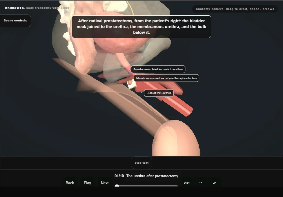
- Mild to moderate stress incontinence after radical prostatectomy.
- From the patient's right: the anastomosis, the membranous urethra and the bulb below it.
- High lithotomy; a urethral catheter is passed to the bladder.
- The landmarks are the ischiopubic rami, the obturator foramina and the adductor longus tendons.
- 02
Perineal exposure
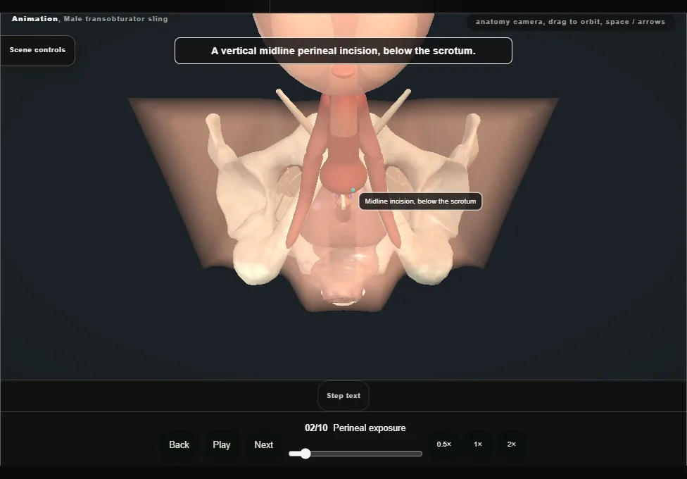
- A vertical midline perineal incision below the scrotum.
- Diathermy through Colles fascia and the subcutaneous tissue to bulbospongiosus; the muscle is divided sharply in the midline and dissected off the corpus spongiosum.
- A self-retaining retractor is set.
- 03
Central tendon
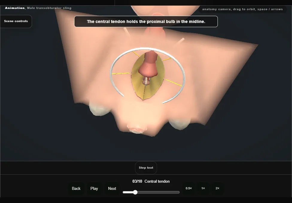
- The central tendon is found in the midline and incised sharply, which mobilises the proximal bulbar urethra.
- A sterile marking pen marks this level on the bulb: the proximal edge of the sling will lie here.
- 04
Passer round the ramus
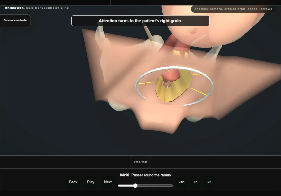
- A stab incision in the groin crease, below the adductor longus tendon; a spinal needle finds the obturator foramen.
- The helical passer goes outside-in: lateral to the ischiopubic ramus, into the foramen, through the fascia, and out between the lateral edge of the spongiosum and the medial border of the corpus cavernosum, with the bulbar urethra held medially.
- 05
Sling arm drawn through
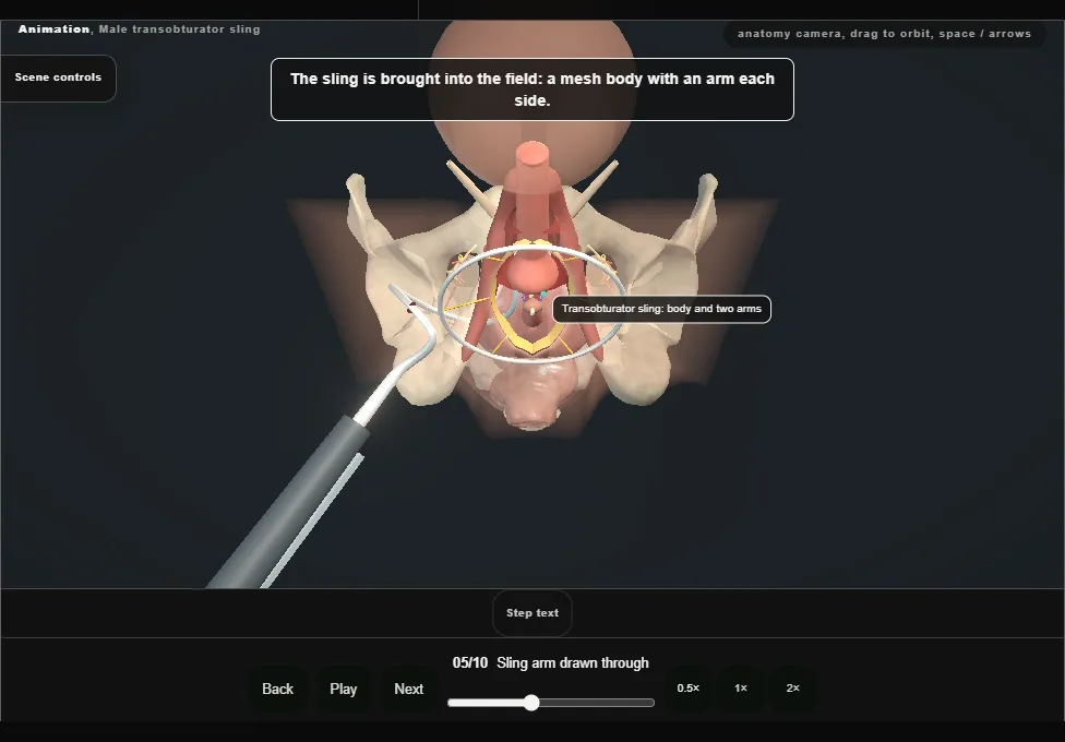
- The sling is brought into the field and connected to the tip of the passer.
- Reverse rotation draws the arm through the obturator foramen and out of the groin incision, loosely.
- The connector and the distal sling are cut above the skin and the end is held in a haemostat.
- 06
The left side
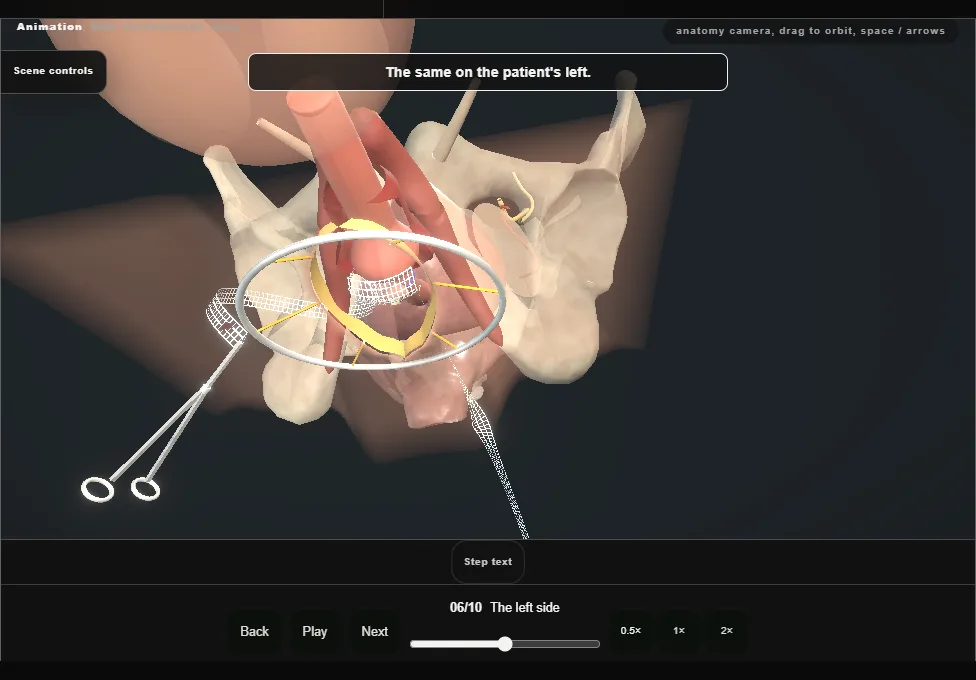
- The same on the other side.
- The centre of the mesh is kept on the urethral midline.
- 07
Fixation on the bulb
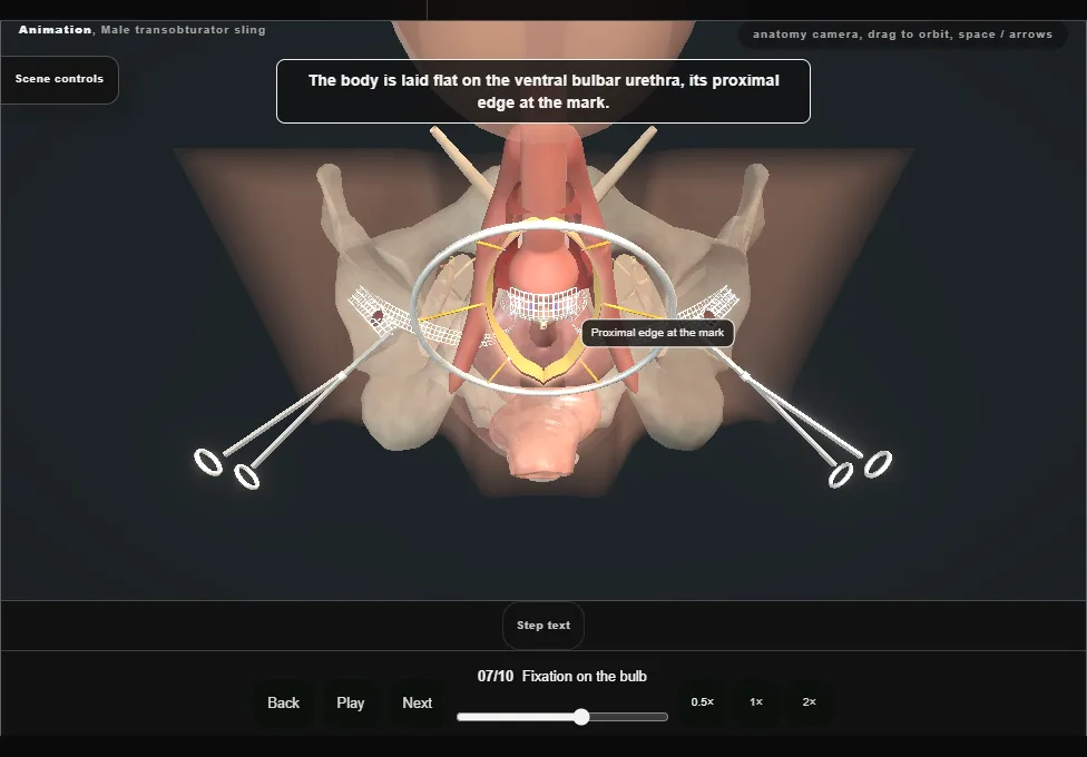
- The sling is laid flat on the ventral surface of the bulbar urethra, its proximal edge at the mark.
- The four corners are fixed to the superficial corpus spongiosum.
- 08
Tension
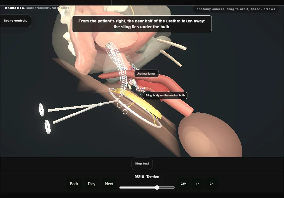
- Both arms are pulled tight, evenly.
- The urethra is displaced proximally.
- 09
Cystoscopy
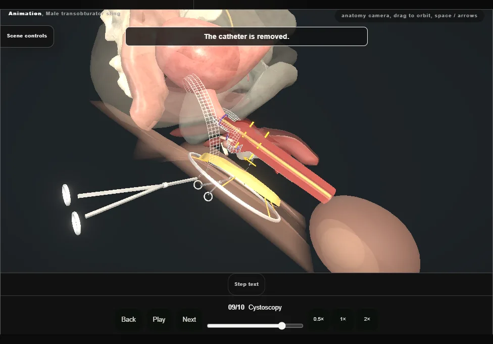
- The catheter is removed and a flexible cystoscope is advanced to the bulbomembranous junction.
- Both arms are lifted under vision: the proximal bulb moves and the urethra is not obstructed.
- The bladder is intact, with efflux from both ureteric orifices.
- The scope comes out and the catheter is replaced.
- 10
Closure
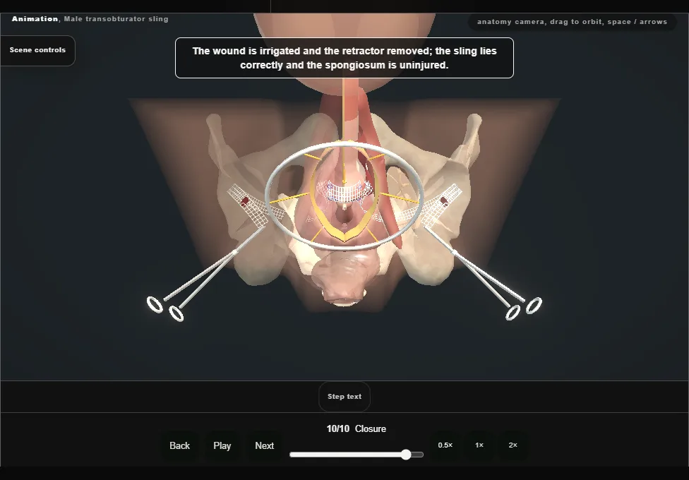
- The wound is irrigated and the retractor removed; the sling is seen to lie correctly with the spongiosum uninjured.
- Bulbospongiosus is closed over the sling, then the subcutaneous tissue and Colles fascia separately so no dead space is left, then skin.
- The groin incisions are closed.
Derived from pelvis/MaleSlingScene.tsx at build time: every step title and caption above is the authored
ProcedureDef text, reproduced verbatim, and this page shares its readable-points renderer with the animation
itself. 10 of 10 steps have a captured frame.
Text fingerprint f34f80daa95f2da0. A caption edit without a re-capture fails the production
check, so the sheet and the animation cannot drift apart silently.