A buckling injury of the erect penis taught as one spatial rule: the tunica tears on the side under tension, opposite the direction of the bend, usually as a transverse defect along the ventrolateral corporal body within the ten to two o'clock arc.
The repair is a subcoronal degloving, haematoma evacuation and absorbable closure of the tear, with the ventral urethra inspected whenever blood sits at the meatus.
Core tierFRCS andrology, genital surgery, emergencyInteractive 3D
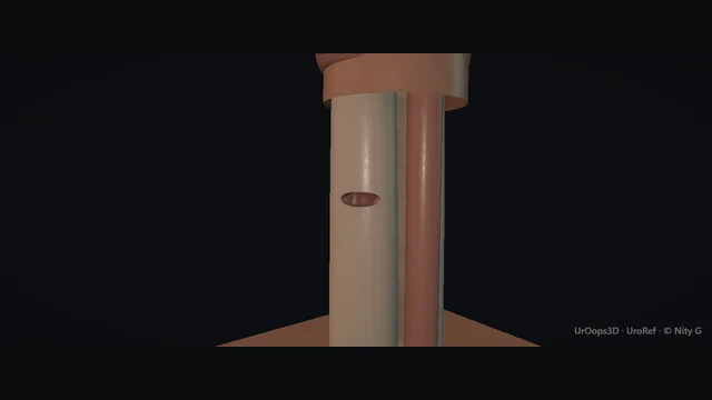
The erect shaft in cross-section, seen from the feet. Schematic, not to scale; relationships only.
Picture the erect shaft in cross-section from the feet: tunica-wrapped corpora side by side, the spongiosum and urethra ventral at 6 o'clock, the neurovascular bundle dorsal at 12.
Buckling bends the shaft and puts the far side of the corpora under tension, so a leftward buckle predicts a right-sided tear and vice versa.
The tear itself is transverse, on the ventrolateral corporal body within the ten to two o'clock arc, and the haematoma beneath the shaft skin is what distorts everything until it is evacuated.
›Tunica albuginea - the white coat of each corporal body, thin when erect; a transverse tear in it is the fracture.
›Corpora cavernosa - paired bodies side by side; the tear runs transversely along one of them, ventrolaterally, within the ten to two o'clock arc.
›Corpus spongiosum + urethra - ventral at 6 o'clock, injured with the buckling in a proportion of cases; blood at the meatus is its signature.
›Dorsal neurovascular bundle - deep dorsal vein, arteries and nerves at 12 o'clock just under the skin; respected at degloving and closure.
›Shaft skin - the degloving sleeve; it rolls to the root and back, and the haematoma spreads beneath it along the whole shaft.
The buckling momentThe erect penis buckles against the perineum or the pubic arch during vigorous intercourse or manipulation, and the story is characteristic. There is an audible or felt crack, sharp pain, immediate loss of the erection and a bruising swelling that tracks along the shaft. While the deep fascia holds, the haematoma is confined to the shaft, which is why the deformity follows the penis rather than spreading to the scrotum and abdominal wall. The direction of the bend identifies the side put under tension, because the tunica tears opposite the bend rather than on it, and that geometry is recorded because it predicts the site the operation will find. where it bites: the tunica tears on the tensile side, opposite the bend; the tear site is predicted from the direction of the injury.
Assessment + selective imagingThe history usually carries the diagnosis and the examination confirms it: a bent, bruised, tender shaft with a classic story needs no imaging before theatre. The meatus is examined for blood at first contact and the patient is asked directly about voiding. Blood at the meatus, a spraying stream or retention means the urethra is injured, and the operative plan changes. Imaging is reserved for atypical stories and the genuinely equivocal examination, where it separates a true corporal tear from a rupture of the dorsal vein alone, and the meatal findings are documented before consent. The animation shows the urethral check as a schematic retrograde urethrogram: a catheter tip in the fossa navicularis with the balloon just inflated (typical), contrast running back to the bladder, and any extravasation at the fracture level declaring the urethral injury before anyone passes an instrument blind. where it bites: blood at the meatus, a spraying stream or retention means the urethra is injured and the operative plan changes.
Subcoronal deglovingA circumferential subcoronal incision is made and the shaft skin is degloved down to the root in one sleeve, lifting skin and subcutaneous tissue together off the deep fascia. The plane stays on Buck's fascia throughout, which keeps the dissection clean and keeps the dorsal neurovascular bundle at 12 o'clock out of harm at both incision and closure. Degloving to the root is the point of the approach: it exposes the whole haematoma for evacuation and both corpora for circumferential inspection, not merely the site the bruise points to. A degloving stopped short of the root hides the proximal tear at the penoscrotal junction, which is the classic miss. where it bites: the degloving plane stays on Buck's fascia; the dorsal neurovascular bundle at 12 o'clock is respected at incision and closure.
Haematoma evacuationClot is evacuated systematically with suction and swabbing until the tunica of both corpora can be seen, because the haematoma distorts everything beneath it and no judgement is made on a dirty field. Each corporal body is then inspected through its full circumference, and the tear presents as a transverse defect on the ventrolateral corporal body within the ten to two o'clock arc. The haematoma is followed to its deepest point, since the tear usually sits at the base of the clot. A second tear is excluded before any repair begins, because stopping at the first defect with the penis partly degloved is the incomplete operation. where it bites: a second tear is excluded before repair begins; the classic miss is stopping at the first defect with the penis partly degloved.
Tunica repair + urethral inspectionThe tear edges are freshened back to healthy tunica and closed with interrupted fine absorbable sutures, taking careful shallow bites so the repair is watertight without bunching. Deep bites are the trap of this step: dorsally they threaten the neurovascular bundle at 12 o'clock, and ventrally they threaten the urethra, which may already be torn. When the meatus showed blood or the history suggests urethral involvement, the urethra is inspected over a catheter or with the scope before any wound is closed. Finding the injury on the table converts an accident into a planned two-structure repair, documented with sites and the catheter plan. where it bites: deep bites dorsally threaten the neurovascular bundle; ventrally they threaten the urethra that may already be torn.
Closure + counsellingThe degloved skin is returned to its natural position, a light non-constricting dressing is applied with the penis elevated, and the dressing is checked to be sure it does not compromise the glans. Haemostasis is confirmed once more before closure, because a re-formed haematoma beneath the flap is the commonest early reason for a return to theatre. Counselling before discharge covers the realistic course: bruising and swelling that settle over the following weeks, a scar at the coronal margin, the possibility of later curvature, and an honest account of erectile recovery. The conversation is documented, because the patient warned at discharge is the patient who does not return angry. where it bites: unrealistic expectations about immediate function do more damage than the repair; the conversation happens before discharge.
The spatial relationships the animation is built to make automatic. Before you read each one, ask yourself: at this point in the operation, what is about to go wrong, and what is my first move? The save for each sits in the Complications tab.
RiskThe ventrolateral corporal body - where the transverse tear sits, within the ten to two o'clock arc; found by evacuating clot, not by blind probing.
RiskVentral urethra at 6 o'clock - torn with the buckling in a proportion of cases; blood at the meatus or a voiding problem must change the plan.
RiskDorsal bundle at 12 o'clock - just under the skin at closure; deep dorsal bites convert a good repair into numbness and deformity.
When the case turns: the problem, then the next move.
ActionNo tear found after degloving - evacuate all clot, inspect circumferentially, extend the degloving proximally to the crura before doubting the diagnosis.
ActionTear approaches the dorsal bundle - take the closure with the bundle visualised and controlled; fine absorbable sutures, minimal bites, no blind passes at 12 o'clock.
ActionTear at the penoscrotal junction or crural origin - extend the incision or counter-incise; the hidden proximal tear is found by following the haematoma, not the surface bruise.
ActionUrethra injured on the same side as the tear - repair the tunica first, then the urethra over a catheter; both repairs are documented with sites and sizes.
The examiner stems this scene prepares you for. Try to answer out loud first, then reveal, and learn the discriminator, the detail that turns a pass into a strong pass.
QIs operative repair or conservative management right for penile fracture?reveal answer
Operative repair, in almost all convincing cases.
The rationale is mechanical: conservative management leaves a transverse tunical defect to heal by fibrosis, and fibrosis shortens the tunica on that side, so erection curves toward the scar, with painful intercourse and a harder secondary reconstruction.
Repair evacuates the haematoma, closes the defect with absorbable sutures and restores the cylindrical integrity of the corporal body, which shortens recovery and lowers the later rates of curvature and erectile difficulty.
Conservative management survives only where the patient refuses surgery, the diagnosis is genuinely in doubt after assessment, or comorbidity makes anaesthesia prohibitive;
even then it is a supervised conservatism with early review, explicit counselling about deformity and erectile function, and a low threshold for delayed reconstruction.
I would also say what repair does not do: it does not guarantee recovery of erectile function, because the injury itself can damage erectile physiology regardless of how well the tunica is closed, and that honesty belongs in the consent.
Strong pass
Say all three
Framing the decision mechanically
Fibrosis shortens the tunica and the erection bends toward the scar
Rather than presenting repair as a general preference
Examiner note
Framing the decision mechanically, fibrosis shortens the tunica and the erection bends toward the scar, rather than presenting repair as a general preference.
QHow does the timing of repair matter?reveal answer
Early repair is the standard: the sooner the tear is closed, the less haematoma dissects the planes, the easier the defect is to define, and the less fibrosis is laid down before the tunica is restored.
Practically, a penile fracture is an emergency but not a race to the knife in the first hour;
repair within the first day or two is the usual window, and a case that presents a few days late is still repaired, just through angrier tissue with more clot to evacuate.
What changes with genuine delay is the pathology: after weeks the defect has fibrosed, the anatomy is scarred rather than simply bruised, and the operation stops being a repair and becomes a reconstruction of the tunica, with grafting considered and results that are more modest.
The counsel of perfection is therefore assessment the same day, repair on the next suitable list, and no interval of watchful waiting for a classic presentation, because the natural history of an untreated tear is curvature, painful erection and a proportion who return for much bigger surgery.
Strong pass
Say both
Distinguishing the early repair window from the delayed reconstruction
Saying that late repair is still offered but through fibrosed planes with different objectives
Examiner note
Distinguishing the early repair window from the delayed reconstruction, and saying that late repair is still offered but through fibrosed planes with different objectives.
QA man reports a cracking injury during intercourse three weeks ago and now has curved, painful erections. What has happened and what do you offer?reveal answer
He describes an untreated penile fracture: the tunica tore at the buckling moment, the defect healed by fibrosis, and the scarred segment is shorter and less compliant than the rest of the shaft, so the erect penis bends toward the injury and aches as the scar stretches.
Assessment is history and examination: the direction and degree of curvature, ideally documented with photographs at home, a palpable plaque or defect at the injury site, and imaging to map the tunical anatomy before anything is offered.
Management is staged rather than immediate: the scar is allowed to mature, since curvature can moderate and the tissues must stabilise before reconstruction is reliable, so several months of observation is legitimate.
If the curvature then interferes with intercourse, options are the reconstructive ladder for Peyronie-like disease: plication on the opposite side for modest curvature, plaque incision or excision with grafting for larger defects.
Erectile function is assessed in parallel, because impaired rigidity changes the choice; a man whose erections are failing may be better served by a prosthesis with straightening.
Counselling is explicit about timeline and expectations.
Strong pass
Say both
Recognising the healed fracture as a Peyronie-type problem with a staged pathway
Rather than offering immediate surgery on immature scar
Examiner note
Recognising the healed fracture as a Peyronie-type problem with a staged pathway, rather than offering immediate surgery on immature scar.
QWhen do you suspect urethral injury in penile fracture, and what changes in theatre?reveal answer
Suspicion rests on findings at the meatus and the voiding history: blood at the meatus, difficulty or inability to pass urine, a spraying stream after the injury, or gross haematuria.
The mechanism raises the index of suspicion further when the bend was ventral or the injury bilateral, since bilateral corporal tears travel with urethral injury more often.
In theatre, three things change.
First, instrumentation: no blind catheterisation through a possible injury; the urethra is assessed with the scope, or a catheter is passed under vision once the injury is defined.
Second, sequence: the tunical tears are repaired, and any urethral tear is then repaired over a catheter with fine absorbable sutures, with the repairs kept independent so each heals in its own plane.
Third, disposition: a catheter stays for the healing period decided by the injury, and follow-up includes a voiding assessment, because the late sequelae of a missed repair are stricture and fistula.
The honest summary is that urethral injury converts a satisfying single-repair operation into a two-structure operation, and the meatus examination in the emergency department is what makes that discovery deliberate rather than accidental.
Strong pass
Say both
Naming the specific triggers at the meatus and the rule against blind catheterisation
Plus the sequencing of the two repairs
Examiner note
Naming the specific triggers at the meatus and the rule against blind catheterisation, plus the sequencing of the two repairs.
Say it aloud, then reveal. Reading is not remembering.
01The relationshipSay the critical spatial relationship out loud, then reveal.reveal
Memory frame
Picture the erect shaft in cross-section from the feet
Why?
Picture the erect shaft in cross-section from the feet: tunica-wrapped corpora side by side, the spongiosum and urethra ventral at 6 o'clock, the neurovascular bundle dorsal at 12. Buckling bends the shaft and puts the far side of the corpora under tension, so a leftward buckle predicts a right-sided tear and vice versa. The tear itself is transverse, on the ventrolateral corporal body within the ten to two o'clock arc, and the haematoma beneath the shaft skin is what distorts everything until it is evacuated.
02The first moveMissed urethral injury. Your first move?reveal
First move
Recognise it early: examine the meatus at first assessment and ask directly about voiding.
03The escalation triggerMissed urethral injury. What makes you call for help?reveal
Escalate when
Retention, extravasation or a delayed fistula → formal imaging and reconstruction planning with specialist input.
04The discriminatorWhat turns a pass into a strong pass?reveal
Strong pass
Say all three
Framing the decision mechanically
Fibrosis shortens the tunica and the erection bends toward the scar
Rather than presenting repair as a general preference
Examiner note
Framing the decision mechanically, fibrosis shortens the tunica and the erection bends toward the scar, rather than presenting repair as a general preference.
05The pitfallWhere does this operation bite first?reveal
Pitfall
The ventrolateral corporal body
Why?
where the transverse tear sits, within the ten to two o'clock arc; found by evacuating clot, not by blind probing.
Revision practice with self-marked answers, not an assessment of clinical competence. Kept in
this browser only; it does not sync between devices.