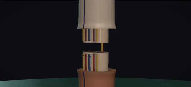
Schematic model; the order of repair, the side of the artery and nerve joined (the patient's left) and the single urethral catheter are authoring decisions taken from the book chapter.
Operative animation, Penile reimplantation
Penile reimplantation after amputation: microsurgical, on a urethral catheter
Microsurgical reimplantation of an amputated penis. The part is kept cold without touching ice and the stump is prepared under a tourniquet.
The two pieces are splinted on a urethral catheter and joined from the inside out: corpora, urethra, deep dorsal vein, a dorsal artery, a dorsal nerve, then dartos and skin.
Check rotation before the first tunical stitch, join the vein before the artery, and warn him that skin loss and stricture are common.
Advanced tierFRCS andrology, genital trauma, microvascular surgeryInteractive 3D
How to use this module. Play through the nine steps, or use Back and Next. Watch the part leave the iced water, the cut face read, the catheter thread both pieces and the dorsal mark line up, then each join in turn. The triggers show a congested part, a pale part, skin loss and a narrowed urethral join.
Prepare the partReimplantation is considered for every patient whose part has been recovered and is not too badly damaged; the EAU guideline asks for it within 24 hours of the amputation. The part is washed with sterile saline, wrapped in saline-soaked gauze, sealed in a sterile bag and the bag is put in iced water. In theatre, under loupes, it is irrigated with warm saline and antibiotic solution and debrided as little as possible, which also confirms that it is viable. The cut face is read before anything is joined. If the injury was self-inflicted, psychiatric assessment is arranged early; it does not delay the reimplantation. where it bites: the part must never lie directly on the ice; and whatever is debrided is length that cannot be put back.
Prepare the stumpBleeding from the stump is controlled first with a pressure dressing or a tourniquet. In theatre a tourniquet goes round the base of the stump and is released at intervals through the case. Skin and dartos are dissected back a short distance from the stump edge to expose the corpora and the dorsal neurovascular bundle, with haemostasis by fine diathermy and ties. The ends of the deep dorsal vein, the dorsal arteries and the dorsal nerves are found and tagged before anything is joined. where it bites: diathermy at the bundle ends burns the vessels that are about to be joined; a tourniquet left on without release adds to the ischaemia.
Align on a catheterThe part is put back in its anatomical position against the stump and a urethral catheter is passed through the meatus, through the part and the stump, into the bladder. The catheter stabilises the two pieces and marks the lumen for the urethral join. It does not control rotation: the part can turn about it. The dorsal midline, the septum and the urethra are lined up by eye before the first stitch. where it bites: a part stitched on with a twist cannot be corrected later without taking the corpora down.
Join the corporaThe corpora cavernosa are aligned and the tunica albuginea of each is approximated all the way round with interrupted absorbable stitches. This is the structural repair, and it takes the tension off everything that follows. The cavernosal arteries inside the corpora are generally too small to join and are left alone. where it bites: a bite that misses the full thickness of the tunica cuts out; the septum must meet the septum.
Join the urethraThe proximal and distal urethral ends are debrided as little as possible and spatulated on opposite sides. A one-layer anastomosis is completed with interrupted absorbable stitches over the catheter. Spatulation makes the join oblique and wider than either end. where it bites: a circular, unspatulated join narrows as it heals; a gap in the suture line leaks and becomes a fistula.
Repair the deep dorsal veinThe operating microscope is draped and brought in. The deep dorsal vein is identified and freed proximally and distally, its ends are spatulated on opposite sides, and the venous anastomosis is completed with fine monofilament, interrupted or as two running halves. The book joins the vein before the artery: the part is given a way out before it is given a way in. where it bites: arterial inflow without venous drainage congests the part; venous congestion is among the complications reported most often.
Repair a dorsal arteryOne dorsal artery is identified, dissected and joined end to end in the same way; when both arteries are found and look usable, both are joined. The tourniquet is removed and both anastomoses are inspected for good pulsatile flow and for bleeding. The part regains colour slowly and incompletely on the table. where it bites: no pulse beyond the join is a technical fault until proven otherwise: a clamp left on, a kink, tension, or thrombus at the suture line.
Repair a dorsal nerveAt least one dorsal nerve is repaired, and both when both are found and usable. The ends are brought together without tension and joined with interrupted fine stitches placed in the epineurium. In the pooled series more nerves joined went with better outcomes, but return of sensation is hoped for and never promised. where it bites: tension pulls a nerve repair apart: the ends must lie together before the first stitch goes in.
Close and counselThe wound is irrigated and haemostasis confirmed again. Dartos and skin are closed as separate layers with interrupted absorbable stitches. Antibiotic ointment and gauze go on the suture line, the penis is wrapped very loosely, the catheter is connected to drainage and the penis is supported against the abdomen to limit swelling. A suprapubic catheter may be added at the end; the EAU guideline places both. The patient is told what to expect: skin loss, stricture, fistula, altered sensation and erectile dysfunction all follow technically sound operations. where it bites: a snug dressing is a tourniquet on a part that depends on one small artery and one vein.
The spatial relationships the animation is built to make automatic. Before you read each one, ask yourself: at this point in the operation, what is about to go wrong, and what is my first move? The save for each sits in the Complications tab.
RiskCold storage - saline gauze, then a sealed bag, then iced water: the tissue never on the ice. The EAU guideline asks for reimplantation within 24 hours of the amputation.
RiskRotation - the catheter splints the pieces and lets the part turn about it. The dorsal midline, the septum and the urethra must all agree before the first tunical stitch.
RiskOutflow - the deep dorsal vein is joined before the artery. A part with a pulse and no drainage turns dark, swollen and tense.
RiskThe arterial join - one small artery carries the part. No pulse beyond the join after the tourniquet is off is a technical fault until proven otherwise.
RiskThe urethral join - spatulated on opposite sides and stitched over the catheter; a circular join strictures and a gap in it fistulates.
RiskThe skin - the layer lost most often, sometimes over a living shaft. The dressing stays loose and the patient is told beforehand.
When the case turns: the problem, then the next move.
ActionThe part is missing or too damaged to use - close the stump as for a partial penectomy. Later options named by the EAU guideline are lengthening by suspensory ligament division with a V-Y plasty, a pseudoglans with a split-thickness graft, and delayed phalloplasty.
ActionNo microsurgeon is available - the EAU guideline prefers a microsurgical repair and allows a non-microsurgical one. Discuss transfer with the part correctly stored before settling for corpora and urethra alone.
ActionOnly one dorsal artery or one nerve can be found - one of each is the least the book describes. Join what is usable and record what was not.
ActionThe pieces will not sit without rotation - take the catheter as the axis and turn the part until the dorsal midline, the septum and the urethra all agree. Do not accept a compromise at the first stitch.
ActionThe part stays pale after the tourniquet is off - check the clamps and the lie of the artery, then treat it as a problem at the arterial join: look, open, revise.
The examiner stems this scene prepares you for. Try to answer out loud first, then reveal, and learn the discriminator, the detail that turns a pass into a strong pass.
QA man in his thirties is brought in two hours after an amputation of the penis at mid-shaft. The part has come with him. What do you do?reveal answer
I resuscitate him and control bleeding from the stump with a pressure dressing or a tourniquet.
I make sure the part is stored correctly: washed with sterile saline, wrapped in saline-soaked gauze, sealed in a sterile bag and the bag placed in iced water, so that the tissue never lies on the ice.
The EAU guideline says reimplantation should be considered in every patient whose part is recovered and not too badly damaged, and performed within 24 hours.
A microsurgical repair is preferred, so I call the microsurgical team, or arrange transfer with the part if there is none.
I give antibiotics and take consent with honest expectations.
If the injury was self-inflicted I arrange psychiatric assessment early; it does not delay the reimplantation.
Strong pass
Say all three
Storing the part correctly without being asked
Gauze then a sealed bag then iced water
Giving the 24-hour window with its source
Examiner note
Storing the part correctly without being asked, gauze then a sealed bag then iced water, and giving the 24-hour window with its source.
QDescribe the operation.reveal answer
Under loupes I irrigate the part and debride it as little as possible.
I put a tourniquet on the stump, release it at intervals, and take skin and dartos back to expose the corpora and the dorsal bundle.
I place the part against the stump and pass a urethral catheter through both into the bladder, and I check rotation against the dorsal midline, the septum and the urethra.
I approximate the tunica albuginea of each corpus all round with interrupted absorbable stitches.
I spatulate the urethral ends on opposite sides and join them in one layer over the catheter.
Under the operating microscope I join the deep dorsal vein, then at least one dorsal artery; the tourniquet comes off and I inspect both joins for flow and bleeding.
I join at least one dorsal nerve with epineurial stitches.
I close dartos and skin in two layers, with a very loose dressing and the catheter on drainage.
Strong pass
Say all four
The order with its reasons: alignment first
Corpora for stability
Vein before artery
One artery and one nerve as the least that is done
Examiner note
The order with its reasons: alignment first, corpora for stability, vein before artery, and one artery and one nerve as the least that is done.
QWhy is the vein joined before the artery, and what do you see when venous drainage fails?reveal answer
The part needs a way out before it is given a way in.
If arterial blood enters and cannot leave, the part becomes dark, swollen and tense.
That is venous congestion, which after skin necrosis is among the complications reported most often.
I would loosen the dressing, look at the venous join under the microscope for a kink, a twist or clot, and take it down and revise it, with the microsurgical lead involved at once.
The cavernosal arteries are generally too small to join, so the dorsal vessels are what the part lives on.
Strong pass
Say both
Treating congestion as a failed venous join that has to be looked at
Saying that inflow without outflow loses the part
Examiner note
Treating congestion as a failed venous join that has to be looked at, and saying that inflow without outflow loses the part.
QWhat do you tell him about the results?reveal answer
Skin necrosis is the commonest complication reported, often over a shaft that survives, and venous congestion, urethral stricture and fistula follow it.
In the published reports most men pass urine through the reimplanted penis, and many, but not all, regain erections and sensation.
Those are collected case reports with unstandardised outcomes, so they are likely to flatter the operation, and the reviews of them conclude that complications remain highly prevalent even with meticulous microsurgery.
So I tell him that he is likely to pass urine through it and may well regain erections and sensation, that none of that is promised, and that further operations for skin or urethra are common.
Strong pass
Say both
Naming skin necrosis as the commonest complication
Saying that what is known of outcomes comes from pooled case reports
Examiner note
Naming skin necrosis as the commonest complication and saying that what is known of outcomes comes from pooled case reports.
Curriculum. Supports learning relevant to the ISCP Urology Curriculum syllabus topics Genitourinary trauma and Andrology. Not a workplace-based assessment and not evidence of competence.
Two minutes
Close the lap: recall it before you leave.
Say it aloud, then reveal. Reading is not remembering.
01The relationshipSay the critical spatial relationship out loud, then reveal.reveal
Memory frame
The cut face is the map.
Why?
Two corpora lie side by side with the septum between them and a cavernosal artery in the centre of each. The urethra sits in its spongiosum in the ventral groove. On the dorsum the deep dorsal vein runs in the midline with a dorsal artery either side of it and a dorsal nerve lateral to each artery. The same map lies on the stump and on the part: the operation brings the two face to face without rotation and joins them from the inside out.
02The first moveVenous congestion. Your first move?reveal
First move
Loosen the dressing and look at the venous join under the microscope for a kink, a twist or clot.
03The escalation triggerVenous congestion. What makes you call for help?reveal
Escalate when
Call the microsurgical lead at once: the decision to re-explore is theirs and it is urgent.
04The discriminatorWhat turns a pass into a strong pass?reveal
Strong pass
Say all three
Storing the part correctly without being asked
Gauze then a sealed bag then iced water
Giving the 24-hour window with its source
Examiner note
Storing the part correctly without being asked, gauze then a sealed bag then iced water, and giving the 24-hour window with its source.
05The pitfallWhere does this operation bite first?reveal
Pitfall
Cold storage
Why?
saline gauze, then a sealed bag, then iced water: the tissue never on the ice. The EAU guideline asks for reimplantation within 24 hours of the amputation.
Revision practice with self-marked answers, not an assessment of clinical competence. Kept in
this browser only; it does not sync between devices.