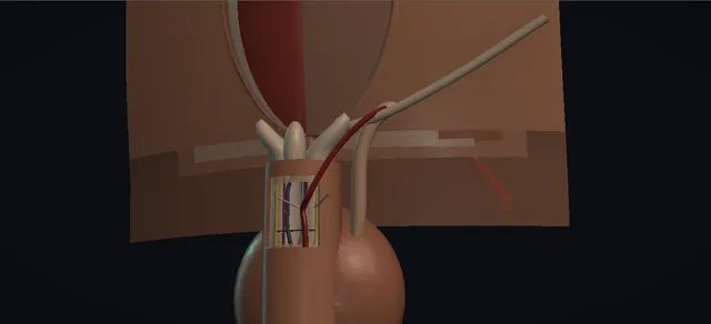
UrOops3D, danger-zone atlas

Schematic model; the lower abdomen is authored procedurally, the laterality (patient's left) and the end-to-side joint are authoring decisions from the book.

Operative animation, Penile revascularisation

Penile arterial revascularisation: inferior epigastric artery to the deep dorsal artery, end to side

A highly selected operation, not routine erectile dysfunction care. A young man with an isolated arterial lesion after pelvic trauma, a patent donor on the arteriogram and no venous leak.

The inferior epigastric artery is harvested on its iliac origin, passed through the floor of the inguinal canal medial to the cord and under the skin to the root of the penis, and joined end to side to the dorsal artery under the microscope.

The lesson is in three places: selection, the route without a twist, and the joint.

Advanced tier FRCS andrology, erectile dysfunction, microvascular surgery Interactive 3D

Operative visualisation

Schematic scene preview: Penile arterial revascularisation
Preparing the 3D anatomy…

UrOops3D · UroRef · © Nity G

How to use this module. Play through the nine steps, or use Back and Next. Watch the artery lift off the rectus, its branches clipped, the pedicle pass medial to the cord, the slit open between two clamps and the stitches go round. The triggers show no flow, a twisted pedicle, a nerve in the sling and a flushed glans.

Click me next Section 2 of 8 Anatomy The structures this operation works between, and the one it must not cross.

The structures in the scene

Penile revascularisation: how the structures relateFrom the lower abdominal wall, down the canal floor, to the dorsum of the penile root. Inferior epigastric pedicle, on its origin: inflow stays attached here External iliac artery: the origin, kept. Inferior epigastric pedicle, on its origin: freed from its bed, branches clipped Rectus: the artery on its posterior face. Inferior epigastric pedicle, on its origin: passes medial; a twist or kink occludes Spermatic cord: the tunnel is medial to it (danger). Inferior epigastric pedicle, on its origin: the free end lies on the slit, no tension Dorsal artery: slit, joined end to side. Inferior epigastric pedicle, on its origin: clamps and stitches stay clear of it Dorsal nerve, lateral, never slung (danger). Inferior epigastric pedicle, on its origin: beside the join, untouched Deep dorsal vein, midline, held aside.Inferior epigastricpedicle, on its originExternal iliacartery: the origin,keptRectus: the arteryon its posteriorface!Spermatic cord: thetunnel is medial toitDorsal artery: slit,joined end to side!Dorsal nerve,lateral, never slungDeep dorsal vein,midline, held asideinflow staysattached herefreed from its bed,branches clippedpasses medial; atwist or kinkoccludesthe free end lieson the slit, notensionclamps and stitchesstay clear of itbeside the join,untouchedTargetDangerLandmarkContext
From the lower abdominal wall, down the canal floor, to the dorsum of the penile root. Schematic, not to scale; relationships only.
  • Two fields joined by one tunnel.
  • In the abdomen the donor artery runs on the back of the rectus from the external iliac to the umbilicus and is lifted on its origin;
  • at the penile root the dorsal artery lies beside the midline vein with the nerve lateral to it.
  • The pedicle passes from the canal floor medial to the cord and under the skin to the root, and the joint sits end to side on the anterior wall of the dorsal artery, between two clamps, clear of the nerve.
  • ›Inferior epigastric artery - the donor: from the external iliac artery, behind the rectus to the umbilicus; harvested on its origin with its branches clipped.
  • ›External iliac artery - the origin of the donor, kept intact; the pedicle's inflow.
  • ›Rectus abdominis and linea alba - opened at the midline, the left rectus lifted laterally; the artery lies on its posterior face.
  • ›Inguinal canal and spermatic cord - the pedicle passes through the canal floor medial to the cord, then subcutaneously to the root; the cord is never caught.
  • ›Fundiform and suspensory ligaments - preserved at the dorsal root incision.
  • ›Deep dorsal artery - the recipient, isolated and mobilised on a sling; the arteriotomy on its anterior wall.
  • ›Deep dorsal vein - midline under Buck's fascia, held aside and untouched here; the target of the arterialisation variants, which are not drawn.
  • ›Dorsal nerves - lateral to the arteries; held aside, never slung; sensation is what they carry.
  • ›Internal pudendal and common penile artery - the occluded segment on the arteriogram, deep to the field and not operated on.
Click me next Section 3 of 8 Steps The operation in order, with the landmark that guards each move.

Operative steps

  1. Select the patient The operation is the selection. A young man with erectile dysfunction after pelvic or perineal trauma, no vascular risk factors, an isolated occlusion of the internal pudendal or common penile artery on pharmaco-arteriography, a patent inferior epigastric artery on the same study, and no veno-occlusive dysfunction on cavernosometry. Alternatives are discussed and recorded: oral therapy, intracavernosal injection, a penile prosthesis and, where the lesion suits it, endovascular treatment. This is not routine erectile dysfunction care.
    where it bites: a venous leak on cavernosometry is a contraindication; the operation revascularises an inflow lesion and nothing else.
  2. Open the abdomen Supine, catheter in, the abdomen and genitalia prepped together. A lower midline incision from below the umbilicus to the pubis, the subcutaneous tissue divided, the rectus aponeurosis opened at the linea alba and the recti separated. The peritoneum stays closed. The left rectus is lifted superolaterally with padded retractors and the inferior epigastric neurovascular bundle lies on its posterior face.
    where it bites: the bundle is on the back of the muscle; a retractor placed under the muscle without looking tears it.
  3. Harvest the donor Under loupes the inferior epigastric artery is dissected from its origin at the external iliac artery to the level of the umbilicus. Every side branch is clipped or tied and divided, never avulsed. Systemic heparin is given by the unit protocol, a microvascular clamp goes on the artery and it is divided at the umbilical level: a live pedicle on its own iliac origin, with its vein kept where it helps.
    where it bites: the origin from the external iliac is the one thing that cannot be re-made; branches are clipped, not torn, because a torn branch bleeds into the pedicle wall.
  4. Expose the recipient A separate dorsal incision at the base of the penis is carried through dartos and Buck's fascia with the fundiform and suspensory ligaments preserved; a self-retaining retractor sets the field. The dorsal neurovascular bundle is identified and the left deep dorsal artery is isolated and circumferentially mobilised for a working length, the deep dorsal vein and the dorsal nerves held aside and untouched.
    where it bites: the dorsal nerves lie lateral to the arteries; a sling that takes the nerve costs sensation, and decreased sensation is a listed complication of this operation.
  5. Route the pedicle A long curved clamp passes from the penile incision up the subcutaneous plane to the floor of the ipsilateral inguinal canal, medial to the spermatic cord, and the harvested artery is drawn down through the canal floor and the tunnel into the penile incision. The pedicle is laid flat and checked along its whole length: no twist, no kink, no tension, and the cord not caught.
    where it bites: a twist in the tunnel is invisible once the skin is closed and thromboses after release; the marker stripe is checked at the ring and at the root before the microscope comes in.
  6. Prepare the joint The operating microscope is draped and brought in. Two microvascular clamps go on the deep dorsal artery clear of the nerve and a short arteriotomy is made on its anterior wall between them. Topical papaverine is applied to both vessels and good flow from the donor is confirmed before it is trimmed to lie on the arteriotomy without tension.
    where it bites: spasm at this stage looks like a bad donor; vasodilator and patience before any decision to abandon.
  7. End-to-side anastomosis The inferior epigastric artery is joined end to side to the deep dorsal artery with fine monofilament, as interrupted sutures or two running halves. Heel and toe first, then the far wall in sequence, the vessel turned, and the near wall; every bite full thickness and the back wall never caught. The donor meets the recipient at an acute angle.
    where it bites: a back-wall stitch closes the lumen; a bite that misses the intima lifts a flap that thromboses.
  8. Release and check All three clamps come off in order and the suture line is watched: a pulse that passes the joint and runs down the dorsal artery, no bleeding beyond what pressure settles. Patency is confirmed with a Doppler on the distal artery. No flow is a technical fault at the joint or in the route until proven otherwise.
    where it bites: bleeding at the suture line is answered with pressure and time, then a single extra stitch; never with a clamp left on.
  9. Close Both incisions irrigated and haemostasis confirmed before the retractors come out. Dartos and the penile skin closed, then the rectus muscles, the aponeurosis, Scarpa's fascia and the skin of the abdominal wound, with the pedicle never compressed by any layer. Adhesive strips and a gauze dressing on both wounds; counts correct.
    where it bites: a tight closure at the ring or at the root is a second tunnel: the pedicle is checked again after every layer.
Click me next Section 4 of 8 Danger zones Where this operation bites, and why it bites there.
Critical relationship

Danger zones

The spatial relationships the animation is built to make automatic. Before you read each one, ask yourself: at this point in the operation, what is about to go wrong, and what is my first move? The save for each sits in the Complications tab.

  • RiskSelection - an isolated arterial lesion in a young man without vascular risk factors and without veno-occlusive dysfunction. Outside that, the operation fails and the patient has had two incisions for nothing.
  • RiskThe origin and the branches - the external iliac origin cannot be re-made; a torn branch bleeds into the pedicle wall. Clips, not traction.
  • RiskThe tunnel - twist, kink and tension are the three ways a patent joint thromboses. The route is the canal floor medial to the cord and the pedicle is laid flat and checked before the microscope comes in.
  • RiskThe dorsal nerves - lateral to the arteries under Buck's fascia; a sling or a clamp that takes the nerve costs sensation.
  • RiskThe joint - full-thickness bites, the intima included, the back wall never caught; no clamp left on; spasm treated before it is called failure.
Practise it Take me into the scene Opens the animation at a danger moment and asks you to predict the mechanism and your first move before it reveals.
Click me next Section 5 of 8 Complications The save for each one: the first move, the escalation trigger, the op-note line.

Complications and immediate saves

Trigger each of these live in the Animation tab, then read the save: the first move, the escalation trigger, and the line that goes in the op note.

No flow after release

  • the clamps are off and there is no pulse past the joint and a flat Doppler.
  • Technical failure is at the joint or in the route, not in the donor: a back-wall stitch, an intimal flap, a twist or kink in the tunnel, or a clamp forgotten.
The save
  1. Check along the whole pedicle for a kink, a twist at the ring or a clamp left on; lay it flat. first move
  2. Topical vasodilator and a minute; spasm looks like thrombosis.
  3. Re-apply the clamps, open the anastomosis, clear it and revise; re-check flow with the Doppler.
  4. Call the microsurgical lead early; a second revision is a shared decision, and so is abandoning the joint. escalation trigger
  5. Op note: the finding at the joint, what was revised, the Doppler result after revision. document

Pedicle twisted or kinked in the tunnel

  • the marker stripe has turned and the pedicle narrows where it passes the ring.
  • A twisted pedicle is patent on the table and thromboses later, when nobody is looking.
The save
  1. Withdraw the pedicle and re-pass it with the stripe in line, before any clamp goes on the recipient. first move
  2. Widen the tunnel and the passage through the canal floor; the pedicle must lie flat with slack at both ends.
  3. A pedicle that will not lie flat in the canal-floor route is a reason to stop and ask the microsurgical lead about the alternative route between the rings and through a scrotal incision, which one series describes and this module does not draw. escalation trigger
  4. Op note: the route taken, the check of the stripe at the ring and at the root. document

Dorsal nerve injury

  • the sling or a clamp has taken a dorsal nerve with the artery during recipient exposure.
  • Decreased penile sensation is a listed complication of the operation.
The save
  1. Release the sling and re-sling the artery alone; the nerve stays lateral under its own fascia. first move
  2. Micro clamps go on the artery only, clear of the nerve.
  3. A nerve clearly divided or crushed is a senior conversation about repair and about what the patient is told. escalation trigger
  4. Op note: the event, the re-sling and the state of the nerve as seen. document

Glans hyperaemia

  • a swollen, flushed glans after revascularisation.
  • The book lists it; the case series describe it mainly with the dorsal vein arterialisation variants, where arterial flow is put into the venous side.
  • With the artery-to-artery joint drawn here it is the less common cause.
The save
  1. Assess the glans: colour, swelling, sensation, the skin over it. first move
  2. Escalate to the operating team; with a vein arterialisation the answer is a senior decision about the flow into the venous side. escalation trigger
  3. Op note and clinic letter: the finding, the type of joint performed, the plan. document
Click me next Section 6 of 8 Troubleshooting When the case turns: the problem, then the next move.

Intra-operative troubleshooting

When the case turns: the problem, then the next move.

  • ActionThe donor looks small or in spasm on the bed - topical papaverine and patience before any judgement; a spastic artery is not a bad artery. The preoperative arteriogram already said it was patent.
  • ActionThe pedicle will not reach the root - the harvest was too short or the route too long: mobilise toward the umbilicus to the full described length, and check that the route is the canal floor medial to the cord and not a detour round it.
  • ActionThe recipient artery is atretic - the plan changes: the dorsal vein arterialisation variants exist for exactly this finding and are a senior decision, not an improvisation; they are not drawn here.
  • ActionBleeding at the suture line after release - pressure and time first, then a single extra stitch at the point; never a clamp left on and never diathermy near the joint.
  • ActionFlat Doppler with a visible pulse at the donor - the fault is at the joint or just beyond: re-clamp and look inside.
Click me next Section 7 of 8 FRCS viva Say it out loud, then mark yourself against the model answer.

FRCS viva questions

The examiner stems this scene prepares you for. Try to answer out loud first, then reveal, and learn the discriminator, the detail that turns a pass into a strong pass.

QA man in his late twenties has erectile dysfunction after a pelvic fracture and asks about a vascular operation. How do you assess him and whom do you offer surgery?reveal answer
  • Penile revascularisation is for a small group: a young man with erectile dysfunction after pelvic or perineal trauma, without the vascular risk factors that make arterial disease generalised, in whom a focal lesion of the internal pudendal or common penile artery can be shown and the veno-occlusive mechanism is intact.
  • I take the history for the trauma and for risk factors, examine him, and arrange penile Doppler after intracavernosal injection;
  • a poor arterial response leads to cavernosometry and cavernosography to exclude a venous leak, and then to pharmaco-arteriography, which I only request in a man who is a candidate for the operation, because its purpose is to show the lesion and to confirm a patent inferior epigastric artery as the donor.
  • He is counselled that this is highly selected surgery with uncertain long-term results, that the alternatives are oral therapy, intracavernosal injection, a prosthesis and, in suitable lesions, endovascular treatment, and that venous ligation surgery is no longer recommended.
  • I would refer him to a centre that does the operation.
Strong pass

Say both

  • Naming the veno-occlusive dysfunction as the contraindication
  • Requesting arteriography only in a candidate for surgery
Examiner note
Naming the veno-occlusive dysfunction as the contraindication and requesting arteriography only in a candidate for surgery.
QDescribe the operation.reveal answer
  • Through a lower midline incision the linea alba is opened and the left rectus lifted laterally;
  • the inferior epigastric artery is dissected from its origin at the external iliac artery to the umbilical level with every branch clipped and divided, heparin given by protocol, a microvascular clamp placed and the artery divided superiorly.
  • Through a separate dorsal incision at the base of the penis, preserving the fundiform and suspensory ligaments, the deep dorsal artery is isolated and mobilised with the vein and nerves protected.
  • The pedicle is tunnelled through the floor of the inguinal canal medial to the cord and subcutaneously into the penile incision, laid flat without twist, kink or tension.
  • Under the operating microscope, with two micro clamps on the dorsal artery, an anterior arteriotomy is made, papaverine applied, and an end-to-side anastomosis fashioned with fine monofilament, interrupted or as two running halves.
  • The clamps come off, flow is confirmed by the pulse and with a Doppler, and both wounds are closed in layers without compressing the pedicle.
Strong pass

Say all four

  • The route medial to the cord
  • The ligaments preserved
  • The Doppler check before closure
  • Said without prompting
Examiner note
The route medial to the cord, the ligaments preserved, and the Doppler check before closure, said without prompting.
QThe clamps are off and there is no flow past the joint. What do you do?reveal answer
  • I treat it as a technical fault until proven otherwise, because the donor was patent on the arteriogram and pulsing before the joint was made.
  • First the whole pedicle: a twist at the ring, a kink in the tunnel, a clamp left on, tension at the root.
  • Then spasm: topical vasodilator and a minute, since a spastic vessel looks thrombosed.
  • If there is still no flow I re-apply the clamps, open the anastomosis, clear it and look for a back-wall stitch or an intimal flap, and revise it.
  • I involve the microsurgical lead early, because a second revision or a decision to abandon the joint is not one to take alone.
  • I document what was found and what was done, and the Doppler result after revision.
Strong pass

Say both

  • Checking the route before the joint
  • Naming spasm as a mimic before re-opening the anastomosis
Examiner note
Checking the route before the joint and naming spasm as a mimic before re-opening the anastomosis.
QWhat are the complications and what do you tell him beforehand?reveal answer
  • Bleeding and infection at two wounds;
  • penile oedema and bruising;
  • penile pain;
  • decreased penile sensation from the dorsal nerves at the root incision;
  • glans hyperaemia, described mainly with the dorsal vein arterialisation variants;
  • penile shortening;
  • priapism;
  • and persistent erectile dysfunction, because the joint may thrombose or the lesion may not have been the whole story.
  • He is told that the long-term results are uncertain, that the operation does not replace the other treatments if it fails, and that a prosthesis remains available afterwards.
  • He is told which route the pedicle will take and that there will be an abdominal scar as well as a penile one.
Strong pass

Say both

  • Listing sensation and hyperaemia alongside the obvious wound risks
  • Saying that failure leaves the other treatments open
Examiner note
Listing sensation and hyperaemia alongside the obvious wound risks, and saying that failure leaves the other treatments open.
Click me next Section 8 of 8 Evidence The guidance each of these answers is built on.

Evidence and guidance

Book, chapter 27
Armenakas NA, Fracchia JA, Golan R. Operative Dictations in Urologic Surgery, first edition. Wiley, 2019. Chapter 27, Penile Arterial Revascularization, printed pages 109 to 111
Indication (erectile dysfunction from isolated occlusion of the internal pudendal artery, for example after pelvic trauma), the six essential steps, the variations (Michel, Virag, Crespo, Hauri and Goldstein procedures; paramedian or transverse abdominal incision; penoscrotal approach; end-to-end anastomosis; Doppler confirmation), the complication list, and the template dictation: a 16 Fr urethral catheter, the left or right rectus retracted with padded Richardson retractors, branches clipped with titanium clips or 4-0 silk ties, 3000 units of intravenous heparin, a 5 cm dorsal incision at the base of the penis, the dorsal artery mobilised for about 3 cm, a 3 mm anterior arteriotomy between two microvascular clamps, topical papaverine, interrupted or two running 10-0 polypropylene or nylon sutures, dartos closed with 3-0 polyglactin and skin with subcuticular 4-0 poliglecaprone, rectus with 2-0 chromic and aponeurosis with 1-0 polydioxanone. These figures are book figures and appear nowhere else on this page.
Open guideline
EAU Guidelines
Sexual and Reproductive Health: management of erectile dysfunction, sections 5.6.11.a (surgery for post-traumatic arteriogenic erectile dysfunction in young men), 5.5.4 (pudendal arteriography only in revascularisation candidates) and 5.6.11.b (venous ligation no longer recommended)
Surgery for arteriogenic erectile dysfunction is reserved for young men with pelvic or perineal trauma and a stenosis confirmed on pharmaco-arteriography; veno-occlusive dysfunction is a contraindication. Read 8 October 2026; the fetched text was truncated and the selection wording is to be confirmed against the full guideline; no graded recommendation on revascularisation was found.
Open guideline

Eplasty 2024
Park YJ et al. Penile revascularization for erectile dysfunction secondary to arterial insufficiency: a case series. Eplasty 2024;24:e23
Read 8 October 2026: dorsal vein arterialisation when the dorsal artery is atretic; glans hyperaemia; a donor route between the rings and through a scrotal incision. Supports the "variants exist" line only; no outcome figure from it is shown.
Open guideline

Curriculum. Supports learning relevant to the ISCP Urology Curriculum syllabus topics Erectile dysfunction and Andrology. Not a workplace-based assessment and not evidence of competence.

Two minutes

Close the lap: recall it before you leave.

Say it aloud, then reveal. Reading is not remembering.

  1. 01 The relationship Say the critical spatial relationship out loud, then reveal. reveal
    Memory frame

    Two fields joined by one tunnel.

    Why?
    In the abdomen the donor artery runs on the back of the rectus from the external iliac to the umbilicus and is lifted on its origin; at the penile root the dorsal artery lies beside the midline vein with the nerve lateral to it. The pedicle passes from the canal floor medial to the cord and under the skin to the root, and the joint sits end to side on the anterior wall of the dorsal artery, between two clamps, clear of the nerve.
  2. 02 The first move No flow after release. Your first move? reveal
    First move

    Check along the whole pedicle for a kink, a twist at the ring or a clamp left on; lay it flat.

  3. 03 The escalation trigger No flow after release. What makes you call for help? reveal
    Escalate when

    Call the microsurgical lead early; a second revision is a shared decision, and so is abandoning the joint.

  4. 04 The discriminator What turns a pass into a strong pass? reveal
    Strong pass

    Say both

    • Naming the veno-occlusive dysfunction as the contraindication
    • Requesting arteriography only in a candidate for surgery
    Examiner note
    Naming the veno-occlusive dysfunction as the contraindication and requesting arteriography only in a candidate for surgery.
  5. 05 The pitfall Where does this operation bite first? reveal
    Pitfall

    Selection

    Why?
    an isolated arterial lesion in a young man without vascular risk factors and without veno-occlusive dysfunction. Outside that, the operation fails and the patient has had two incisions for nothing.

Revision practice with self-marked answers, not an assessment of clinical competence. Kept in this browser only; it does not sync between devices.