
UrOops3D, danger-zone atlas
Operative animation, Transplant nephrectomy

Transplant nephrectomy of the failed allograft

The kidney in the iliac fossa that has stopped working and now hurts, bleeds, runs a fever or has thrombosed. Most failed grafts are left alone;

the one that is removed is taken early through the plane around it, or late inside its own capsule with the hilum clamped and oversewn, because the external iliac vessels lie a centimetre beyond the scar.

This page is the indication, the two planes, the vascular stump and the bleeding that makes this operation dangerous.

Advanced tier FRCS transplantation, the failed graft Interactive 3D

Operative visualisation

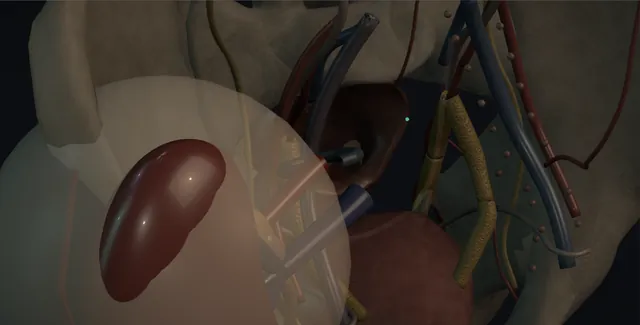
Schematic scene preview: Transplant nephrectomy
Preparing the 3D anatomy…

UrOops3D · UroRef · © Nity G

Click me next Section 2 of 8 Anatomy The structures this operation works between, and the one it must not cross.

The structures in the scene

Transplant nephrectomy: how the structures relateThe iliac fossa reopened through the old Gibson scar. Graft in the iliac fossa: shelled out inside it in the late graft The capsule, the safe plane. Graft in the iliac fossa: the capsule cuffs the pedicle for the clamp Hilum within the capsule. Hilum within the capsule: vein fixed by scar behind; tears on traction External iliac artery and vein (danger). Graft in the iliac fossa: leaves the lower pole medially to the dome Transplant ureter. Graft in the iliac fossa: stuck medially: fill it, never open blind Bladder (danger). Graft in the iliac fossa: fused in the late graft; a diathermy stroke away Peritoneum and the bowel behind it (danger).Graft in the iliacfossaThe capsule, thesafe planeHilum within thecapsule!External iliacartery and veinTransplant ureter!Bladder!Peritoneum and thebowel behind itshelled out insideit in the lategraftthe capsule cuffsthe pedicle for theclampvein fixed by scarbehind; tears ontractionleaves the lowerpole medially tothe domestuck medially:fill it, never openblindfused in the lategraft; a diathermystroke awayTargetDangerLandmarkContext
The iliac fossa reopened through the old Gibson scar. Schematic, not to scale; relationships only.
  • The field is the right or left iliac fossa reopened through the old Gibson scar, the graft lying on the psoas with its hilum medial and posterior, and the external iliac artery and vein passing behind and medial to it under a layer of scar.
  • In the late graft the capsule is opened along the lateral border and the kidney is shelled out toward the hilum, where the capsule cuffs the pedicle above the iliac vessels.
  • The ureter leaves the lower pole and runs medially across the pelvis to the bladder, which lies against the medial surface of the kidney.
  • ›The graft in the iliac fossa - extraperitoneal, on the psoas, with the abdominal wall and peritoneum fused to its front in the late graft.
  • ›The external iliac artery and vein - a centimetre behind the scar at the hilum, with the anastomoses on their anterior or lateral wall.
  • ›The capsule - opened along the convex border and left on the bed; the plane inside it is the safe plane in the late graft.
  • ›The hilum within the capsule - the cuff of capsule around artery, vein and pelvis where the clamp goes.
  • ›The transplant ureter - from the lower pole across the pelvis to the dome; divided low and ligated, or excised with a bladder cuff if it is infected.
  • ›The bladder - stuck to the medial surface of the late graft; identified by filling through the catheter, never opened blind.
  • ›The peritoneum and the bowel behind it - swept medially in the early graft, fused to the kidney in the late one, with bowel a diathermy stroke away.
Click me next Section 3 of 8 Steps The operation in order, with the landmark that guards each move.

Operative steps

  1. Who: the failed graft that hurts, bleeds or infects A failed graft that is quiet is left in place: it keeps some urine output, its removal sensitises the patient and the operation has a real mortality. The graft that is removed is the one that declares itself. Graft intolerance after immunosuppression is withdrawn, in about a third of failed grafts: pain over the kidney, fever, haematuria, a raised CRP and an anaemia that will not correct with erythropoietin. Infection in the graft or a pyelonephritis that does not settle. Bleeding or rupture. Early thrombosis of the artery or the vein, when the kidney is dead and will become an abscess. Malignancy in the graft. And occasionally to make room, though a second kidney usually goes in the other fossa.
  2. Early versus late The timing decides the plane. An early nephrectomy, within the first weeks after transplantation for thrombosis, primary non-function or an early rupture, is done extracapsularly: the planes are fresh, the graft is dissected around to its anastomoses and the vessels are taken at the iliac wall with the patch, the iliac artery and vein repaired under vision. A late nephrectomy, months or years after the transplant, meets a graft fused to the peritoneum, the psoas, the bladder and the iliac vessels by scar. It is done subcapsularly: the capsule is opened, the kidney is shelled out inside it, and the hilum is clamped and oversewn within the capsule, leaving the anastomoses undisturbed.
    where it bites: a late graft dissected extracapsularly toward the iliac vessels is the commonest route to a torn external iliac vein.
  3. Immunosuppression and the timing The patient is back on dialysis, so the operation is timed the day after a session with the potassium and the fluid balance checked, and the nephrologist plans the immunosuppression: the calcineurin inhibitor and the antimetabolite are usually already stopped or stopping, but a patient who has been on prednisolone for years needs hydrocortisone cover through the operation and a taper afterwards, not an abrupt stop. Cross-match and four units available. Antibiotics to cover an infected graft, guided by cultures if they exist. The patient is told the mortality of the operation, small but real, the transfusion risk, the risk to the leg from the iliac vessels, and that removing the graft may raise their antibody levels and make the next kidney harder to find.
  4. Through the old scar The old Gibson or Rutherford Morison incision is reopened along its length, the muscle layers taken through the scar and the retroperitoneal space entered with care, because the graft may lie directly under the wound and the peritoneum is often fused to it. In the early graft the fossa is opened and the kidney is found in its bed with fresh oedematous tissue around it. In the late graft there is no fossa: the kidney is a hard mass with the abdominal wall stuck to its anterior surface, and the dissection stays on the kidney until the capsule is reached. A self-retaining retractor is set, the peritoneum swept medially if it will move, and the surgeon confirms which side the iliac vessels lie before anything is cut.
  5. The subcapsular plane For the late graft the capsule is incised along the convex border with the diathermy and the plane between capsule and cortex is developed with a finger and a swab on a stick, working round the upper pole, the posterior surface and the lower pole in turn, the kidney delivered up into the wound as it frees. The capsule is left behind on the psoas, the peritoneum, the bladder and the vessels, which is the point: everything dangerous stays on the far side of it. The plane bleeds from the cortex and is packed as it is developed. At the hilum the capsule is reflected over the vessels and the pelvis like a cuff, and the dissection stops there.
    where it bites: the capsule stays down on the iliac vessels and the bladder; a finger that leaves the subcapsular plane at the hilum enters scar with the external iliac vein inside it.
  6. The hilum: clamped and oversewn With the kidney freed to its hilum the pedicle is a cuff of capsule around artery, vein and pelvis, well away from the iliac anastomoses. A large curved clamp, or two, is placed across the whole pedicle inside the capsule and the kidney is cut off distal to it. The stump is then oversewn under the clamp with a running 2-0 Prolene, over and over, and the clamp released slowly; a second running layer is placed if there is any ooze. Individual ligation of the artery and vein is better if they can be separated but is not attempted through dense scar. In the early extracapsular graft, by contrast, the artery is taken at the iliac wall and the arteriotomy closed with 5-0 Prolene, then the vein likewise.
    where it bites: the clamp goes across the pedicle within the capsule; a clamp placed wide of it takes the external iliac artery or vein with it.
  7. The ureter and the bladder The transplant ureter runs from the lower pole across the pelvis to the dome of the bladder. In the late graft it is divided as low as it can be reached and ligated with an absorbable tie; the stump and its reimplant are left in the bladder wall and cause no trouble in a bladder that is otherwise healthy. In an infected graft, or where the reimplant is the source, the ureter is followed to the bladder, excised with a cuff and the bladder closed in two layers with a catheter for seven to ten days. The bladder is never opened blindly: it is stuck to the medial surface of the late graft and is identified by filling it through the catheter.
  8. Bleeding: the danger This operation kills by bleeding, from three places. The subcapsular plane oozes from the whole raw cortex and is controlled by packing and by speed, not by chasing points. The pedicle stump bleeds if the clamp slips or the oversew fails, and the answer is pressure with a finger, a second clamp placed under vision and a second layer of suture. And the external iliac vein, thin, fixed and immediately behind the scar, tears when it is pulled or when a clamp bites it: the first move is pressure, proximal and distal control by a vascular surgeon if the tear is not tiny, and a lateral repair with 5-0 Prolene. Blood is in the room before the incision, not sent for after the tear.
    where it bites: the external iliac vein tears on traction; pressure, then control above and below, then repair, never a blind clamp in a pool of blood.
  9. After A drain in the fossa, the wound closed in layers with a non-absorbable or slowly absorbable suture because the patient is immunosuppressed and the wound will not heal quickly. Dialysis the next day through the existing access. The haemoglobin is checked that evening and the next morning, because the subcapsular bed oozes. The leg is watched for swelling and pulses on the first day. The immunosuppression is tapered by the nephrologist, and the antibody screen is repeated at about a month and again before relisting, since the calculated reaction frequency often rises. The patient goes back to the transplant clinic for relisting, and the histology of the graft is read at the multidisciplinary meeting.
Click me next Section 4 of 8 Danger zones Where this operation bites, and why it bites there.
Critical relationship

Danger zones

The spatial relationships the animation is built to make automatic. Before you read each one, ask yourself: at this point in the operation, what is about to go wrong, and what is my first move? The save for each sits in the Complications tab.

  • RiskThe external iliac vein - thin, fixed by scar and immediately behind the pedicle; it tears on traction and bleeds without warning.
  • RiskThe pedicle stump - a clamp that slips or an oversew that fails bleeds into a closed fossa; two layers of Prolene, released slowly.
  • RiskThe arterial anastomosis in infection - an infected arterial stump can blow out days later; the patch is excised and the iliac artery repaired or bypassed with a vascular surgeon.
  • RiskThe bladder and bowel on the graft - stuck to the medial surface and the peritoneal side of a late graft; a diathermy plane that leaves the capsule finds them.
Practise it Take me into the scene Opens the animation at a danger moment and asks you to predict the mechanism and your first move before it reveals.
Click me next Section 5 of 8 Complications The save for each one: the first move, the escalation trigger, the op-note line.

Complications and immediate saves

Trigger each of these live in the Animation tab, then read the save: the first move, the escalation trigger, and the line that goes in the op note.

Iliac vessel haemorrhage

  • a tear in the external iliac vein on traction, or the artery caught by the clamp: sudden dark or bright bleeding filling the fossa from behind the pedicle.
The save
  1. Pressure with a finger or a swab on the bleeding point and the anaesthetist told; do not clamp blind. first move
  2. Blood in the room and the cell saver if there is no infection; a second suction and a better retractor.
  3. Control above and below by exposing the external iliac vessels away from the scar, then a lateral repair with 5-0 Prolene under vision.
  4. A vein that cannot be repaired, or an arterial injury in a scarred field: a vascular surgeon now; the artery may need a patch, an interposition graft or a femorofemoral crossover. escalation trigger
  5. Op note: the vessel, the repair, the units transfused, the distal pulses at the end and the plan for the leg. document

Infected stump and pelvic abscess

  • capsule and vascular stumps left behind in an infected fossa: fever, a collection on ultrasound, and the feared late haemorrhage from an infected arterial stump.
The save
  1. Cultures from the graft and the fossa at the time; antibiotics continued and adjusted to the results. first move
  2. A collection on ultrasound or CT: drained percutaneously and the drain left until it is dry.
  3. In an infected graft the arterial patch is excised and the iliac artery closed primarily or with a vein patch, never left as an infected stump.
  4. Bleeding from the wound, a pulsatile mass or a fall in haemoglobin with fever: an infected pseudoaneurysm; CT angiogram and a vascular surgeon the same day. escalation trigger
  5. Op note: what was left behind, the state of the arterial stump, the cultures sent and the drain plan. document

Bladder or bowel injury

  • the bladder fused to the medial surface of the late graft, or bowel stuck to the peritoneal side, opened by a plane that left the capsule.
The save
  1. Fill the bladder through the catheter to define it before the medial dissection; look for urine or bowel content in the field. first move
  2. A bladder hole: close in two layers with absorbable suture and a catheter for ten days with a cystogram before removal.
  3. A small bowel or colonic injury: repaired primarily by a general surgeon, with antibiotics and the fossa drained separately.
  4. Faecal contamination in an immunosuppressed patient, or a colonic injury in an infected field: resection or a defunctioning stoma, decided in theatre. escalation trigger
  5. Op note: the organ, the repair, the catheter and drain plan and who was called. document

Limb ischaemia and iliac vein thrombosis

  • a narrowed or thrombosed external iliac artery after repair, or a compressed and injured iliac vein: a cold leg, or a swollen one, on the first day.
The save
  1. Feel and Doppler the femoral and pedal pulses at the end of the operation and on the ward at each round for the first day. first move
  2. A swollen leg: duplex for deep vein thrombosis and anticoagulate, remembering the raw fossa.
  3. A leg that was warm and is now cold or painful: CT angiogram, or straight to theatre if the pulse has gone.
  4. Absent pulses with a threatened limb: a vascular surgeon for thrombectomy, patch or bypass within hours. escalation trigger
  5. Note: the pulses at the end of the operation, the anticoagulation plan and the leg observations ordered. document

Sensitisation, the wound and the next transplant

  • antibody levels that rise after the graft is removed and the immunosuppression stops, a wound that will not heal in a steroid-treated patient, and a longer wait for the next kidney.
The save
  1. The immunosuppression taper written by the nephrologist, with hydrocortisone cover and a plan to stop, not an abrupt withdrawal. first move
  2. HLA antibody screen at about a month and before relisting; the calculated reaction frequency is expected to rise.
  3. The wound closed with a durable suture, the drain kept until dry, and the patient warned about hernia and slow healing.
  4. Wound dehiscence, or a collection under the wound with fever: reopen, wash out and lay open or close over a drain. escalation trigger
  5. Note: the indication for nephrectomy, the immunosuppression plan and that the patient was told about sensitisation before the operation. document
Click me next Section 6 of 8 Troubleshooting When the case turns: the problem, then the next move.

Intra-operative troubleshooting

When the case turns: the problem, then the next move.

  • ActionThe capsule will not separate from the kidney - a late graft with cortical infarction fused to its capsule; open the capsule in a second place and work from the pole, packing as you go, or accept a piece of capsule on the kidney where it will not come.
  • ActionThe subcapsular bed will not stop oozing - pack it firmly for ten minutes by the clock, warm the patient, correct the clotting, and leave a haemostatic agent under the pack; do not chase points in the cortex.
  • ActionThe clamp will not sit across the pedicle - the cuff of capsule is too short; free the kidney further at the poles and place two clamps in series from above and below rather than one that reaches around blind.
  • ActionThe oversewn stump oozes when the clamp is released - reapply the clamp under vision, a second running layer of 2-0 Prolene over the first, and release slowly again; a finger on it while the suture is loaded.
  • ActionThe early graft is thrombosed and soft - extracapsular plane, the vessels to the iliac wall, the patches excised and the arteriotomy and venotomy closed with 5-0 Prolene under a side-biting clamp; send the graft for histology.
  • ActionUrine in the field from the bladder - fill the bladder to find the hole, close it in two layers, catheter for ten days and a cystogram before removal.
Click me next Section 7 of 8 FRCS viva Say it out loud, then mark yourself against the model answer.

FRCS viva questions

The examiner stems this scene prepares you for. Try to answer out loud first, then reveal, and learn the discriminator, the detail that turns a pass into a strong pass.

QWhen would you remove a failed renal transplant, and when would you leave it in?reveal answer
  • I would leave a failed graft in place when it is asymptomatic, because retained grafts keep some residual urine output, avoid an operation with a real mortality, and their removal, together with the withdrawal of immunosuppression, is associated with a rise in HLA antibodies that makes the next transplant harder to match.
  • I would remove it when it declares itself.
  • The commonest indication is the graft intolerance syndrome after immunosuppression is stopped, in about a third of failed grafts: pain over the graft, fever, haematuria, a raised CRP and an anaemia resistant to erythropoietin, where nephrectomy or embolisation resolves the inflammation.
  • Other indications are infection in the graft that does not settle on antibiotics, bleeding or rupture, early arterial or venous thrombosis where the kidney is dead and will become an abscess, malignancy in the graft, and rarely the need for space.
  • I would consent for bleeding and transfusion, injury to the iliac vessels with risk to the leg, injury to bladder or bowel, wound problems, a mortality that is small but real, and sensitisation.
  • The decision is made with the nephrologist, who plans the immunosuppression withdrawal and the dialysis around it.
Strong pass

Say both

  • Graft intolerance syndrome named with its features and its frequency
  • Sensitisation given as the reason the asymptomatic graft is left in
Examiner note
Graft intolerance syndrome named with its features and its frequency, and sensitisation given as the reason the asymptomatic graft is left in.
QDescribe a late transplant nephrectomy and explain the plane you would use.reveal answer
  • A late graft is fused by scar to the abdominal wall, peritoneum, psoas, bladder and, at the hilum, the external iliac vessels, so I would use the subcapsular technique, which keeps all of those on the far side of the capsule.
  • After dialysis the day before, with blood cross-matched and hydrocortisone cover, I would reopen the old Gibson incision and dissect onto the kidney rather than around it.
  • I would incise the capsule along the convex border and develop the plane between capsule and cortex with a finger and a swab on a stick around both poles and the posterior surface, packing the raw bed as I went, until the kidney was attached only at the hilum by a cuff of capsule around the vessels and pelvis.
  • I would clamp the whole pedicle inside the capsule, divide the kidney off distal to the clamp, and oversew the stump with running 2-0 Prolene in two layers, releasing the clamp slowly under vision.
  • I would divide and ligate the ureter low, or excise it with a bladder cuff if it was the source of infection.
  • I would drain the fossa, close in layers, check the femoral and pedal pulses, and arrange dialysis and an antibody screen.
Strong pass

Say both

  • The subcapsular plane justified by what it leaves behind
  • The clamp placed inside the capsule with the anastomoses left undisturbed
Examiner note
The subcapsular plane justified by what it leaves behind, and the clamp placed inside the capsule with the anastomoses left undisturbed.
QHow does an early transplant nephrectomy differ from a late one?reveal answer
  • The difference is the plane and what is done at the vessels, and it follows from the state of the tissues.
  • In the first weeks after transplantation, for thrombosis, primary non-function or an early rupture, the planes are fresh and the graft can be dissected extracapsularly around to its anastomoses.
  • I would then take the artery and the vein at the iliac wall, excising the patches under side-biting clamps and closing the arteriotomy and venotomy with 5-0 Prolene, which removes all donor tissue and leaves no stump, and I would excise the ureter with a bladder cuff and close the bladder.
  • That matters in infection, where a retained arterial patch can become an infected pseudoaneurysm.
  • A late graft, months or years on, is encased in scar with the external iliac vein directly behind the hilum, and extracapsular dissection there is the commonest route to a major venous injury;
  • so I would stay inside the capsule, leave capsule and anastomoses in place, and clamp and oversew the pedicle within the cuff.
  • The trade is that the late technique leaves donor vessel and capsule behind, which is acceptable in a sterile field and unacceptable in an infected one, where a vascular surgeon may be needed to deal with the anastomosis.
Strong pass

Say both

  • The infected arterial patch as the reason the extracapsular technique matters
  • The external iliac vein as the reason the subcapsular technique exists
Examiner note
The infected arterial patch as the reason the extracapsular technique matters, and the external iliac vein as the reason the subcapsular technique exists.
QWhat are the complications of transplant nephrectomy, and how do you reduce them?reveal answer
  • The operation carries a morbidity of a fifth to a half in the published series and a mortality of a few per cent, both higher than for a native nephrectomy, and the figures I would quote are of that order rather than precise.
  • The dominant complication is haemorrhage: from the raw subcapsular bed, from a pedicle stump that was not oversewn securely, and from the external iliac vein torn by traction or caught by a clamp.
  • I reduce it by having blood in the room, by packing rather than chasing the bed, by two layers of Prolene on the stump released slowly, and by staying inside the capsule in the late graft.
  • Infection: retained capsule or an infected arterial stump can form an abscess or a pseudoaneurysm, reduced by cultures, drainage and excising the patch when the field is infected.
  • Injury to the bladder or bowel fused to the graft, reduced by filling the bladder and staying on the kidney.
  • Limb ischaemia or iliac vein thrombosis, detected by checking pulses and the leg.
  • Wound dehiscence and hernia in an immunosuppressed patient.
  • And sensitisation, with a rise in HLA antibodies that lengthens the wait for the next kidney, which is why the asymptomatic failed graft is left alone.
Strong pass

Say both

  • The three sources of bleeding each paired with the measure that prevents it
  • Sensitisation named as a complication with a consequence for relisting
Examiner note
The three sources of bleeding each paired with the measure that prevents it, and sensitisation named as a complication with a consequence for relisting.
Click me next Section 8 of 8 Evidence The guidance each of these answers is built on.

Evidence and guidance

Two minutes

Close the lap: recall it before you leave.

Say it aloud, then reveal. Reading is not remembering.

  1. 01 The relationship Say the critical spatial relationship out loud, then reveal. reveal
    Memory frame

    The field is the right or left iliac fossa reopened through the old Gibson scar, the graft lying on the psoas with its hilum medial and posterior, and the external iliac artery and vein passing behind and medial to it under a layer of scar.

    Why?
    In the late graft the capsule is opened along the lateral border and the kidney is shelled out toward the hilum, where the capsule cuffs the pedicle above the iliac vessels. The ureter leaves the lower pole and runs medially across the pelvis to the bladder, which lies against the medial surface of the kidney.
  2. 02 The first move Iliac vessel haemorrhage. Your first move? reveal
    First move

    Pressure with a finger or a swab on the bleeding point and the anaesthetist told; do not clamp blind.

  3. 03 The escalation trigger Iliac vessel haemorrhage. What makes you call for help? reveal
    Escalate when

    A vein that cannot be repaired, or an arterial injury in a scarred field

    More detail
    A vein that cannot be repaired, or an arterial injury in a scarred field: a vascular surgeon now; the artery may need a patch, an interposition graft or a femorofemoral crossover.
  4. 04 The discriminator What turns a pass into a strong pass? reveal
    Strong pass

    Say both

    • Graft intolerance syndrome named with its features and its frequency
    • Sensitisation given as the reason the asymptomatic graft is left in
    Examiner note
    Graft intolerance syndrome named with its features and its frequency, and sensitisation given as the reason the asymptomatic graft is left in.
  5. 05 The pitfall Where does this operation bite first? reveal
    Pitfall

    The external iliac vein

    Why?
    thin, fixed by scar and immediately behind the pedicle; it tears on traction and bleeds without warning.

Revision practice with self-marked answers, not an assessment of clinical competence. Kept in this browser only; it does not sync between devices.