
Revision sheet
Bladder rupture: pelvic fracture + cystogram
7 steps, each one the authored title and caption from the interactive animation, with the frame captured from that step. Read it before the case, or print it and work through the steps in order.
Teaching only, not for diagnosis or treatment decisions. The text on this sheet is the animation's own step copy, reproduced verbatim.
UrOops3D · UroRef · © Nity G
- 01
The mechanism
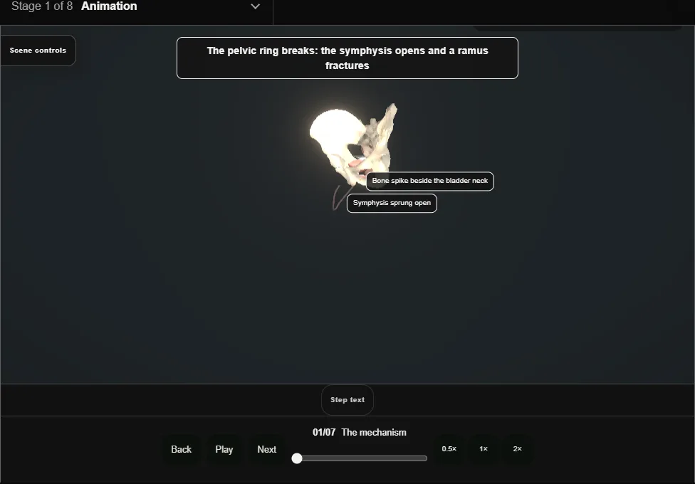
- Blunt trauma breaks the pelvic ring: the symphysis springs open, rami fracture, and the bladder is torn either against the bone at its neck or burst at the peritonealised dome by pressure.
- The same force decides both patterns.
- 02
Assessment first
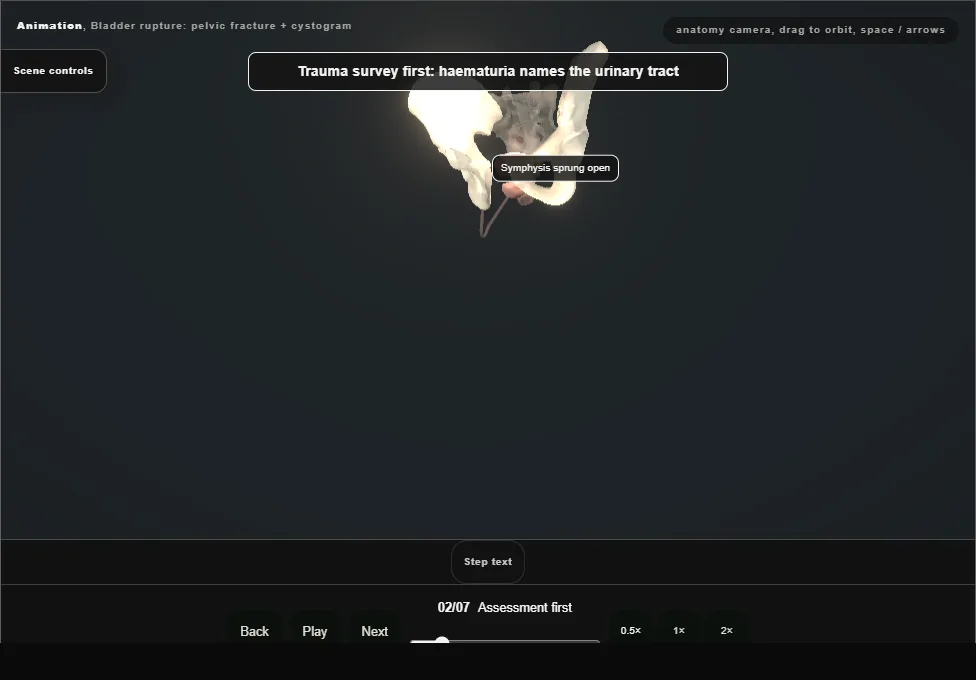
- Trauma survey before urology: gross haematuria names the urinary tract, but bleeding and shock are managed by the whole team.
- Blood at the meatus changes the plan to a urethral study before any catheter.
- 03
CT cystogram, properly filled
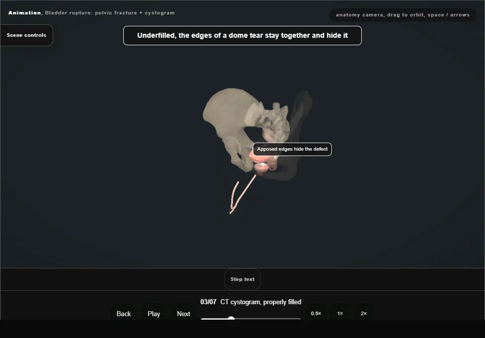
- Retrograde contrast with the bladder filled to discomfort and the catheter clamped.
- An underfilled bladder wall apposes the tear and hides it: the underfilled cystogram is a false-negative machine.
- 04
Read the leak
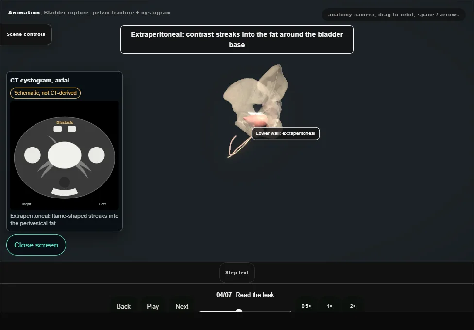
- Extraperitoneal contrast stripes into the pelvic haematoma around the neck.
- Intraperitoneal contrast outlines bowel loops: the dome tear has burst into the peritoneal cavity.
- The two leaks go to two different theatres.
- 05
Extraperitoneal: catheter drainage
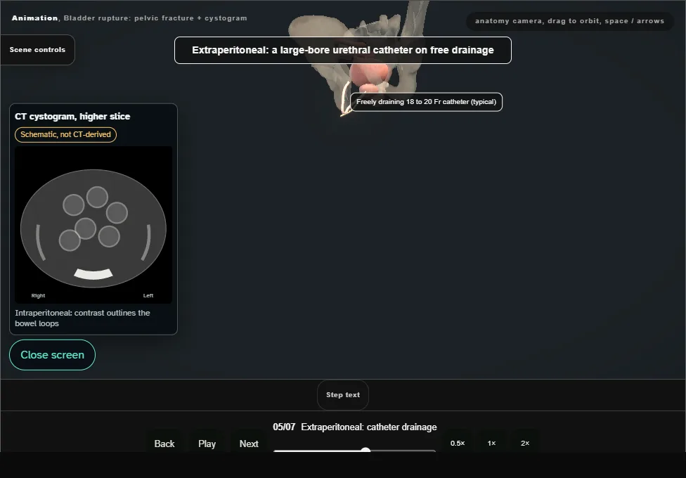
- Extraperitoneal rupture heals on a freely draining catheter.
- The bladder is closed by the body, not the surgeon, provided the urine is diverted and the catheter never blocks.
- The exceptions go to theatre: bladder neck involvement, a bone fragment in the wall, a rectal or vaginal injury, or a pelvis already being opened for fixation.
- 06
Intraperitoneal: repair at laparotomy
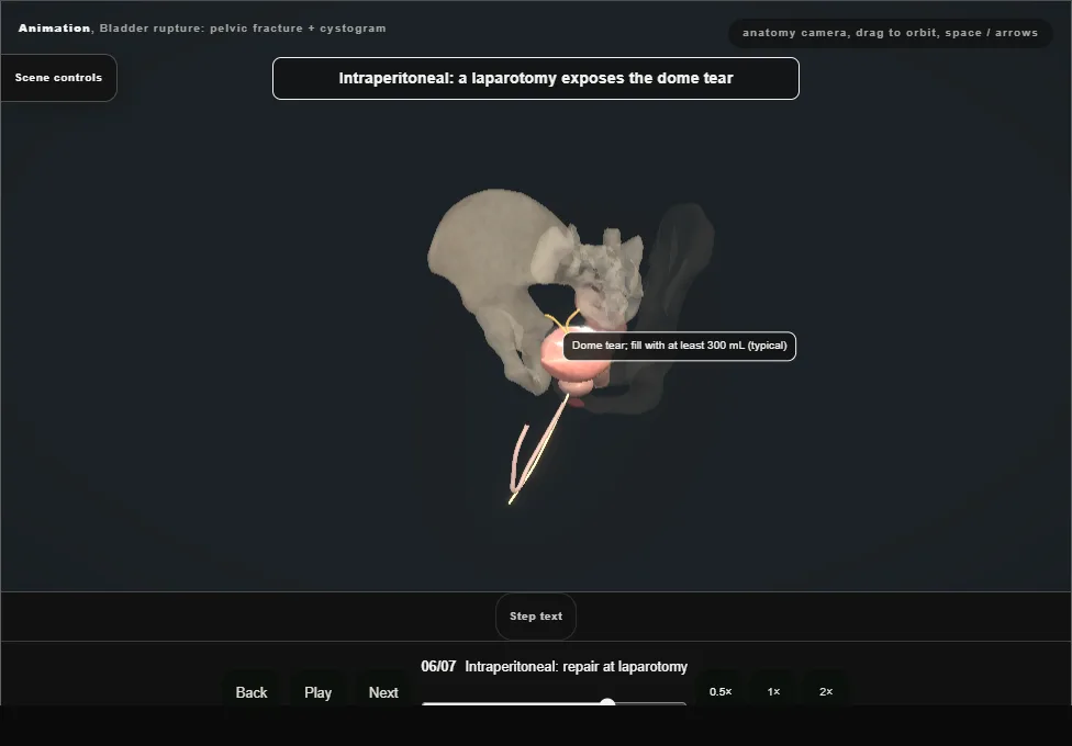
- Intraperitoneal rupture is a laparotomy: the dome tear is closed in two absorbable layers, the bladder neck is inspected, and a catheter keeps the repair at rest.
- 07
Coordination with orthopaedics
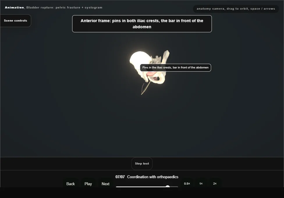
- The external fixator pins and bar own the same lower abdominal wall as the suprapubic corridor.
- Fixator position and catheter route are decided in one conversation, not two.
Derived from BladderTraumaScene.tsx at build time: every step title and caption above is the authored
ProcedureDef text, reproduced verbatim, and this page shares its readable-points renderer with the animation
itself. 7 of 7 steps have a captured frame.
Text fingerprint a41f35e09a043f27. A caption edit without a re-capture fails the production
check, so the sheet and the animation cannot drift apart silently.