
Revision sheet
Epididymal cyst excision and epididymectomy
8 steps, each one the authored title and caption from the interactive animation, with the frame captured from that step. Read it before the case, or print it and work through the steps in order.
Teaching only, not for diagnosis or treatment decisions. The text on this sheet is the animation's own step copy, reproduced verbatim.
UrOops3D · UroRef · © Nity G
- 01
Examination and consent
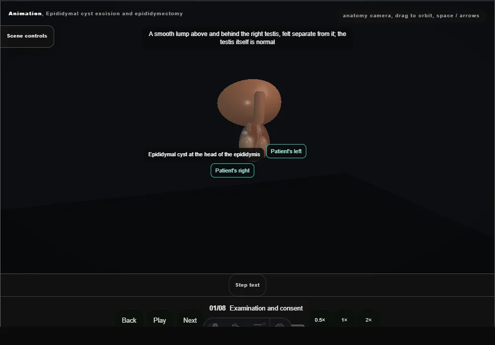
- A smooth lump above and behind the testis, separate from it, that transilluminates; ultrasound confirms an epididymal cyst.
- Consent covers recurrence, pain and the risk to fertility on that side.
- 02
Incision
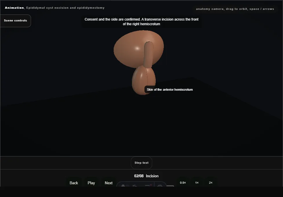
- A transverse incision across the front of the right hemiscrotum, and the dartos divided with diathermy down to the tunica vaginalis.
- 03
Delivery of the testis
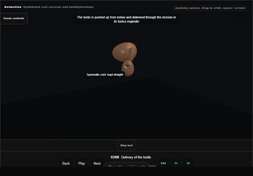
- The testis is pushed up from below and delivered through the incision in its tunica vaginalis, the cord kept straight.
- 04
Tunica opened, cyst found
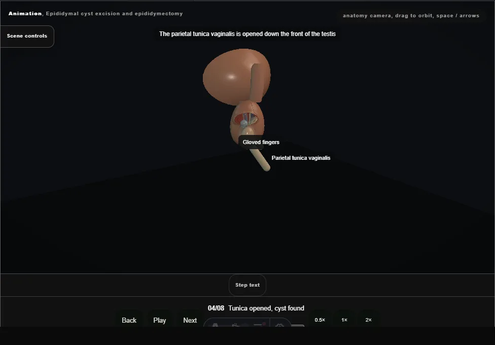
- The parietal tunica vaginalis is opened.
- The cyst sits on the head of the epididymis; the tubules lie under its base and the testicular vessels enter the upper pole beside it.
- 05
Dissection off the epididymis
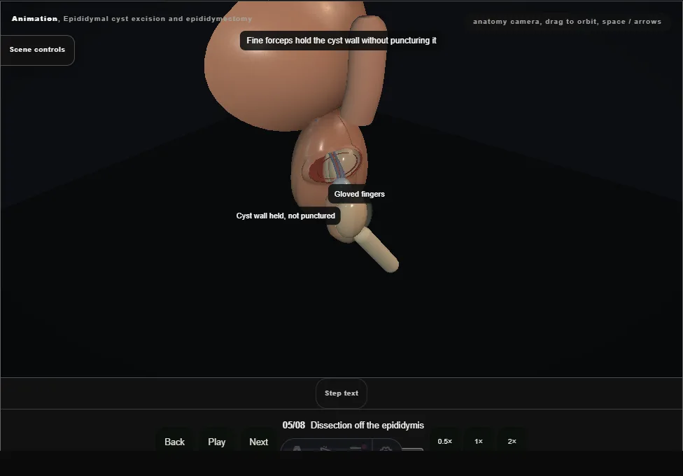
- The cyst wall is held without puncturing it and dissected off the head with fine scissors, staying on the cyst, until its base narrows to a single neck.
- 06
Neck tied, cyst removed intact
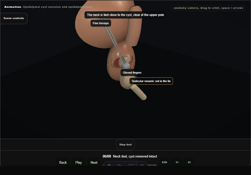
- The neck is tied close to the cyst, clear of the upper pole, and divided.
- The cyst comes away intact and goes to pathology.
- 07
Base cauterised, head covered
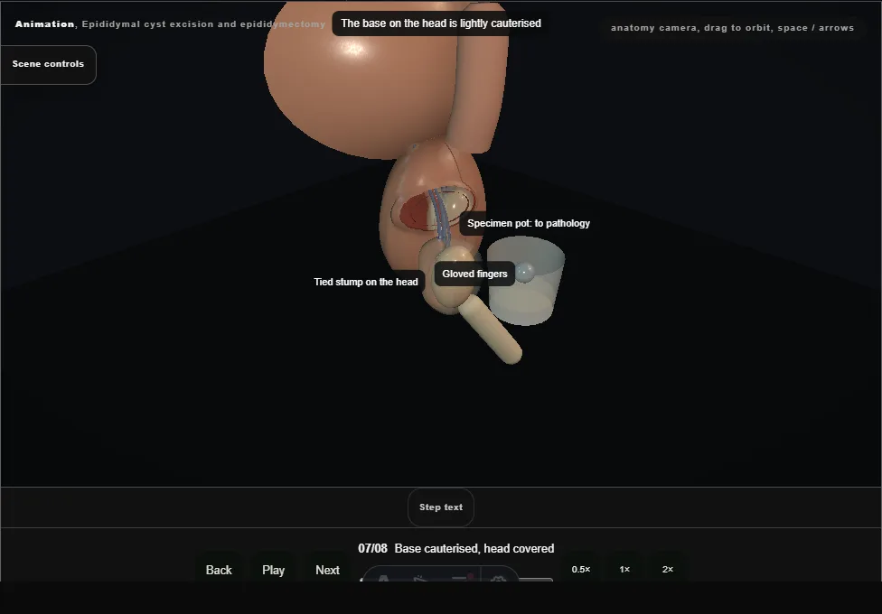
- The base is lightly cauterised and the covering of the head closed over it.
- Epididymis, testis and cord are inspected: intact, the testis pink.
- 08
Return and closure
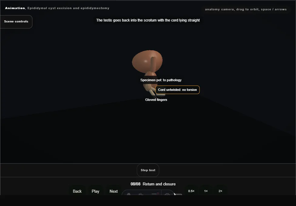
- The testis goes back with the cord untwisted.
- Dartos and skin are closed with absorbable stitches, and a dressing and a scrotal support applied.
Derived from scrotal/EpididymalSurgeryScene.tsx at build time: every step title and caption above is the authored
ProcedureDef text, reproduced verbatim, and this page shares its readable-points renderer with the animation
itself. 8 of 8 steps have a captured frame.
Text fingerprint b607a8705ea159a0. A caption edit without a re-capture fails the production
check, so the sheet and the animation cannot drift apart silently.