
Revision sheet
Hydrocele repair: Jaboulay repair
8 steps, each one the authored title and caption from the interactive animation, with the frame captured from that step. Read it before the case, or print it and work through the steps in order.
Teaching only, not for diagnosis or treatment decisions. The text on this sheet is the animation's own step copy, reproduced verbatim.
UrOops3D · UroRef · © Nity G
- 01
Examination: transilluminate + examine the testis
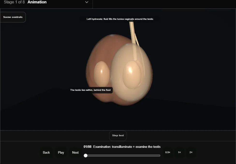
- The swelling may transilluminate and you can get above it, but the testis itself still needs examination.
- Use ultrasound when the testis cannot be confidently palpated, the mass is atypical or non-transilluminating, or underlying pathology is suspected.
- 02
Anterior scrotal incision
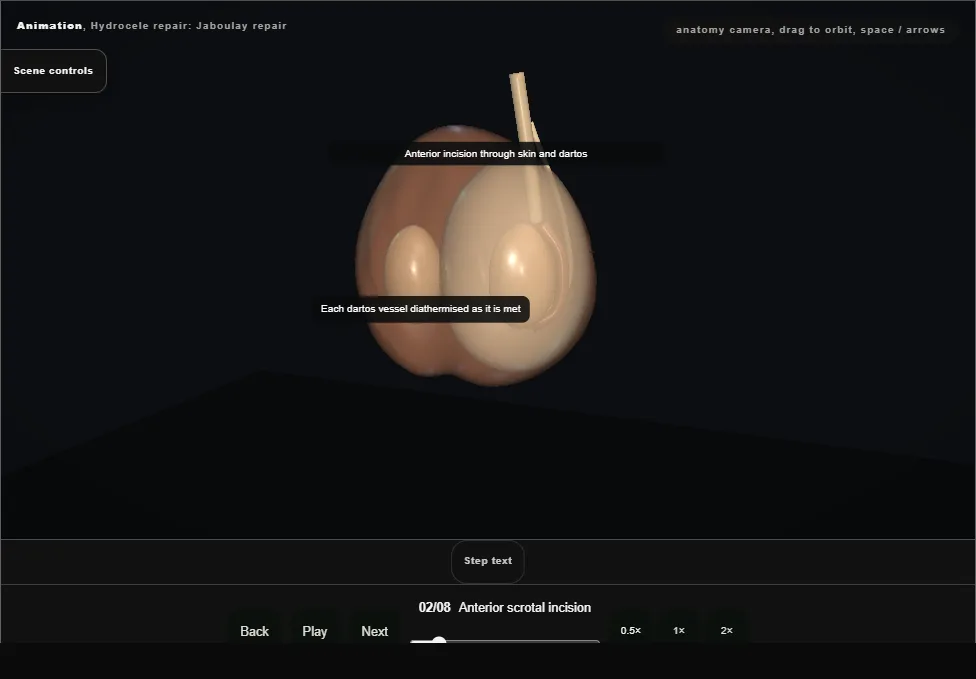
- A vertical incision over the fullest part of the sac, anterior scrotum, through skin and dartos.
- The dartos vessels bleed freely: haemostasis starts here, not at the end.
- 03
Delivery of the testis in its tunica
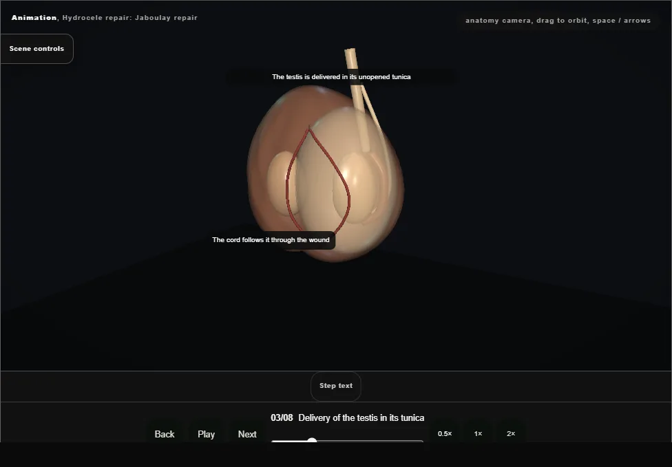
- The shiny parietal tunica is freed from the dartos and the testis is delivered in its unopened tunica through the wound, the cord following it.
- 04
Opening + draining the tunica vaginalis
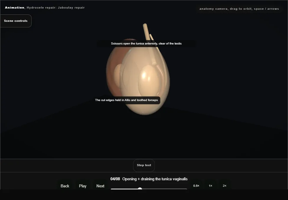
- The tunica vaginalis is opened anteriorly, clear of the testis behind it, and the fluid drained.
- The cut edge is held as it opens; testis and epididymis are then inspected.
- 05
Eversion of the tunica
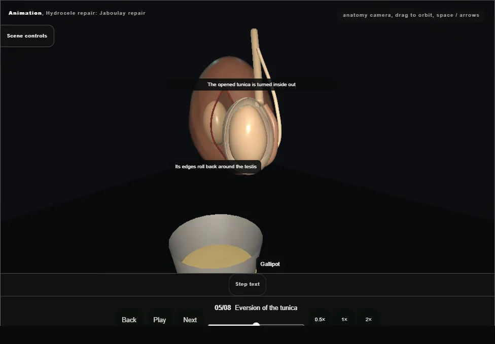
- The opened tunica is everted: turned inside out around the testis, so its lining faces outwards and both cut edges come to lie behind the testis and cord.
- A very large sac may first have its surplus trimmed.
- 06
Continuous closure behind the testis and cord
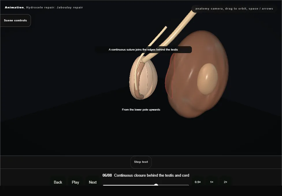
- The edges are closed with a continuous suture behind the testis and cord, from the lower pole upwards.
- It must not strangle the cord: a little finger should pass along the cord into the everted tunica.
- 07
Diathermy + haemostasis
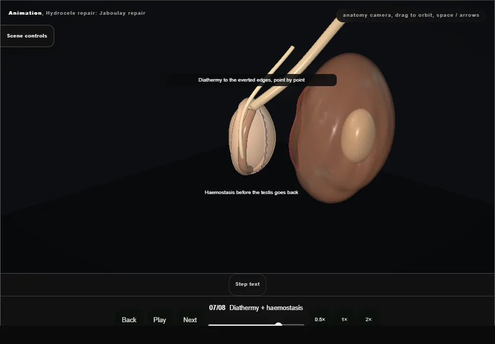
- Diathermy to the everted edges and every bleeding point until the field is dry.
- The scrotum is dependent: a vessel left now fills the space with blood.
- 08
Return, layered closure + support
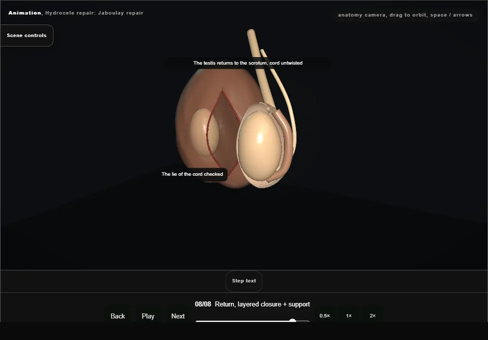
- The testis goes back into the scrotum with the cord untwisted.
- Dartos and skin are closed in layers, then a scrotal-support dressing and elevation.
Derived from HydroceleScene.tsx at build time: every step title and caption above is the authored
ProcedureDef text, reproduced verbatim, and this page shares its readable-points renderer with the animation
itself. 8 of 8 steps have a captured frame.
Text fingerprint 0fd8d4f9a426ab9e. A caption edit without a re-capture fails the production
check, so the sheet and the animation cannot drift apart silently.