
Revision sheet
Ileal ureter
9 steps, each one the authored title and caption from the interactive animation, with the frame captured from that step. Read it before the case, or print it and work through the steps in order.
Teaching only, not for diagnosis or treatment decisions. The text on this sheet is the animation's own step copy, reproduced verbatim.
UrOops3D · UroRef · © Nity G
- 01
Open and expose
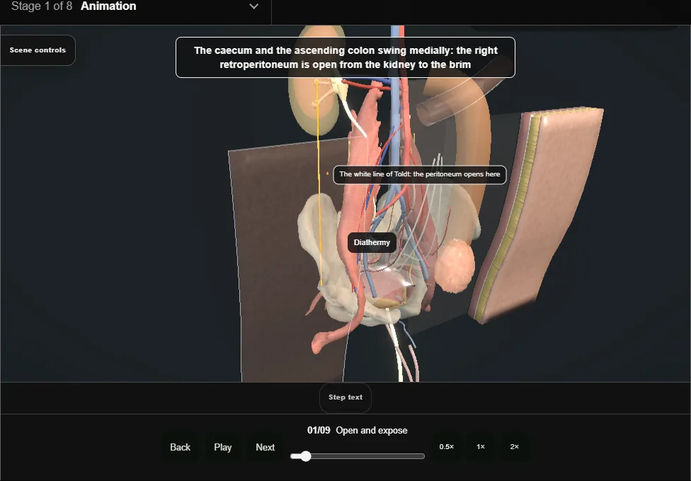
- Midline laparotomy.
- The white line of Toldt is opened with diathermy and the caecum and ascending colon are reflected medially.
- The scarred ureter is seen pale and narrow from below the junction to the brim, with the cava medial to it and the duodenum above.
- 02
Choose the segment
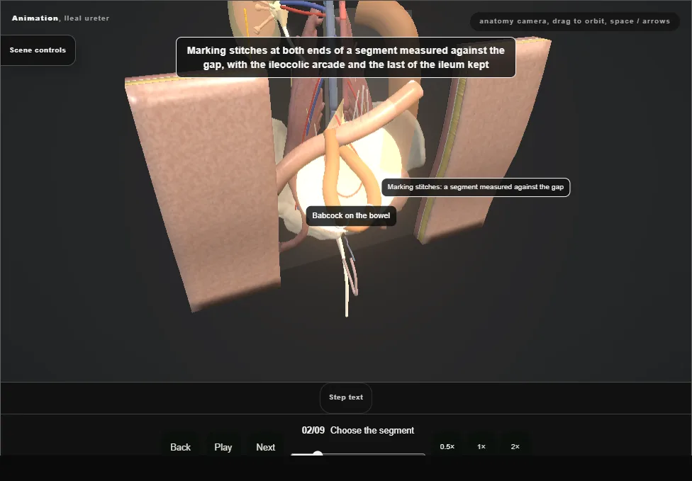
- The terminal ileum followed from the caecum.
- A loop is held against the light and the arcades show through the mesentery; marking stitches at both ends of a segment measured against the gap, well back from the valve.
- 03
Isolate on its pedicle
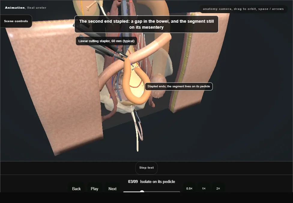
- The sealing device opens a mesenteric window at each end between the arcades; a linear cutting stapler divides the bowel at each end.
- The segment is free on its pedicle, both ends stapled shut.
- A window too deep devascularises the segment.
- 04
Restore continuity
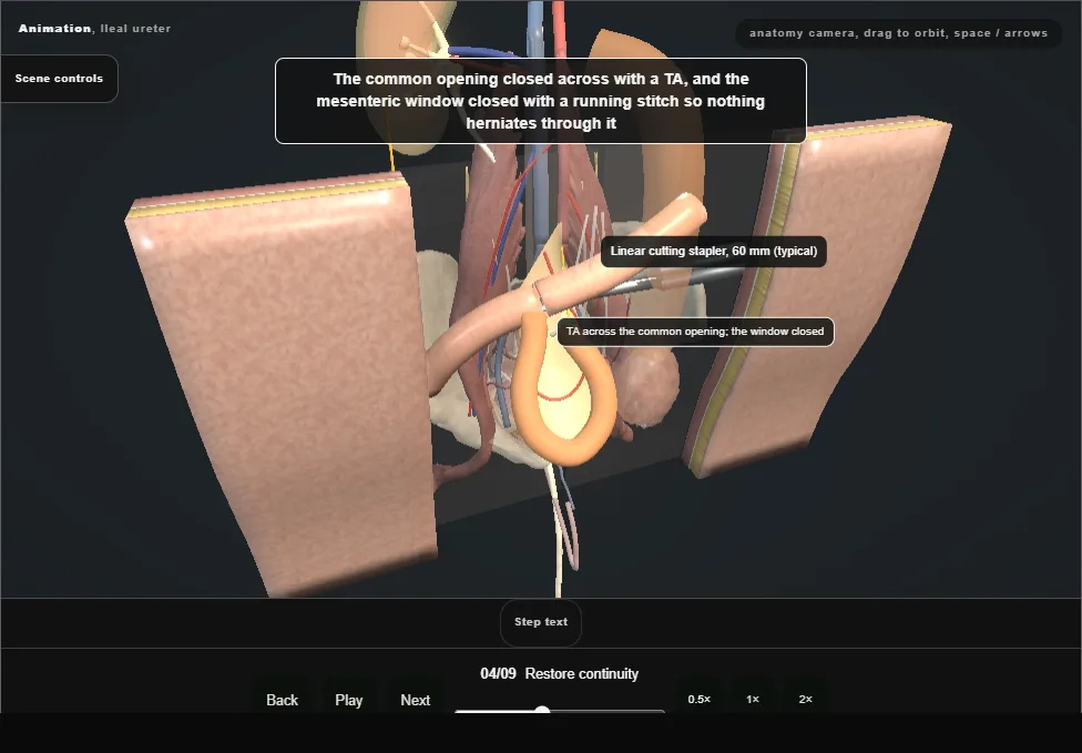
- Antimesenteric corners trimmed; the stapler limbs into both ends and fired side to side; a TA across the common opening; the mesenteric window closed with a running stitch against an internal hernia.
- The segment lies caudal to the join.
- 05
Open, wash and orient
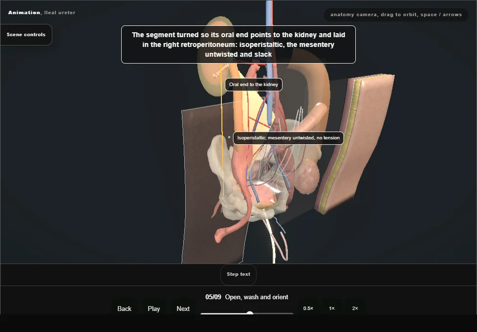
- The staple lines cut off, the lumen washed through with saline.
- The segment is turned so its oral end points to the kidney and laid in the right retroperitoneum: isoperistaltic, the mesentery untwisted and without tension.
- 06
Ileopyelostomy
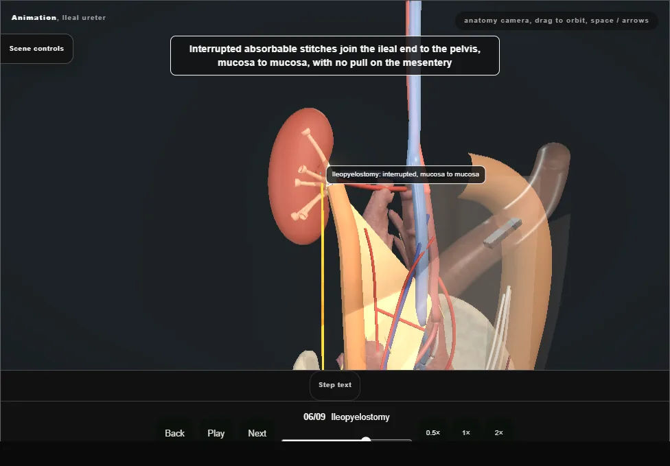
- Gerota's fascia opened over the renal pelvis; stays above and below; an oblique pyelotomy as long as the bowel opening.
- The native ureter is divided below the junction and its stump tied.
- Interrupted absorbable stitches join the ileal end to the pelvis, with no pull on the mesentery and the hilar vessels behind.
- 07
Bladder window
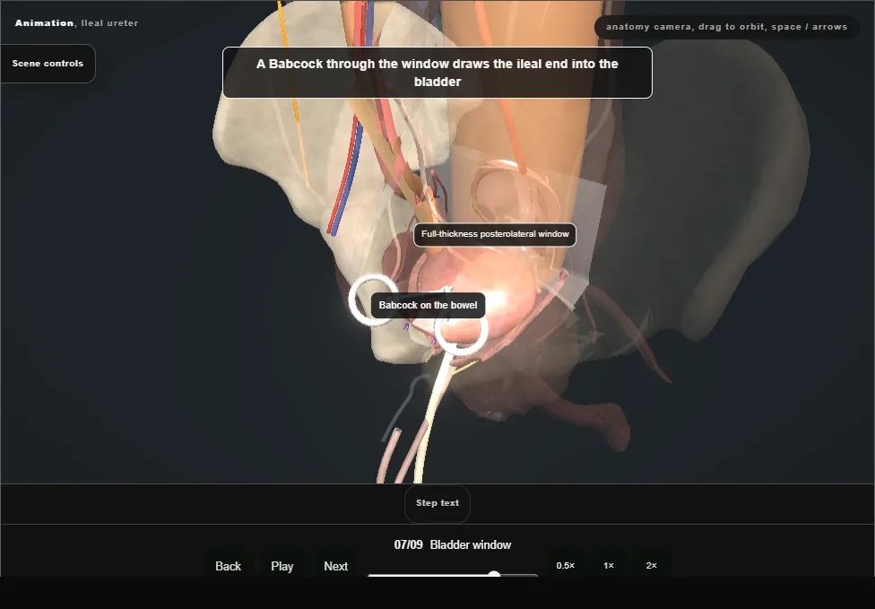
- Stays on the anterior bladder and a midline cystotomy; the halves part.
- A full-thickness disc is excised from the posterolateral wall, clear of the right orifice and the trigone, and a Babcock through it draws the ileal end into the bladder.
- 08
Ileovesical join and test
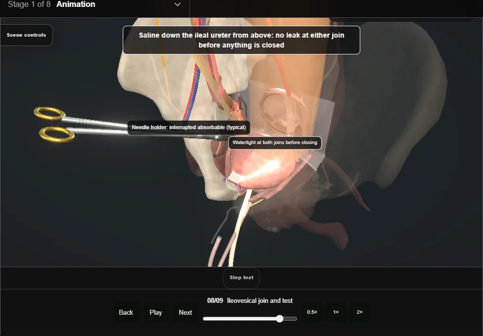
- An outer seromuscular row and an inner mucosal row of interrupted stitches, refluxing by design.
- Saline down the ileal ureter: watertight at both joins before the cystotomy is closed in two layers over the catheter.
- 09
Drain and close
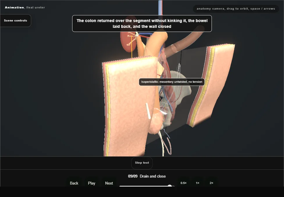
- A drain to the retroperitoneum beside the joins, the colon returned over the segment without kinking it, the bowel laid back and the wall closed.
- One continuous lumen from the renal pelvis to the bladder.
Derived from pelvis/IlealUreterScene.tsx at build time: every step title and caption above is the authored
ProcedureDef text, reproduced verbatim, and this page shares its readable-points renderer with the animation
itself. 9 of 9 steps have a captured frame.
Text fingerprint ab3ea11d3db9e1ed. A caption edit without a re-capture fails the production
check, so the sheet and the animation cannot drift apart silently.