
Revision sheet
Deceased-donor kidney retrieval
9 steps, each one the authored title and caption from the interactive animation, with the frame captured from that step. Read it before the case, or print it and work through the steps in order.
Teaching only, not for diagnosis or treatment decisions. The text on this sheet is the animation's own step copy, reproduced verbatim.
UrOops3D · UroRef · © Nity G
- 01
Who: the donor
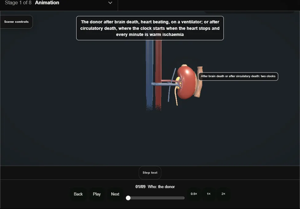
- After brain death, heart beating on a ventilator; or after circulatory death, where the clock starts when the heart stops and every minute is warm ischaemia.
- The kidneys tolerate the most and go last.
- 02
The team, the consent and the clock
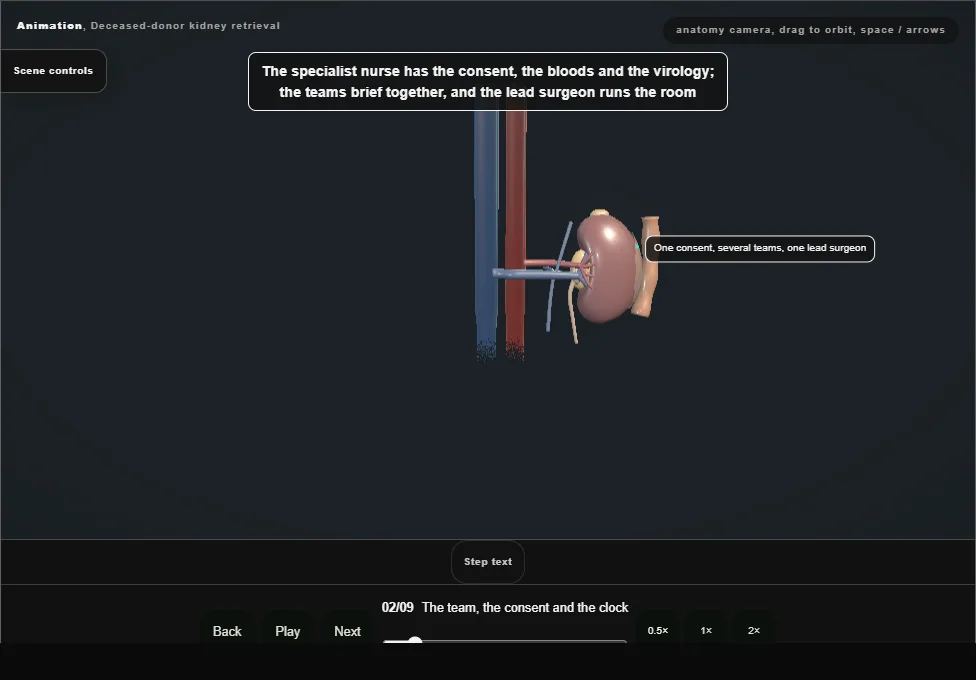
- The specialist nurse has the consent, the bloods and the virology; the teams brief together and the lead surgeon runs the room.
- In circulatory death the teams wait outside for the five minutes after asystole.
- 03
The incision and the exploration
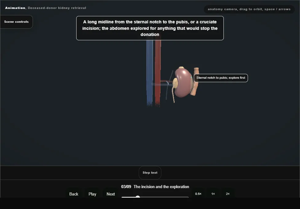
- A long midline from the sternal notch to the pubis; the abdomen explored for anything that would stop the donation.
- The right colon and small bowel mobilised up and to the left off the retroperitoneum.
- 04
The aorta and the cava
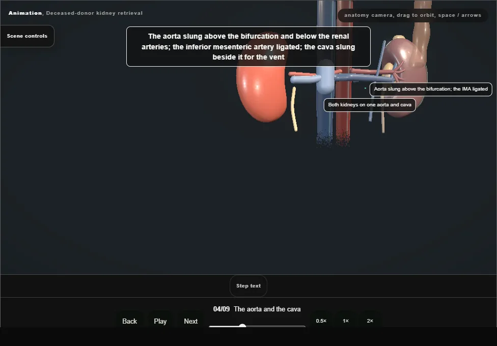
- The aorta slung above the bifurcation and below the renal arteries, the inferior mesenteric artery ligated, the cava slung for the vent; the supracoeliac aorta exposed through the crus for the cross-clamp.
- 05
Cannulation
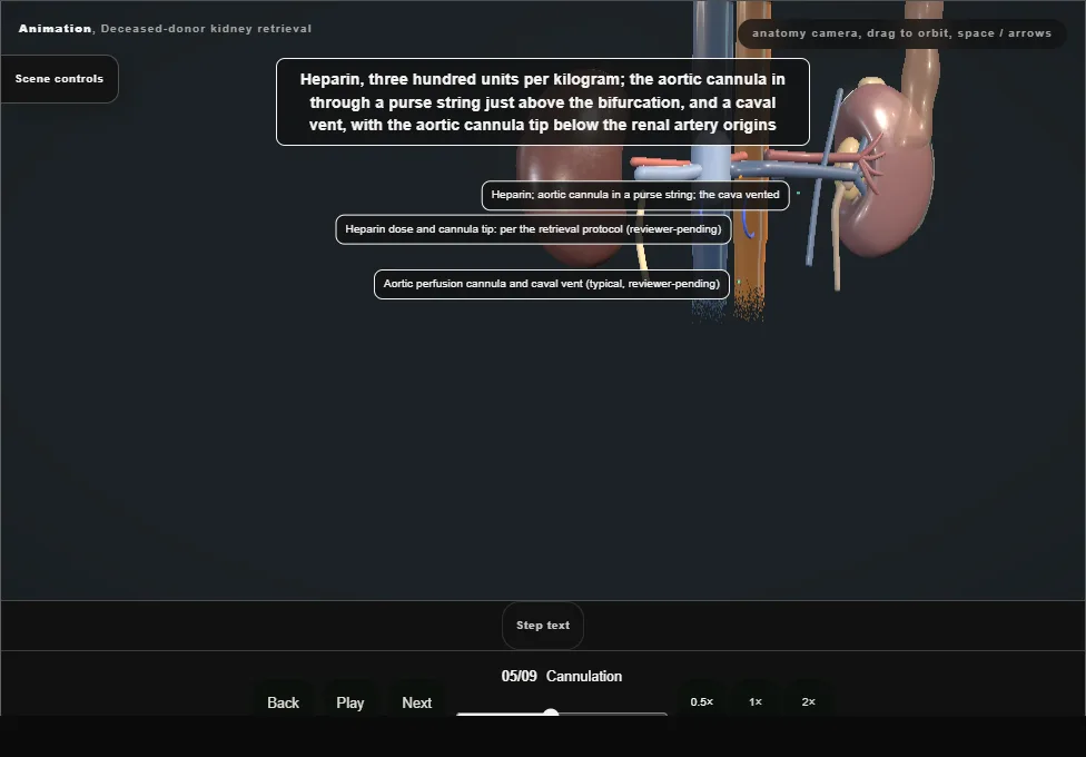
- Heparin, three hundred units per kilogram; the aortic cannula through a purse string just above the bifurcation, a caval vent, with the aortic cannula tip below the renal artery origins.
- In circulatory death this is done first and at speed.
- 06
Cross-clamp, cold perfusion and ice
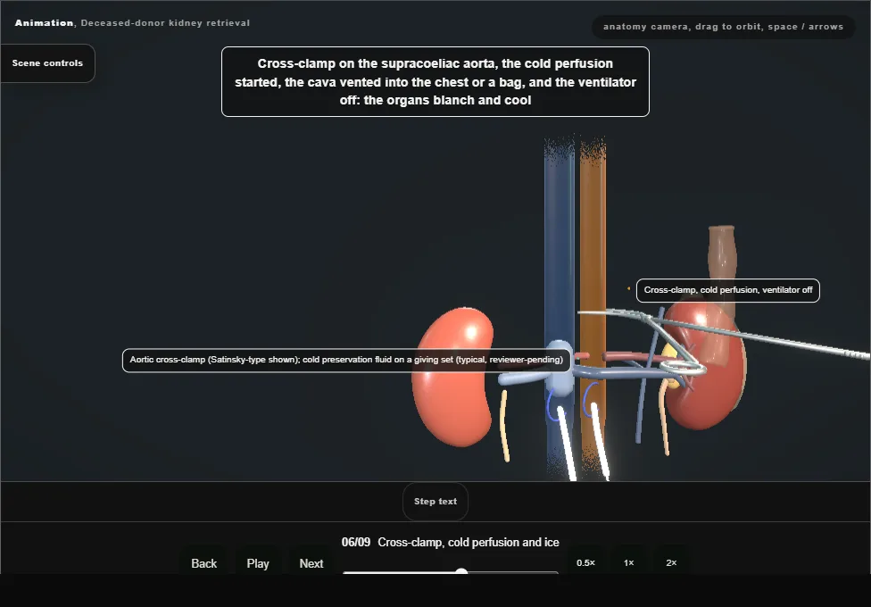
- Cross-clamp on the supracoeliac aorta, cold perfusion started, the cava vented, the ventilator off: the organs blanch and cool.
- Slush ice into the abdomen around every organ.
- 07
The kidneys en bloc
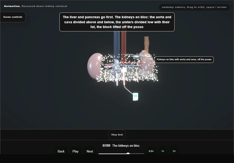
- After the liver and pancreas: the aorta and cava divided above and below, the ureters divided low with their fat, the block lifted off the psoas.
- Gerota on, ureter long, nothing dissected close to the kidney in the donor.
- 08
The bench and the packing
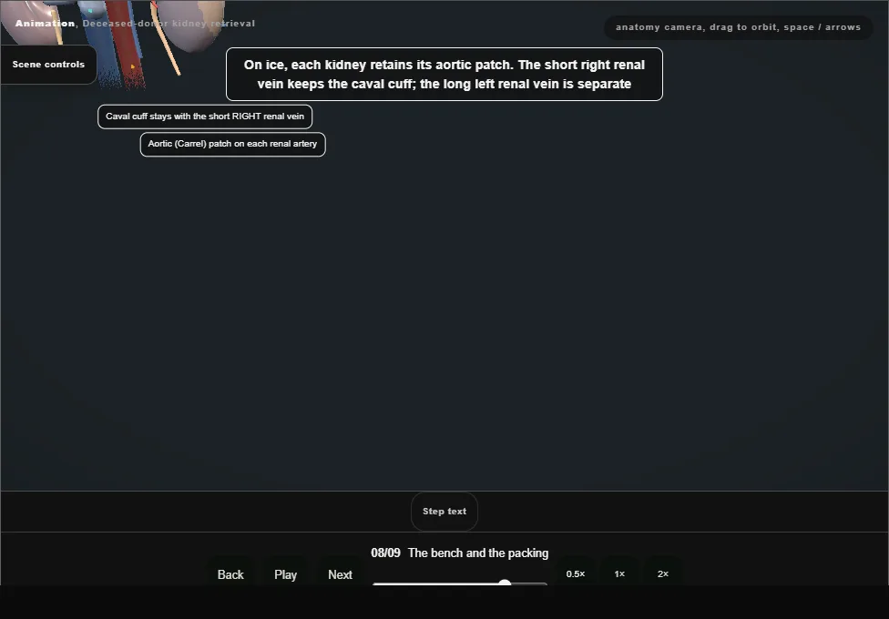
- On the bench in ice the aorta opened along its back wall between the ostia and the kidneys separated with a patch of aorta each; the caval cuff retained with the short right renal vein.
- Both kidneys inspected, the arteries counted, the ureter checked; three bags, cold solution, ice.
- 09
Documentation and the recipient centres
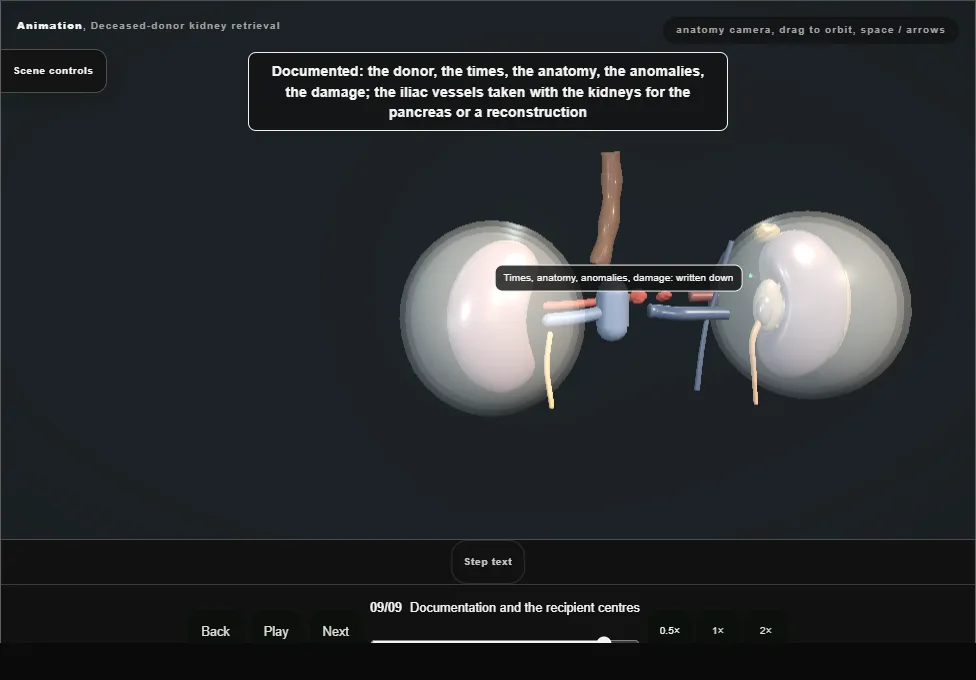
- The donor, the times, the anatomy, the anomalies, the damage, written down; the iliac vessels taken with the kidneys.
- The donor closed with care, and the kidneys to their centres with the paperwork that lets the surgeon there trust the box.
Derived from kidney/OrganRetrievalScene.tsx at build time: every step title and caption above is the authored
ProcedureDef text, reproduced verbatim, and this page shares its readable-points renderer with the animation
itself. 9 of 9 steps have a captured frame.
Text fingerprint da4e80a5de4d21a4. A caption edit without a re-capture fails the production
check, so the sheet and the animation cannot drift apart silently.