
Revision sheet
Paediatric urinary stone procedures
10 steps, each one the authored title and caption from the interactive animation, with the frame captured from that step. Read it before the case, or print it and work through the steps in order.
Teaching only, not for diagnosis or treatment decisions. The text on this sheet is the animation's own step copy, reproduced verbatim.
UrOops3D · UroRef · © Nity G
- 01
The child and the work-up
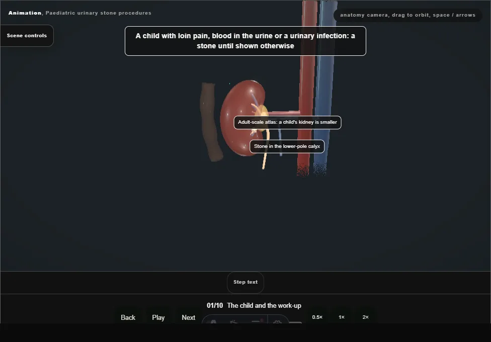
- Loin pain, blood in the urine or a urinary infection.
- Ultrasound first, with a plain film; low-dose CT only when they leave doubt.
- Urine cultured, and blood and urine sent for the metabolic screen from the start.
- 02
The route by size and site
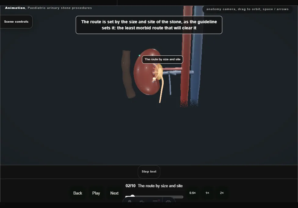
- Shock waves first for most renal stones; ureteroscopy for the lower ureter; mini-PCNL for the larger stone and the lower pole, where shock-wave fragments clear poorly.
- This child has a larger lower-pole stone.
- 03
Anaesthetic, catheter, prone
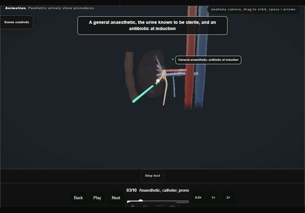
- A general anaesthetic with sterile urine and an antibiotic at induction.
- A ureteric catheter placed cystoscopically, then prone on chest rolls, padded and kept warm.
- Contrast fills the system; screening pulsed and collimated.
- 04
Ultrasound-guided puncture
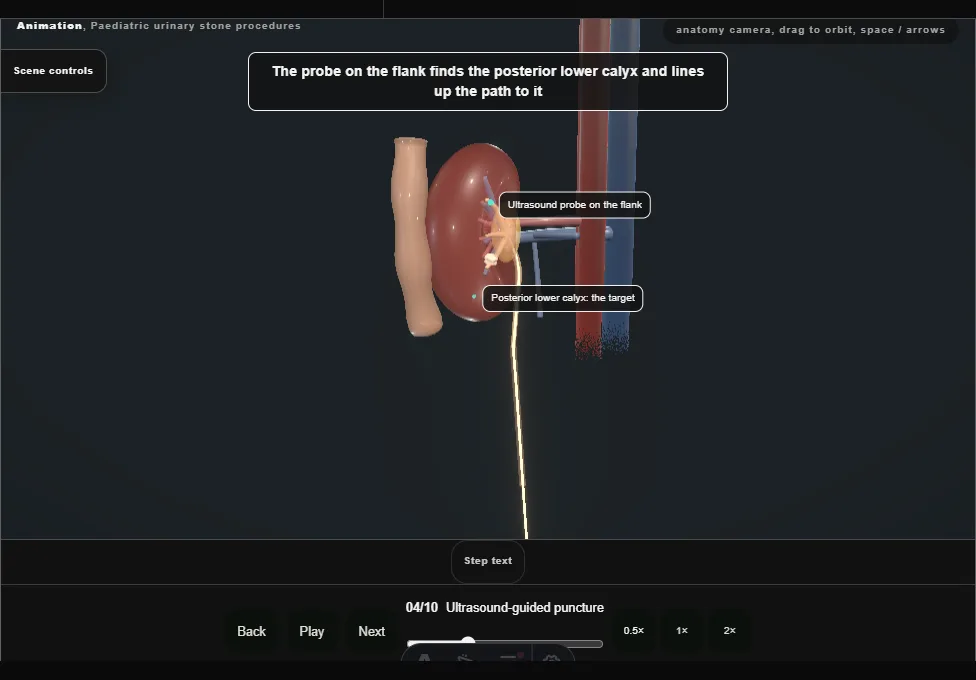
- The probe on the flank finds the posterior lower calyx; the needle is advanced in the plane of the beam, through the papilla into the calyx, watched on the screen the whole way.
- Urine at the hub confirms it; pus at the hub stops the operation.
- 05
One dilation to a small sheath
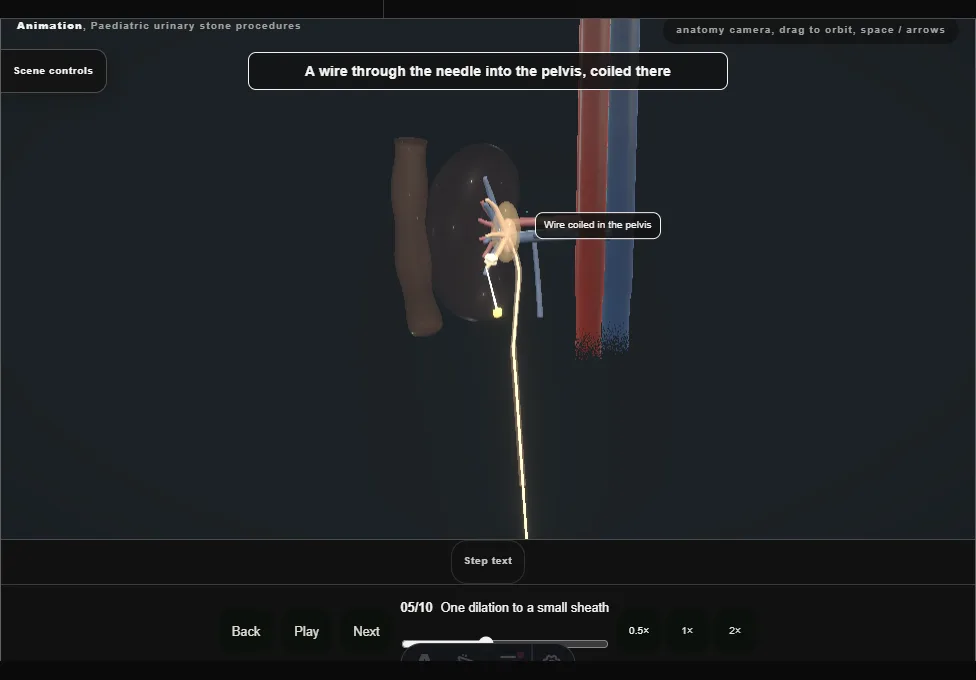
- A wire through the needle, coiled in the pelvis; a small skin cut; the tract dilated once, in a single pass, to the small sheath, whose mouth is seated in the calyx.
- Less kidney is torn than with a full-size tract.
- 06
The miniature nephroscope and the laser
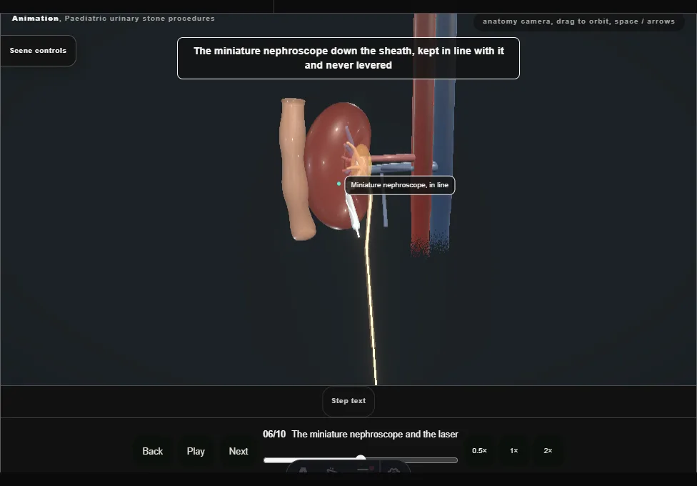
- The miniature nephroscope goes down the sheath, kept in line with it and never levered.
- The laser breaks the stone into pieces small enough to leave through the sheath, with the irrigation kept at a low pressure.
- 07
Washing the fragments out
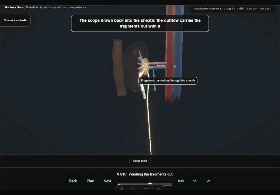
- The scope is drawn back into the sheath and the outflow carries the fragments out with it, one after another; a larger piece is caught with a grasper.
- Every calyx is looked into, and the stone goes for analysis.
- 08
Nephrostomy or tubeless
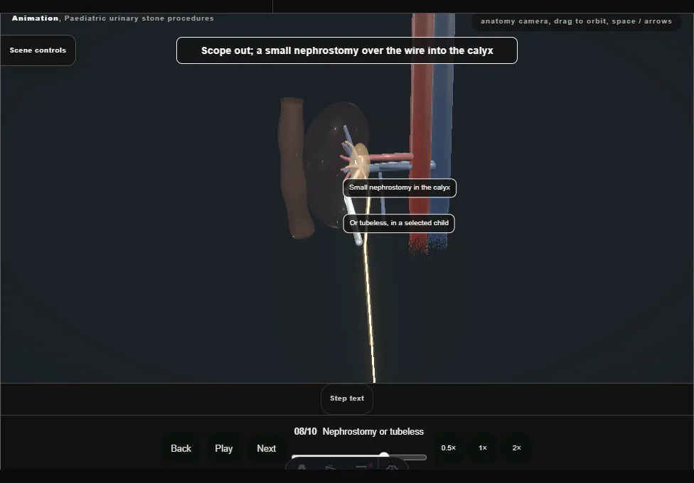
- A small nephrostomy over the wire into the calyx, the sheath out over it and the catheter out; tubeless, with or without a stent, in a selected child after an uncomplicated clearance.
- The child turned back and woken.
- 09
The second path: ureteroscopy
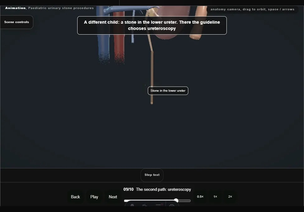
- For a stone in the lower ureter: a wire beyond the stone, the smallest semirigid ureteroscope beside it with no access sheath, the stone dusted with the laser, the ureter inspected on the way out, and a stent only when it is needed.
- 10
Analysis, work-up and follow-up
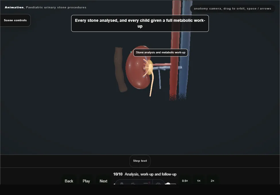
- Every stone analysed and every child given a full metabolic work-up: timed urine and blood chemistry.
- Plenty to drink, less salt, normal calcium, treatment of the cause, and ultrasound to follow for fragments and regrowth.
Derived from kidney/PaediatricStonesScene.tsx at build time: every step title and caption above is the authored
ProcedureDef text, reproduced verbatim, and this page shares its readable-points renderer with the animation
itself. 10 of 10 steps have a captured frame.
Text fingerprint 1caa162dcc2bf151. A caption edit without a re-capture fails the production
check, so the sheet and the animation cannot drift apart silently.