
Revision sheet
Scrotal trauma: exploration and tunical repair
8 steps, each one the authored title and caption from the interactive animation, with the frame captured from that step. Read it before the case, or print it and work through the steps in order.
Teaching only, not for diagnosis or treatment decisions. The text on this sheet is the animation's own step copy, reproduced verbatim.
UrOops3D · UroRef · © Nity G
- 01
Assessment and ultrasound
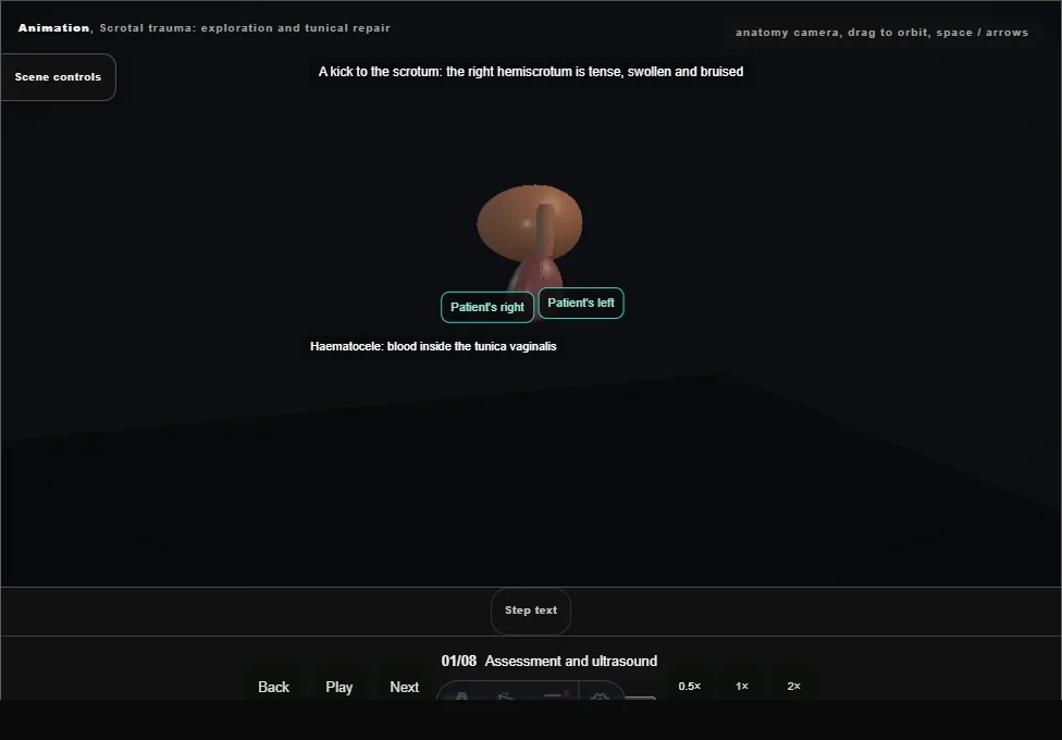
- A tense, bruised right hemiscrotum after a blunt blow.
- The urethra and perineum are checked.
- Ultrasound shows a heterogeneous testis that has lost its outline: rupture, so the testis is explored.
- 02
Midline raphe incision
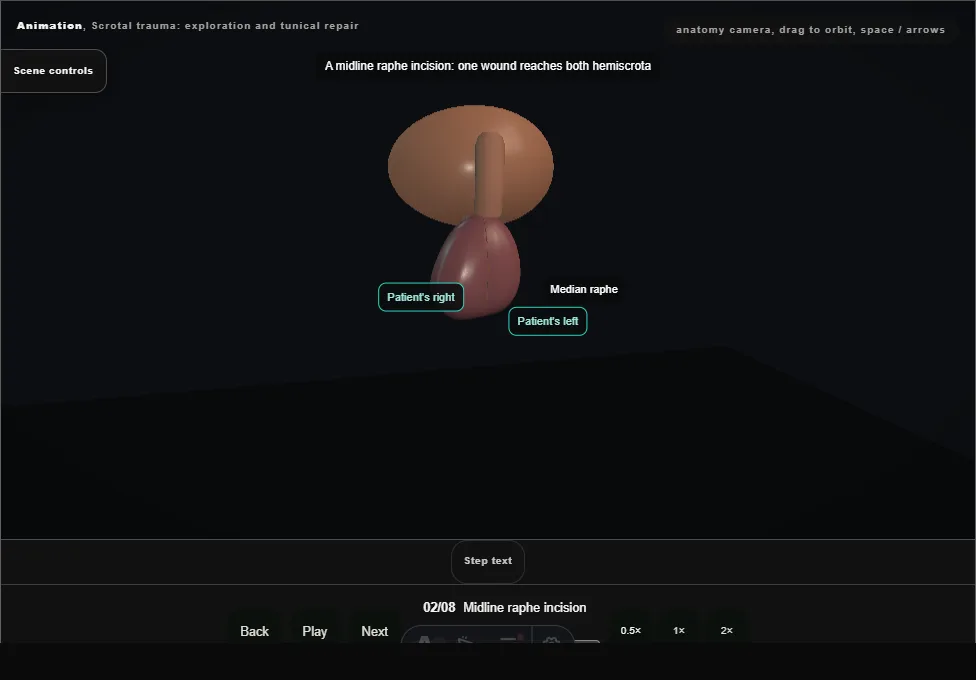
- A midline raphe incision through skin and dartos, which reaches both hemiscrota.
- The tunica vaginalis beneath is dark with blood.
- 03
Open the tunica vaginalis, evacuate the haematocele
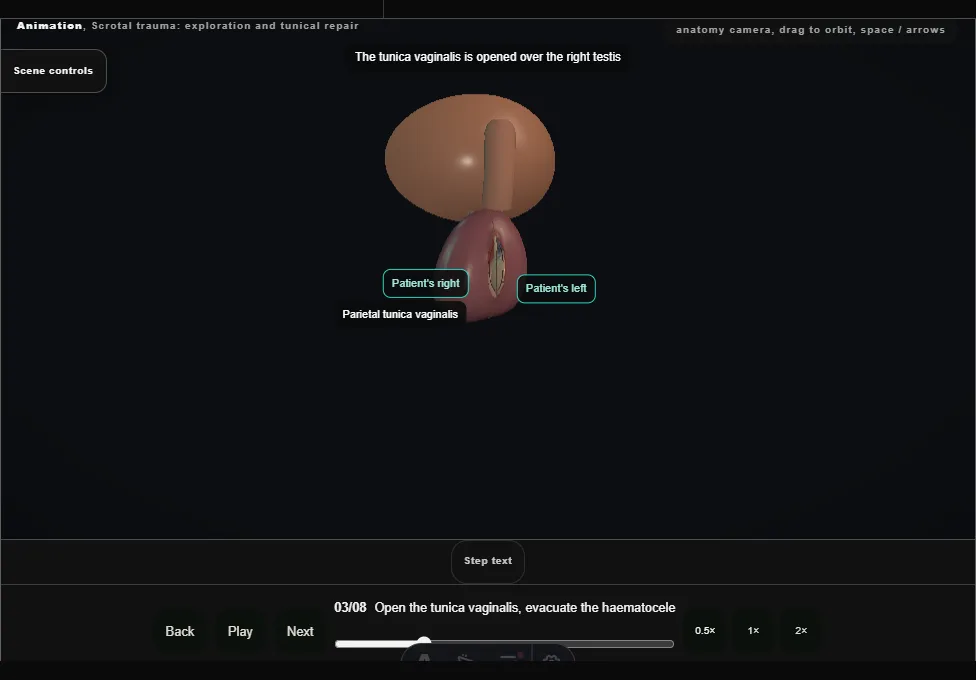
- The tunica vaginalis is opened and the haematocele evacuated: clot sucked out and the cavity washed, sparing the cord.
- 04
Deliver and inspect
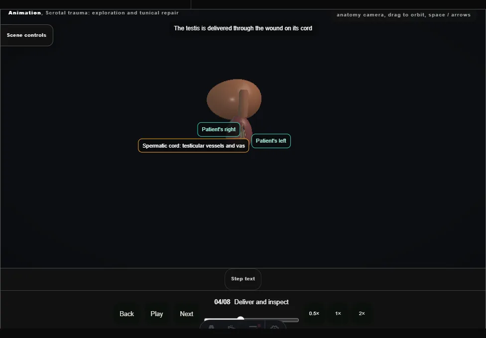
- The testis is delivered on its cord.
- The tear in the tunica albuginea is seen, with seminiferous tubules extruded through it.
- 05
Debride non-viable tubules
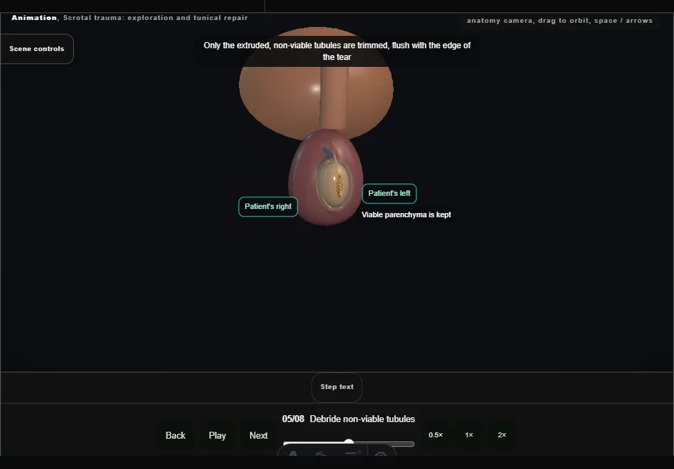
- Only the extruded, non-viable tubules are trimmed, flush with the edge of the tear.
- Viable parenchyma is kept and bleeding points are controlled.
- 06
Close the tunica albuginea
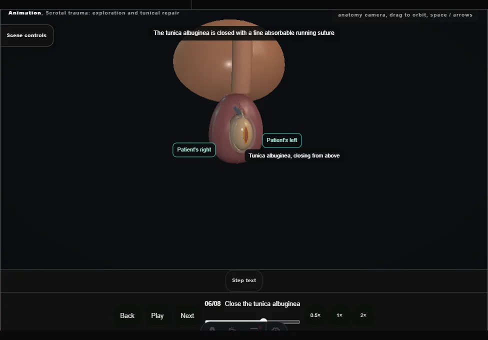
- The tunica albuginea is closed with a fine absorbable running suture.
- If the defect cannot be closed without tension, a tunica vaginalis flap covers it.
- 07
Check cord and epididymis, return
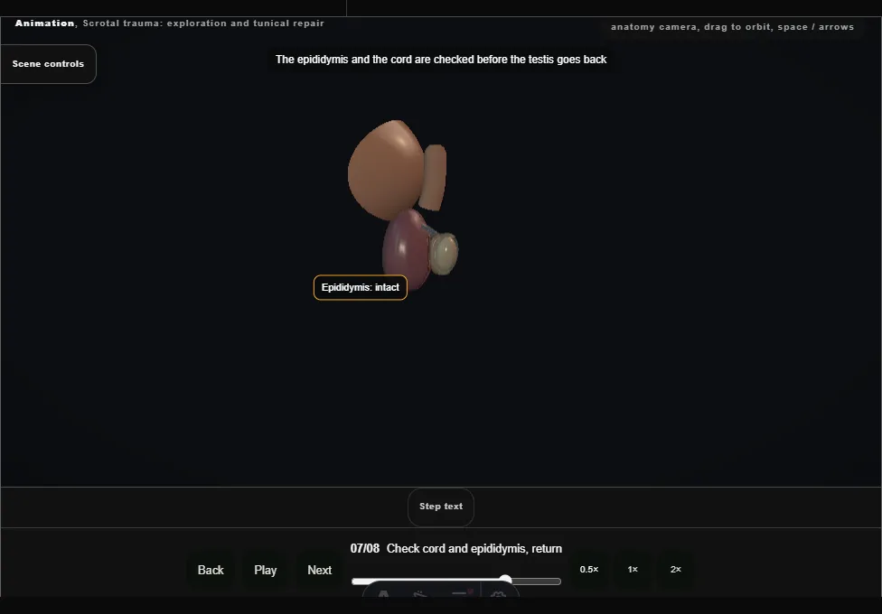
- The epididymis and the cord are checked, the testis is returned the right way round, and the other testis is inspected through the same wound.
- 08
Layered closure and follow-up
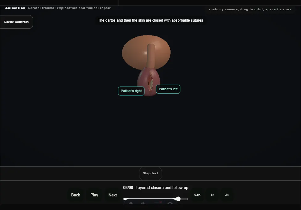
- The tunica vaginalis loosely, then dartos and skin with absorbable sutures.
- No drain; a support.
- Follow-up covers fertility and hormone function.
Derived from scrotal/ScrotalTraumaScene.tsx at build time: every step title and caption above is the authored
ProcedureDef text, reproduced verbatim, and this page shares its readable-points renderer with the animation
itself. 8 of 8 steps have a captured frame.
Text fingerprint e65e20dfc3e56e53. A caption edit without a re-capture fails the production
check, so the sheet and the animation cannot drift apart silently.