
Revision sheet
Treatment of a transplant lymphocele
9 steps, each one the authored title and caption from the interactive animation, with the frame captured from that step. Read it before the case, or print it and work through the steps in order.
Teaching only, not for diagnosis or treatment decisions. The text on this sheet is the animation's own step copy, reproduced verbatim.
UrOops3D · UroRef · © Nity G
- 01
The collection
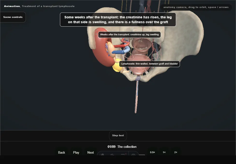
- Weeks after the transplant: a rising creatinine, a swollen leg, a fullness over the graft.
- A thin-walled lymphocele lies between the lower pole and the bladder, on the iliac vein, with the graft ureter stretched over its medial wall.
- 02
Ultrasound and the differential
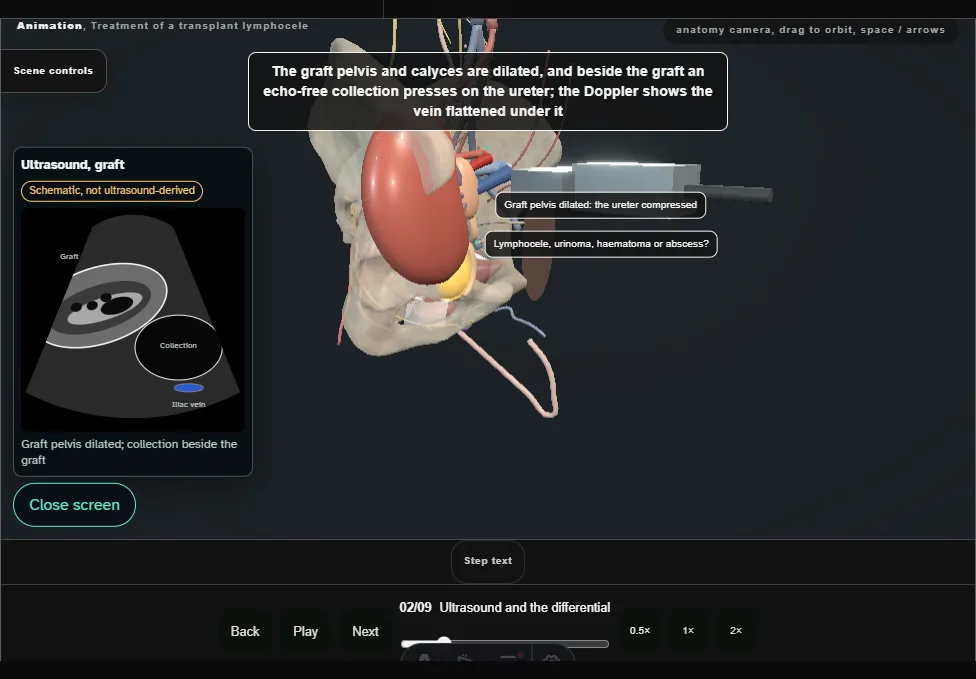
- The graft pelvis and calyces are dilated and an echo-free collection lies beside the graft, pressing on the ureter and the vein.
- Lymphocele, urinoma, haematoma or abscess: the fluid tells them apart.
- 03
Aspiration and the fluid creatinine
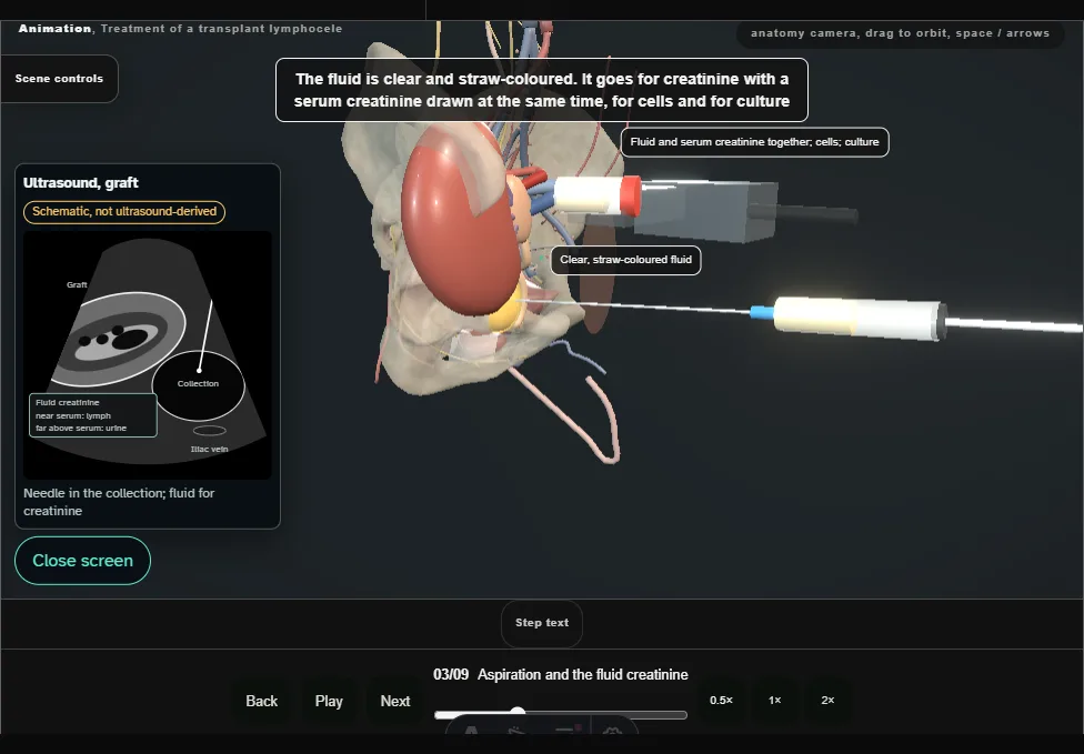
- Under ultrasound, a needle onto the dome from in front and lateral, away from the ureter and the vein.
- Clear straw fluid goes for creatinine with a serum sample: close to the serum value, it is lymph; far above it, urine.
- 04
Percutaneous drain
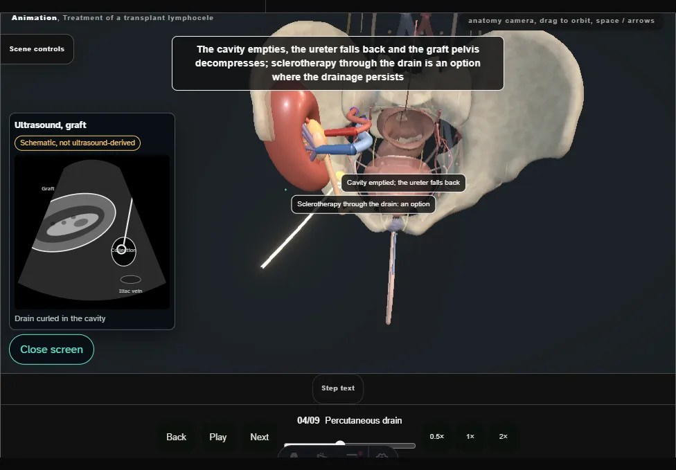
- A drain along the same track, curled in the cavity: the collection empties, the ureter falls back and the graft decompresses.
- Sclerotherapy through the drain is an option for persistent drainage; a collection that comes back goes to a window.
- 05
Ports and the bulge
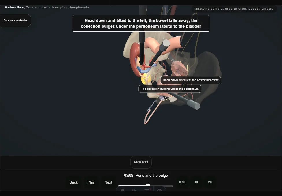
- The camera at the umbilicus and two working ports on the left, each placed under vision.
- Head down and tilted left, the bowel falls away, and the collection bulges under the peritoneum lateral to the bladder.
- 06
The ureter first
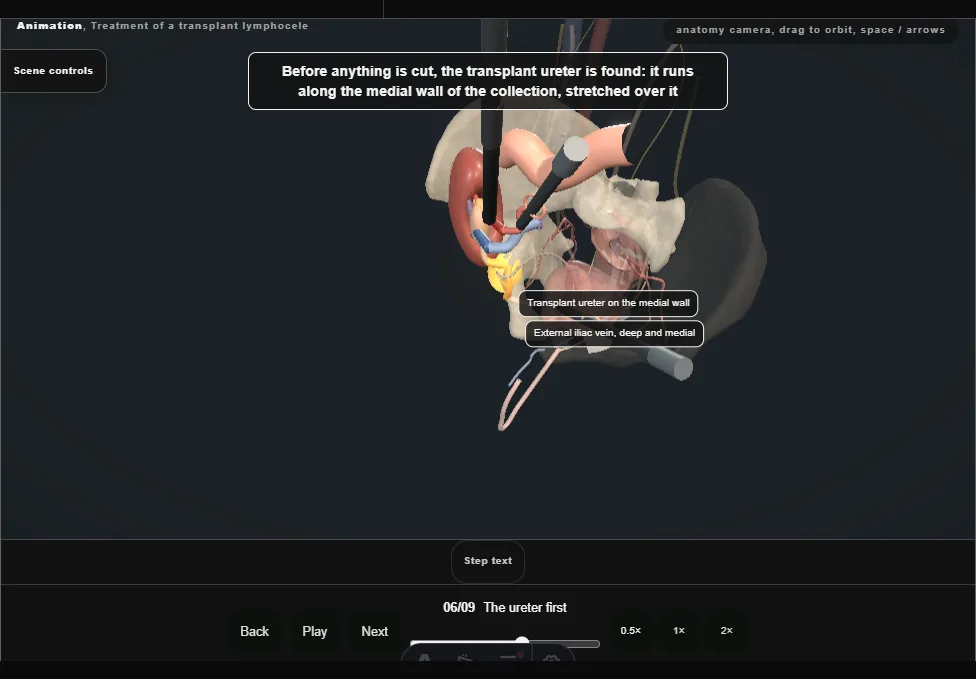
- Before anything is cut, the transplant ureter is found on the medial wall of the collection, with the iliac vein deep and medial to it.
- The window is planned on the dome, wide and away from both.
- 07
The window
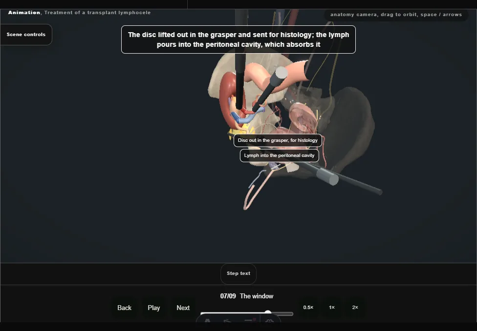
- The dome held up and the wall cut round with scissors, peritoneum and lymphocele wall together.
- The disc comes out in the grasper for histology, and the lymph drains into the peritoneal cavity.
- 08
Edges secured and the check
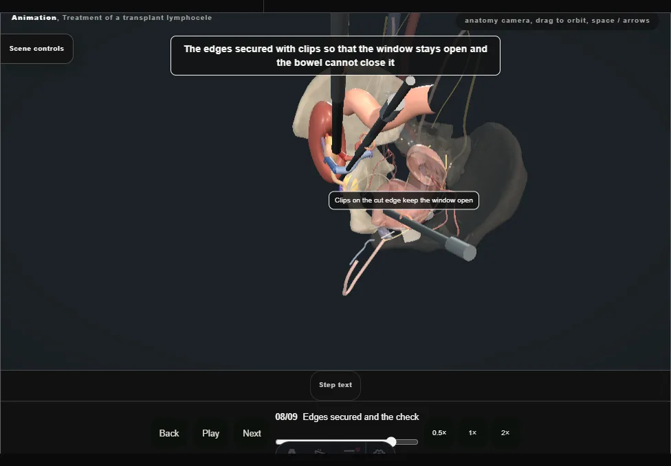
- Clips on the cut edge keep the window open.
- The check: the ureter whole on the medial wall, no urine in the field, no bleeding when the insufflation pressure is lowered.
- 09
After
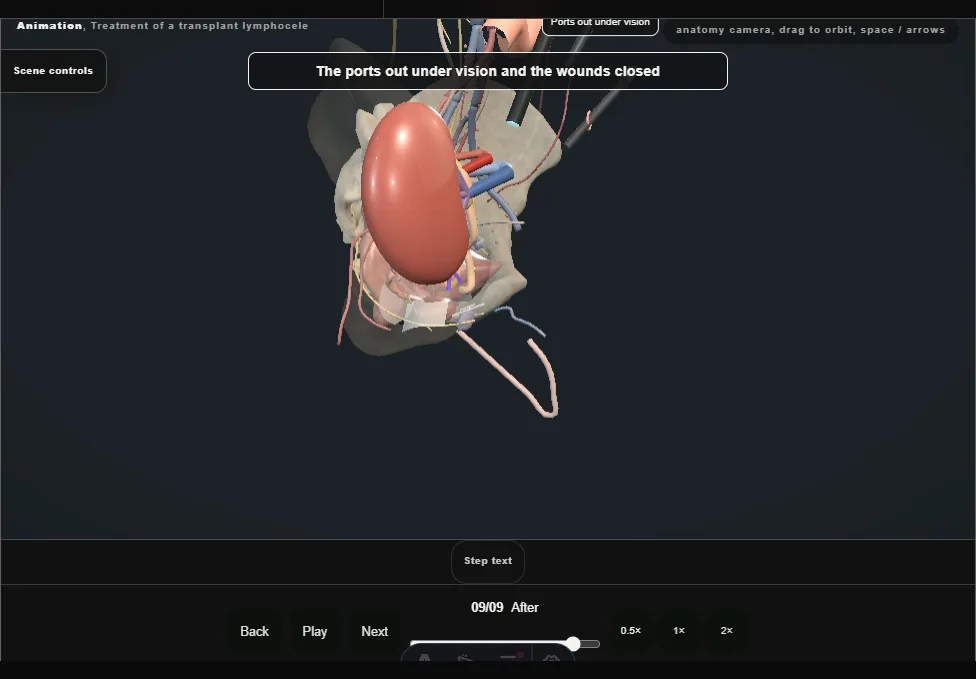
- Ports out under vision.
- Ultrasound before discharge and at follow-up: no collection, the graft pelvis back to normal, the creatinine falling.
- A recurrence is rare after a wide window, and is imaged again.
Derived from pelvis/TransplantLymphoceleScene.tsx at build time: every step title and caption above is the authored
ProcedureDef text, reproduced verbatim, and this page shares its readable-points renderer with the animation
itself. 9 of 9 steps have a captured frame.
Text fingerprint c8ee6537c97e3e2e. A caption edit without a re-capture fails the production
check, so the sheet and the animation cannot drift apart silently.