
Revision sheet
Ureterolysis with omental wrapping
9 steps, each one the authored title and caption from the interactive animation, with the frame captured from that step. Read it before the case, or print it and work through the steps in order.
Teaching only, not for diagnosis or treatment decisions. The text on this sheet is the animation's own step copy, reproduced verbatim.
UrOops3D · UroRef · © Nity G
- 01
Stents, then the plaque
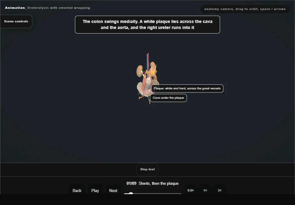
- A stent is placed in each ureter from below.
- Midline laparotomy; the white line of Toldt is opened and the right colon reflected medially.
- A white plaque covers the cava and the aorta, and the ureter runs into it.
- 02
Find the ureter above
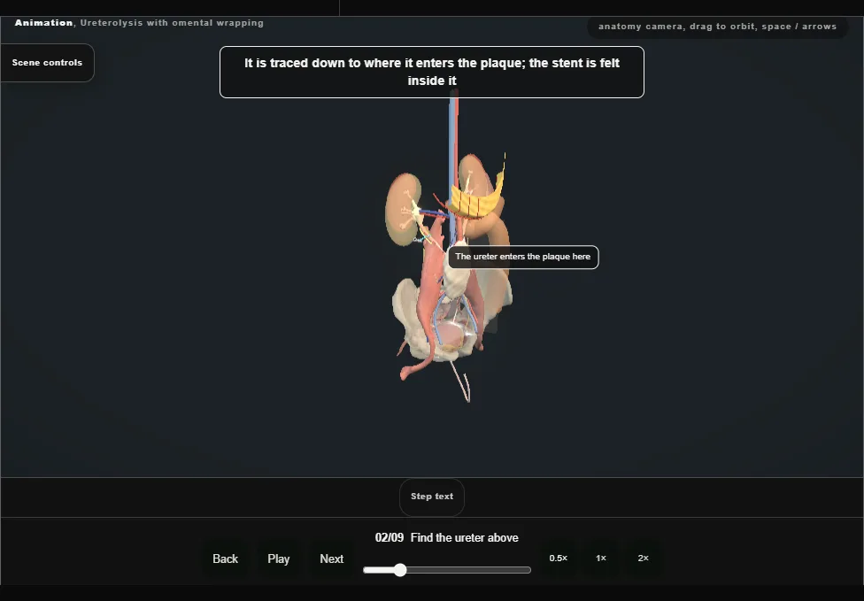
- The ureter is found above the plaque, where it is wide.
- It is dissected with its adventitia left on it and a vessel loop is passed round it.
- It is then traced down to the plaque with the stent inside it as the guide.
- 03
Biopsy
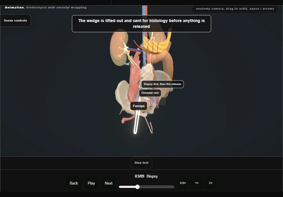
- Before any release a biopsy of the fibrotic tissue beside the ureter is taken and sent for histology, and the operation goes on.
- The wedge is taken clear of the cava and of the ureter.
- 04
Release, from above downwards
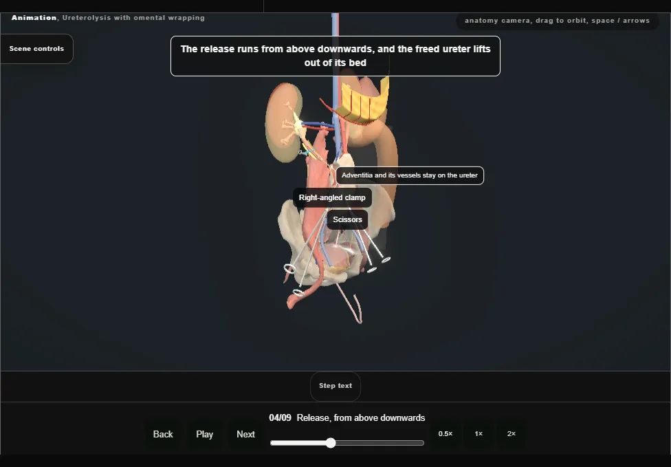
- The ureter is released from the plaque by sharp and blunt dissection, from above downwards, and lifts out of its bed with its adventitia on it.
- The cava, then the right common iliac vessels, lie deep and medial.
- 05
A living ureter
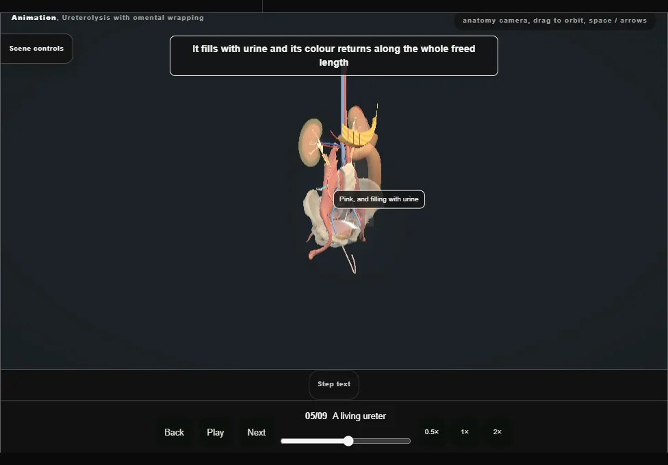
- Once it is free the ureter is laid lateral without tension.
- It fills with urine and stays pink along the whole freed length.
- 06
Intraperitonealise
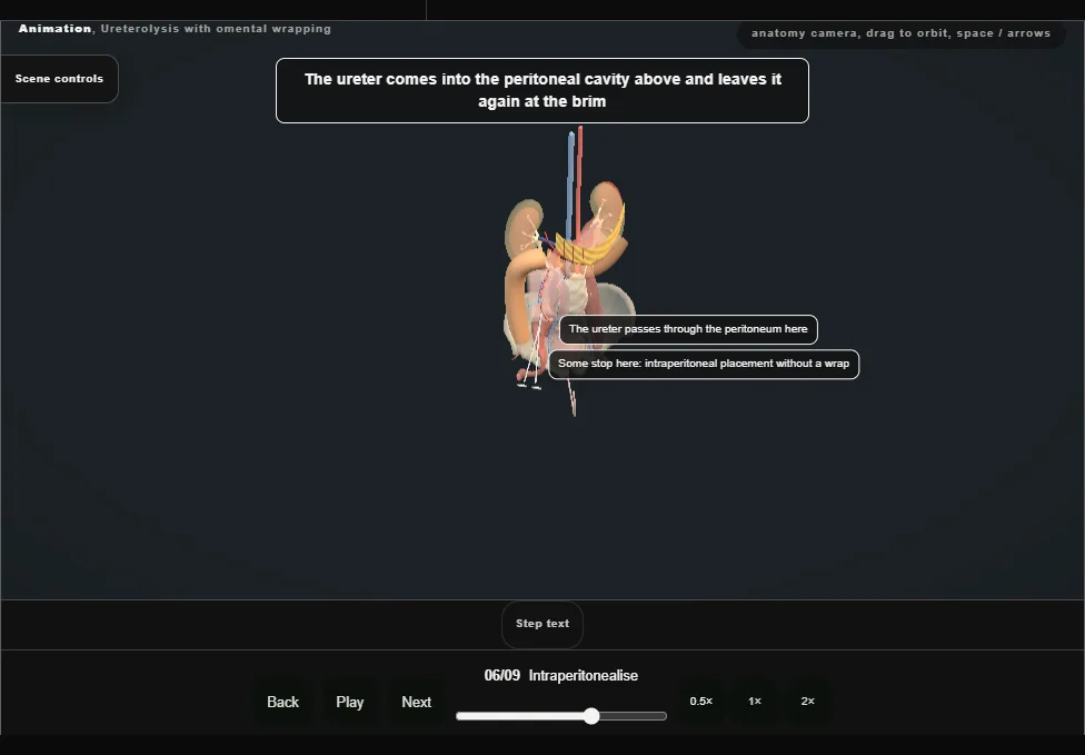
- The ureter is positioned laterally and the posterior peritoneum is closed behind it with a running stitch.
- The ureter now lies in the peritoneal cavity, off the plaque.
- 07
Raise the omental flap
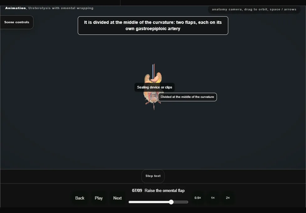
- The omentum is mobilised off the greater curvature of the stomach with the gastroepiploic arteries kept on it, and divided at the middle of the curvature into two flaps.
- The right flap is brought down to the right ureter.
- 08
Wrap and drain
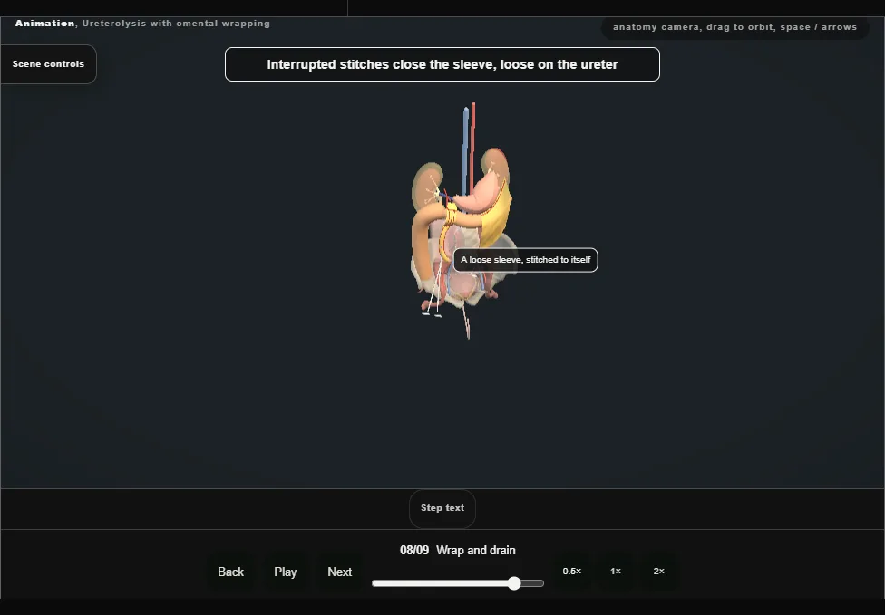
- The ureter is wrapped round its whole circumference in the flap along the freed length, and the flap is secured with interrupted stitches.
- A drain is left in the retroperitoneum.
- 09
The other side, and after
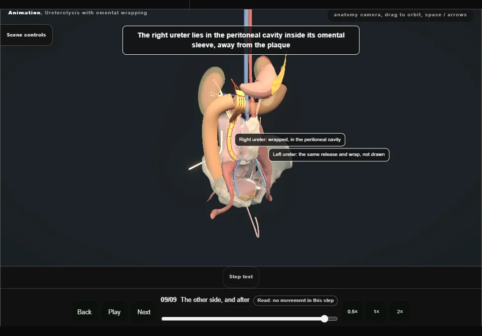
- The right ureter lies lateral, in the peritoneal cavity, inside its omental flap.
- The left ureter is released, placed and wrapped in the same way at the same operation.
- The disease still needs its medical treatment and follow-up imaging.
Derived from pelvis/UreterolysisScene.tsx at build time: every step title and caption above is the authored
ProcedureDef text, reproduced verbatim, and this page shares its readable-points renderer with the animation
itself. 9 of 9 steps have a captured frame.
Text fingerprint a822d0d64280c34a. A caption edit without a re-capture fails the production
check, so the sheet and the animation cannot drift apart silently.