
UrOops3D, danger-zone atlas

Schematic model.

Operative animation, Anterior vaginal prolapse repair

Anterior vaginal prolapse repair: midline plication and the lateral defect

Native-tissue repair of an anterior vaginal wall prolapse with a central and a lateral defect.

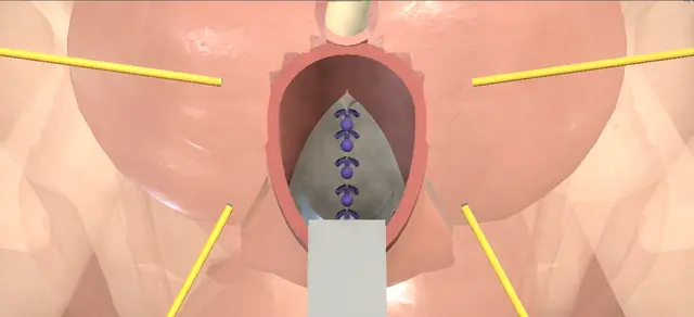
Through a midline incision the vaginal flaps are lifted off the pubocervical fascia, the fascia is plicated in the midline over the bladder, and on each side its lateral edge is sutured back to the arcus tendineus fasciae pelvis.

Cystoscopy then checks the bladder, the ureters and the stitches before the wall is closed.

Advanced tier FRCS female and reconstructive urology, prolapse Interactive 3D

Operative visualisation

Schematic scene preview: Anterior vaginal prolapse repair
Preparing the 3D anatomy…

UrOops3D · UroRef · © Nity G

How to use this module. Play through the nine steps, or use Back and Next. Watch the flaps come off the fascia, the bulge fall tie by tie as the midline is plicated, the fascia brought back to the arcus on each side, and both ureters jet at cystoscopy. The triggers show a bladder entered and a silent ureter.

Click me next Section 2 of 8 Anatomy The structures this operation works between, and the one it must not cross.

The structures in the scene

Anterior vaginal prolapse repair: how the structures relateThe pubocervical fascia under the anterior vaginal wall, and what lies round it. Pubocervical fascia: plicated and reattached: lies under it; the flaps are lifted off it Anterior vaginal epithelium: the flaps. Pubocervical fascia: plicated and reattached: covers it; bites stay in the fascia Bladder base, inside the bulge (danger). Pubocervical fascia: plicated and reattached: its lateral edge is sutured back to it Arcus tendineus fasciae pelvis. Pubocervical fascia: plicated and reattached: a deep apical bite can catch one Ureters, above the apex of the defect (danger). Pubocervical fascia: plicated and reattached: continues under it as periurethral fascia Urethra and bladder neck (danger).Pubocervical fascia:plicated and reattachedAnterior vaginalepithelium: theflaps!Bladder base, insidethe bulgeArcus tendineusfasciae pelvis!Ureters, above theapex of the defect!Urethra and bladdernecklies under it; theflaps are liftedoff itcovers it; bitesstay in the fasciaits lateral edge issutured back to ita deep apical bitecan catch onecontinues under itas periurethralfasciaTargetDangerLandmark
The pubocervical fascia under the anterior vaginal wall, and what lies round it. Schematic, not to scale; relationships only.
  • In lithotomy the anterior vaginal wall lies between the speculum and the urethral meatus, and in this prolapse it bulges down toward the introitus with the bladder base inside it.
  • Under the epithelium is the pubocervical fascia, thinned in the midline over the bladder.
  • Out on each side the fascia should reach the arcus tendineus on the pelvic side wall; here it has come away from it.
  • The ureters enter the bladder above the apex of the defect.
  • ›Anterior vaginal wall - its epithelium is opened in the midline and raised as two flaps.
  • ›Pubocervical fascia - the vaginal muscularis and adventitia under the epithelium; plicated in the midline and sutured laterally.
  • ›Periurethral fascia - the distal continuation of the same layer under the urethra; the flaps come off it too.
  • ›Bladder base - the content of the bulge; reduced into the pelvis by the plication.
  • ›Urethra and bladder neck - the catheter marks them; the incision starts just below the meatus.
  • ›Arcus tendineus fasciae pelvis - the white line on the pelvic side wall from the pubis to the ischial spine; the lateral sutures are anchored to it.
  • ›Ureters - enter the bladder base above and lateral to the apex of the plication.
  • ›Ureteric orifices - watched for efflux at cystoscopy.
Click me next Section 3 of 8 Steps The operation in order, with the landmark that guards each move.

Operative steps

  1. The prolapse Antibiotics and anaesthesia, then dorsal lithotomy on padded stirrups. A urethral catheter drains the bladder and keeps it empty throughout. A weighted speculum goes on the posterior wall and a self-retaining retractor is set. The anterior wall bulges toward the introitus, with the bladder base inside the bulge.
    where it bites: the bulge is bladder; everything that follows is done on its outer surface.
  2. Midline incision The anterior vaginal epithelium is infiltrated with local anaesthetic and adrenaline to aid the dissection and haemostasis. With the bladder decompressed, a midline incision is made in the anterior wall from just below the urethral meatus to the apex of the defect.
    where it bites: the incision is through epithelium only; the fascia and the bladder lie directly beneath it.
  3. Vaginal flaps By sharp and blunt dissection each lateral vaginal flap is lifted off the periurethral and pubocervical fascia, which exposes the central defect.
    where it bites: too deep a plane goes through thin fascia into the bladder, most easily where the defect is widest.
  4. Plication stitches The bladder is reduced into the pelvis. Interrupted plication stitches are placed across the midline, from the apex of the defect toward the bladder neck, each taking the vaginal muscularis and adventitia on either side over the prolapsed bladder.
    where it bites: each bite is in the fascia only; a deeper bite enters the bladder or, near the apex, catches a ureter.
  5. Tie the plication The plication stitches are tied in turn. Each draws the fascia together in the midline over the bladder and the bladder base rises with every tie. This is the anterior colporrhaphy, and it closes the central defect.
    where it bites: tying does not correct a bite already in the bladder; that is found at cystoscopy.
  6. Lateral defect, first side The bladder is retracted medially and the dissection is carried laterally to the arcus tendineus fasciae pelvis, exposing the lateral defect. Five sutures are placed through the arcus, the lateral edge of the pubocervical fascia and the inner vaginal flap. Once all are placed they are tied one at a time.
    where it bites: the bladder lies medial to this dissection; the retractor keeps it there.
  7. Lateral defect, other side The identical procedure is performed at the contralateral arcus tendineus fasciae pelvis. Throughout the dissection the bladder and the urethra are not entered, and haemostasis is achieved with diathermy.
    where it bites: the urethra lies close to the distal end of the lateral dissection, and is not entered.
  8. Cystourethroscopy The catheter is removed and cystourethroscopy is performed. The proximal urethra and the bladder are intact, clear ureteric efflux is seen from both orifices and there is no suture in the bladder. The cystoscope is removed and the catheter replaced.
    where it bites: a stitch in the bladder or an orifice without efflux is dealt with now, before the wall is closed.
  9. Trim and close The wound is irrigated and haemostasis confirmed again. The edges of the vaginal epithelium are trimmed where they are redundant, and the wall is closed in the midline with interrupted absorbable sutures. The retractor and the speculum are removed and the catheter is left on drainage.
    where it bites: epithelium trimmed too generously narrows the vagina, and dyspareunia is among the chapter's complications.
Click me next Section 4 of 8 Danger zones Where this operation bites, and why it bites there.
Critical relationship

Danger zones

The spatial relationships the animation is built to make automatic. Before you read each one, ask yourself: at this point in the operation, what is about to go wrong, and what is my first move? The save for each sits in the Complications tab.

  • RiskBladder behind the fascia - directly behind thin fascia in the central defect; entered by too deep a dissection or a bite.
  • RiskUreters near the apex - a deep or far lateral bite at the apex of the plication can catch or kink one.
  • RiskUrethra and bladder neck - under the distal end of the incision; the chapter cautions against entering the urethra.
  • RiskLateral dissection to the arcus - the bladder is held medially while the side wall is exposed.
  • RiskOver-trimming - redundant epithelium only; a narrowed vagina is the cost of taking more.
Practise it Take me into the scene Opens the animation at a danger moment and asks you to predict the mechanism and your first move before it reveals.
Click me next Section 5 of 8 Complications The save for each one: the first move, the escalation trigger, the op-note line.

Complications and immediate saves

Trigger each of these live in the Animation tab, then read the save: the first move, the escalation trigger, and the line that goes in the op note.

Bladder perforation

  • the dissection under a flap goes through the fascia into the bladder; bladder perforation is a listed complication of this operation.
The save
  1. Stop dissecting. Identify the whole edge of the hole, its distance from the trigone and both ureteric orifices. first move
  2. Close the bladder in layers clear of the orifices, then continue the repair over it.
  3. A hole close to the trigone or an orifice, or one whose edge cannot be defined, needs senior help. escalation trigger
  4. Cystoscopy confirms the closure is clear of the orifices and that efflux is present; the catheter stays for longer.
  5. Op note: the site and size of the hole, the closure, the cystoscopic finding and the catheter plan. document

Ureteric obstruction

  • one ureteric orifice gives no efflux at cystoscopy because a stitch has caught or kinked that ureter; ureteral obstruction or laceration is a listed complication.
The save
  1. Watch both orifices for longer. If one stays silent, remove the stitch most likely to be responsible, usually at the apex of the plication on that side. first move
  2. Watch again for efflux; an intravenous dye can make a weak jet easier to see.
  3. If efflux does not return, attempt a ureteric catheter and call senior help in theatre. escalation trigger
  4. A ureter that will not take a catheter needs imaging and a plan agreed before the patient leaves theatre.
  5. Op note: which side, which stitch was removed, what restored efflux and the follow-up arranged. document

Urinary retention

  • the woman cannot empty her bladder when the catheter is removed after the repair; the chapter lists urinary retention.
The save
  1. Measure the residual by bladder scan or catheter before assuming she is emptying. first move
  2. Replace the catheter, or teach intermittent self-catheterisation, and plan a further trial without catheter.
  3. Retention that persists, or pain and swelling at the repair, needs senior review and examination of the wound. escalation trigger
  4. A suprapubic catheter placed at the operation is one of the chapter's variations for easing the voiding trial.
  5. Notes: the residual volumes, the catheter plan and the date of the next trial. document
Click me next Section 6 of 8 Troubleshooting When the case turns: the problem, then the next move.

Intra-operative troubleshooting

When the case turns: the problem, then the next move.

  • ActionThe defect is central only - the chapter allows an isolated central defect to be managed by the anterior colporrhaphy alone, without the lateral sutures.
  • ActionThe exposure through a midline incision is poor - an inverted-U midline incision is the chapter's alternative approach to the same repair.
  • ActionThere is stress urinary incontinence as well - the chapter allows a mid-urethral sling at the same operation. NICE NG123 says consider concurrent surgery and explain the uncertainty about its benefit beyond a year.
  • ActionA ureteric jet is hard to see - an intravenous dye given before cystoscopy, one of the chapter's variations, confirms ureteric patency.
  • ActionOozing from the dissected bed - diathermy for haemostasis during the dissection; the chapter allows a vaginal pack after the operation.
  • ActionA graft or mesh is asked about - the chapter lists mesh over the plication as a variation. NICE NG123 recommends anterior repair without mesh, and quotes IPG599 that transvaginal mesh repair should be used only in research.
Click me next Section 7 of 8 FRCS viva Say it out loud, then mark yourself against the model answer.

FRCS viva questions

The examiner stems this scene prepares you for. Try to answer out loud first, then reveal, and learn the discriminator, the detail that turns a pass into a strong pass.

QA woman has a symptomatic anterior vaginal wall prolapse. How do you counsel her about surgery?reveal answer
  • I would first discuss all the options with her, including no treatment, pelvic floor muscle training and a pessary, using a decision aid as NICE NG123 recommends.
  • If she chooses surgery I would offer an anterior repair without mesh, which is the NG123 recommendation for anterior wall prolapse.
  • I would explain that NICE interventional procedures guidance 599 says transvaginal mesh for anterior or posterior wall prolapse has serious but well-recognised safety concerns and inadequate evidence of long-term efficacy, and should only be used in research.
  • If she has no incontinence I would not offer a continence operation, but I would tell her there is a risk of new incontinence after anterior or apical surgery that may need further treatment.
  • I would go through the complications the operative text lists: bleeding, infection, bladder perforation, ureteric obstruction or laceration, retention, new or worse urgency or stress incontinence, dyspareunia, fistula and recurrence.
Strong pass

Native tissue for anterior prolapse, with the reason, and counselling about de novo incontinence without offering a prophylactic continence procedure.

QTalk me through an anterior repair with a central and a lateral defect.reveal answer
  • In lithotomy, I would pass a urethral catheter so that the bladder stays empty throughout, place a weighted speculum and a self-retaining retractor, and infiltrate the anterior epithelium.
  • I make a midline incision from just below the meatus to the apex of the defect and dissect the flaps off the periurethral and pubocervical fascia by sharp and blunt dissection, which shows the central defect.
  • I reduce the bladder and plicate the fascia in the midline with interrupted stitches.
  • Then, holding the bladder medially, I take the dissection out to the arcus tendineus on one side, place five sutures through the arcus, the lateral edge of the fascia and the inner flap, and tie them in turn once all are placed;
  • then the same on the other side.
  • I remove the catheter and do a cystourethroscopy for an intact bladder and urethra, efflux from both orifices and no intravesical suture.
  • I replace the catheter, irrigate, trim only redundant epithelium and close the wall with interrupted stitches.
Strong pass

Say both

  • Placing all the lateral sutures before tying any
  • The three findings wanted at cystoscopy
Examiner note
Placing all the lateral sutures before tying any, and the three findings wanted at cystoscopy.
QAt cystoscopy after the repair, one ureteric orifice does not jet. What do you do?reveal answer
  • I would watch both orifices for longer first, and ask for an intravenous dye if the jets are hard to judge, which the operative text allows.
  • If the orifice stays silent I assume a stitch has caught or kinked that ureter.
  • I would take out the stitch most likely to be responsible, usually the apical plication stitch or the deepest lateral one on that side, and look again.
  • If efflux returns I would replace the stitch more superficially and recheck.
  • If it does not, I would try to pass a ureteric catheter and call senior help, and the ureter needs imaging and a plan before she leaves theatre.
  • I would record the side, the stitch, what restored efflux and the follow-up.
Strong pass

Say both

  • Removing the suspect stitch before anything else
  • Not closing the wall until efflux is seen on both sides
Examiner note
Removing the suspect stitch before anything else, and not closing the wall until efflux is seen on both sides.
QWhat is the difference between a central and a lateral defect, and why does it matter?reveal answer
  • In a central defect the pubocervical fascia has thinned or parted in the midline and the bladder bulges through it.
  • In a lateral defect the fascia has come away from its attachment to the arcus tendineus fasciae pelvis on the side wall.
  • I would repair the central defect by plicating the fascia in the midline, and the lateral defect by suturing the lateral edge of the fascia and the inner vaginal flap back to the arcus.
  • The operative text repairs both in one operation for a prolapse with both defects, and it allows an isolated central defect to be treated by the midline plication alone.
  • It matters because the repair has to match the defect: a midline plication does not reattach fascia that has come off the side wall.
  • I would review her at six months with a vaginal examination, as NICE NG123 recommends after prolapse surgery.
Strong pass

Say both

  • Naming the arcus tendineus as the lateral attachment
  • Matching the repair to the defect
Examiner note
Naming the arcus tendineus as the lateral attachment and matching the repair to the defect.
Click me next Section 8 of 8 Evidence The guidance each of these answers is built on.

Evidence and guidance

Operative Dictations in Urologic Surgery
Armenakas NA, Fracchia JA, Golan R. Anterior vaginal prolapse repair (cystocele). Chapter 91. Wiley, 2019
The indication (a central and a lateral defect), the six essential steps and their order, the template dictation followed step by step here, the variations (an inverted-U incision, colporrhaphy alone for a central defect, mesh over the plication, a concurrent sling, an intravenous dye before cystoscopy, a suprapubic catheter, a vaginal pack) and the complication list. Its sizes are not on the page. The chapter lists complications without their management: the first, escalate and document steps are general surgical practice, and so are the danger lines that do not restate its own cautions.
Open guideline
NICE
Urinary incontinence and pelvic organ prolapse in women: management (NG123), recommendations 1.8.1 to 1.8.4, read 14 October 2026
Offer surgery when symptoms have not improved with, or the woman has declined, non-surgical treatment; use a decision aid and discuss options including no treatment; do not offer surgery to prevent incontinence in women without it; explain the risk of postoperative incontinence after anterior or apical surgery.
Open guideline
NICE
NG123 recommendations 1.8.19 to 1.8.22 and 1.8.24, read 14 October 2026
Discuss non-surgical and surgical options for anterior prolapse; offer anterior repair without mesh. Recommendations 1.8.21 and 1.8.22 on mesh for recurrent anterior prolapse were withdrawn in June 2019 and replaced by a pointer to IPG599. Offer a review at 6 months with a vaginal examination.
Open guideline
NICE
NG123 recommendations 1.9.1 and 1.9.2, read 14 October 2026
Consider concurrent surgery for stress incontinence and prolapse in women with anterior or apical prolapse and stress incontinence, and explain the uncertainty about its effect beyond 1 year and the relative risk of combined against sequential surgery.
Open guideline
NICE
Transvaginal mesh repair of anterior or posterior vaginal wall prolapse (IPG599)
Read as quoted in NG123: serious but well-recognised safety concerns, inadequate evidence of long-term efficacy, to be used only in the context of research. The basis for drawing no mesh.
Open guideline

Curriculum. Supports learning relevant to the ISCP Urology Curriculum syllabus topic Female urology. Not a workplace-based assessment and not evidence of competence.

Two minutes

Close the lap: recall it before you leave.

Say it aloud, then reveal. Reading is not remembering.

  1. 01 The relationship Say the critical spatial relationship out loud, then reveal. reveal
    Memory frame

    In lithotomy the anterior vaginal wall lies between the speculum and the urethral meatus, and in this prolapse it bulges down toward the introitus with the bladder base inside it.

    Why?
    Under the epithelium is the pubocervical fascia, thinned in the midline over the bladder. Out on each side the fascia should reach the arcus tendineus on the pelvic side wall; here it has come away from it. The ureters enter the bladder above the apex of the defect.
  2. 02 The first move Bladder perforation. Your first move? reveal
    First move

    Stop dissecting.

    More detail
    Identify the whole edge of the hole, its distance from the trigone and both ureteric orifices.
  3. 03 The escalation trigger Bladder perforation. What makes you call for help? reveal
    Escalate when

    A hole close to the trigone or an orifice, or one whose edge cannot be defined, needs senior help.

  4. 04 The discriminator What turns a pass into a strong pass? reveal
    Strong pass

    Native tissue for anterior prolapse, with the reason, and counselling about de novo incontinence without offering a prophylactic continence procedure.

  5. 05 The pitfall Where does this operation bite first? reveal
    Pitfall

    Bladder behind the fascia

    Why?
    directly behind thin fascia in the central defect; entered by too deep a dissection or a bite.

Revision practice with self-marked answers, not an assessment of clinical competence. Kept in this browser only; it does not sync between devices.