
UrOops3D, danger-zone atlas
Operative animation, Renal autotransplantation

Renal autotransplantation with ex vivo repair

The kidney is taken out, repaired on ice where the surgeon can see and there is no bleeding and no clock running warm, and put back into the same patient's iliac fossa next to the bladder, so that a ureter that has been lost or an artery that cannot be fixed in place no longer matters.

This page is the rare indication, the planning, the nephrectomy with its vessels kept long, the bench, the two anastomoses to the iliac vessels, the reperfusion, the short new route to the bladder, and the thrombosis that loses the kidney in the first day.

Advanced tier FRCS transplantation and reconstructive urology Interactive 3D

Operative visualisation

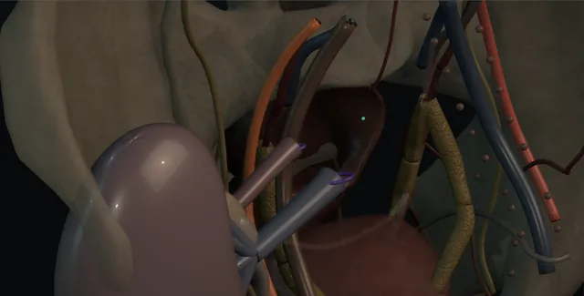
Schematic scene preview: Renal autotransplantation
Preparing the 3D anatomy…

UrOops3D · UroRef · © Nity G

Click me next Section 2 of 8 Anatomy The structures this operation works between, and the one it must not cross.

The structures in the scene

Renal autotransplantation: how the structures relateExtraperitoneal iliac fossa, the kidney put back and its three joins. Kidney in the iliac fossa, hilum medial: a tied branch is a silent infarct Hilar branches repaired on the bench (danger). Kidney in the iliac fossa, hilum medial: renal vein end-to-side, done first External iliac vein (danger). Kidney in the iliac fossa, hilum medial: end-to-side, or end-to-end if the artery is short External or internal iliac artery (danger). Kidney in the iliac fossa, hilum medial: stripped of adventitia, it necroses and leaks Renal pelvis or ureter with its fat (danger). Renal pelvis or ureter with its fat: short route; hitched to the psoas if needed Bladder. Kidney in the iliac fossa, hilum medial: ligated, not cut, or a lymphocele forms beside it Iliac lymphatics.Kidney in the iliacfossa, hilum medial!Hilar branchesrepaired on thebench!External iliac vein!External or internaliliac artery!Renal pelvis orureter with its fatBladderIliac lymphaticsa tied branch is asilent infarctrenal veinend-to-side, donefirstend-to-side, orend-to-end if theartery is shortstripped ofadventitia, itnecroses and leaksshort route; hitched tothe psoas if neededligated, not cut,or a lymphoceleforms beside itTargetDangerLandmarkContext
Extraperitoneal iliac fossa, the kidney put back and its three joins. Schematic, not to scale; relationships only.
  • The kidney has left the loin and sits in the iliac fossa outside the peritoneum, its pole turned so that the hilum faces medially and the pelvis lies low, a few centimetres above the bladder.
  • The renal vein runs to the external iliac vein and the artery to the external iliac artery beside it, both end-to-side, and the short route from the pelvis or ureter to the bladder crosses that gap without tension.
  • Above, the empty renal bed in the loin is out of the picture; below, the bladder can be hitched up toward the kidney if it needs to reach.
  • ›The renal artery and vein, kept long - taken at the aorta and the cava; the length is what makes the reimplantation possible.
  • ›The bench - a bowl of ice slush at 4 degrees, the kidney flushed with preservation solution, the repair done under loupes with hours to spare.
  • ›The external iliac vein - the first anastomosis, end-to-side, with the kidney under slush.
  • ›The external iliac artery, or the internal iliac - end-to-side to the external, or end-to-end to the internal when the renal artery is short.
  • ›The renal pelvis or the remaining ureter - kept with its fat; joined to the bladder by a tunnel or directly as a pyelovesicostomy over a stent.
  • ›The bladder - assessed for capacity and pressure beforehand; hitched to the psoas if the pelvis will not reach.
  • ›The iliac lymphatics - ligated, not cut, along the vessels, or a lymphocele forms beside the kidney.
Click me next Section 3 of 8 Steps The operation in order, with the landmark that guards each move.

Operative steps

  1. Who: the ureter that cannot be replaced, the artery that cannot be repaired in place Four patients, all rare. The long ureteric loss: avulsion at ureteroscopy, a stricture after radiotherapy or retroperitoneal fibrosis, or a failed previous repair, where a Boari flap will not reach and an ileal ureter is unattractive because of bowel disease or a poor kidney. The complex renal artery disease: a branch aneurysm, fibromuscular dysplasia into the segmental vessels, or an injury, that cannot be reconstructed in situ without prolonged warm ischaemia. The exceptional central or multifocal tumour in a solitary kidney where a bench partial nephrectomy in the cold is the only nephron-sparing option. And, in highly selected patients after a pain MDT, loin pain haematuria syndrome, where the operation denervates the kidney and the outcome is uncertain. In every case nephrectomy is stated plainly as the alternative.
  2. Planning: function, the vessels, the bladder The kidney must be worth the operation: split function on a MAG3 or DMSA above about a fifth, and a total GFR that makes preservation matter, above all the solitary kidney. CT angiography maps the arteries and veins and the length available, because early branching or a short right vein changes the bench plan. The ureter is mapped by antegrade or retrograde study. The bladder is assessed for capacity and compliance, because the kidney will drain into it by a short route, and a small, high-pressure or irradiated bladder needs augmentation or a different plan. The iliac vessels are checked for calcification. Consent names thrombosis and loss of the kidney at a few per cent, bleeding, urine leak, delayed function, and, for pain, that the pain may not go.
  3. Nephrectomy with the vessels long The kidney is removed as for a living donor, open or laparoscopically, with the renal artery taken at the aorta and the vein at the cava, on the left with the adrenal, gonadal and lumbar veins divided for the whole length, and on the right with a cuff of cava if the vein is short and the approach is open. The ureter is taken as long as its healthy length allows with its adventitia intact, or, where it is gone, the pelvis is kept with its fat. The patient is heparinised before the vessels are clamped, mannitol given, and the artery divided first and the vein second, the kidney lifted straight out and onto the bench in the ice with the warm ischaemia measured in seconds.
    where it bites: a short artery or a vein divided flush with the kidney to make the nephrectomy easy makes the reimplantation impossible; the length is the graft.
  4. The bench: cold, and the repair The kidney lies in ice slush at 4 degrees and the artery is flushed with cold preservation solution until the effluent runs clear; then the clock is cold and the surgeon has hours. The repair is done under loupes or the microscope in the slush: an aneurysm excised and the branches reimplanted or joined to a saphenous vein or internal iliac graft with 7-0 polypropylene, a stenosis resected and joined end-to-end, a central tumour excised with a margin and the collecting system closed with the vessels seen and no bleeding, or the ureter trimmed to healthy tissue. The repair is tested by flushing again; a leak of perfusate from a branch or the collecting system is fixed now. A short vein is lengthened with gonadal vein or a caval cuff.
    where it bites: the bench is bloodless and calm, and the mistake it invites is a branch tied or a suture placed through the collecting system that no one sees until reperfusion.
  5. Into the iliac fossa: vein then artery A Gibson or a lower midline incision into the iliac fossa, usually the same side, or the other if that fossa has been used before. The peritoneum is swept up and the external iliac artery and vein exposed with the lymphatics ligated, not cut, to prevent a lymphocele. The kidney comes from the ice with its pole orientated so that the pelvis lies medially and low, close to the bladder. The vein is done first: a Satinsky on the external iliac vein, a venotomy, and an end-to-side anastomosis with running 5-0 or 6-0 polypropylene, then the artery end-to-side to the external iliac with 6-0, or end-to-end to the internal iliac if the artery is short, with the kidney kept cold under slush while the sutures go in.
    where it bites: a kidney that warms on the field during a long anastomosis is a second warm ischaemia; it is kept under ice slush and the clock is spoken aloud.
  6. Reperfusion and the clock With both anastomoses complete the venous clamp is released first and then the arterial, and the kidney should pink up within seconds and pass urine within minutes. The anaesthetist has the patient filled and the pressure kept up for this moment, and mannitol or frusemide is given. A kidney that stays mottled or soft is examined for a kink in the vessels, a twist, spasm from cold or handling, or a technical fault at the anastomosis; a flow probe or Doppler is used if there is doubt. Bleeding from the anastomoses is controlled with single sutures, from the parenchyma with pressure, and from the bench repair with the same. The total warm time, the cold time and the anastomosis time are recorded.
    where it bites: a kidney that does not pink up is a technical problem in the artery or the vein until proved otherwise; the anastomosis is taken down and redone in the first minutes, not watched.
  7. The ureter or the pelvis to the bladder The kidney now sits a few centimetres from the bladder and the route is short. Where a ureter remains, it is joined to the bladder as in a transplant, an extravesical Lich-Gregoir tunnel over a stent, with the spatulated ureter sewn to the mucosa and the muscle closed over it. Where the ureter has gone, the renal pelvis is joined directly to the bladder, a pyelovesicostomy, with a wide spatulated anastomosis over a stent and the bladder hitched to the psoas if it needs to reach; some units join the graft ureter to the patient's own distal ureter if a healthy stump remains. The stent stays for four to six weeks. A urethral catheter drains the bladder, and a drain is left beside the kidney.
  8. After A Doppler on the first day confirms flow in the artery and the vein and the resistive index; another the day the creatinine rises, the urine output falls or new pain appears. Fluid balance is matched to the urine output hour by hour in the first day. The drain fluid creatinine is checked if the drain runs high. The catheter comes out at about five days, the stent at four to six weeks, and a renogram at three months measures the kidney in its new home. Blood pressure is followed, because a stenosis at the arterial anastomosis appears as hypertension and a bruit. The pain patient is reassessed by the pain team; the tumour patient is surveyed as after any partial nephrectomy.
Click me next Section 4 of 8 Danger zones Where this operation bites, and why it bites there.
Critical relationship

Danger zones

The spatial relationships the animation is built to make automatic. Before you read each one, ask yourself: at this point in the operation, what is about to go wrong, and what is my first move? The save for each sits in the Complications tab.

  • RiskThe hilar branches on the bench - a segmental branch tied or a suture through a calyx in a bloodless field is a silent infarct or a leak found at reperfusion.
  • RiskThe iliac vessels - a calcified external iliac artery that will not take a suture, and a vein torn by the Satinsky; the anastomoses have to be perfect or the kidney thromboses.
  • RiskThe pelvis and ureter and their fat - stripped of adventitia they necrose; the anastomosis to the bladder leaks and strictures.
  • RiskThe two fields - an upper abdominal nephrectomy and a pelvic transplant in one operation: the bowel, the spleen or the duodenum in the first, the bladder and the peritoneum in the second.
Practise it Take me into the scene Opens the animation at a danger moment and asks you to predict the mechanism and your first move before it reveals.
Click me next Section 5 of 8 Complications The save for each one: the first move, the escalation trigger, the op-note line.

Complications and immediate saves

Trigger each of these live in the Animation tab, then read the save: the first move, the escalation trigger, and the line that goes in the op note.

Arterial or venous thrombosis and loss of the kidney

  • a kidney that produced urine and then stops in the first day, or a Doppler with no flow: the artery clotted at the anastomosis or a reconstructed branch, or the vein kinked or compressed; the kidney has hours.
The save
  1. A fall in urine output or a rising creatinine in the first days: Doppler the same hour, not the next morning. first move
  2. A venous thrombosis or an arterial occlusion on Doppler: straight back to theatre; a kidney explored within hours may be saved, one explored the next day is lost.
  3. Prevention is on the bench and at the anastomosis: no kink, no twist, a wide venous anastomosis, the kidney orientated before it is sewn, and heparin as the unit uses it.
  4. A kidney that is black at re-exploration, or a branch reconstruction that has failed: nephrectomy, and the patient told that the operation was lost. escalation trigger
  5. Op note: the anastomoses, the orientation, the reperfusion appearance, the times, and the anticoagulation plan; the Doppler findings each day. document

Haemorrhage and anastomotic disruption

  • bleeding from the nephrectomy bed in the loin, the arterial or venous anastomosis, a bench repair that opens at reperfusion, or the parenchyma after a bench partial nephrectomy.
The save
  1. At reperfusion: pressure on the point, single 6-0 sutures at the anastomosis with the clamps reapplied if needed, and the pressure kept up by the anaesthetist. first move
  2. On the ward: a haemoglobin and a Doppler; a stable haematoma beside the kidney is watched, an expanding one is explored.
  3. A late bleed with fever is a mycotic pseudoaneurysm at the arterial anastomosis until proven otherwise: CT angiography.
  4. Haemodynamic instability or an expanding haematoma compressing the vein: return to theatre, with vascular help for an anastomosis that has disrupted. escalation trigger
  5. Op note: the bleeding point, the repair, the blood loss and products, and the drain output. document

Urinary leak and obstruction

  • urine in the drain from the pyelovesicostomy, the ureteric anastomosis or a bench collecting system repair in the first fortnight; or a stricture at the join months later with hydronephrosis and a falling function.
The save
  1. Drain fluid creatinine against the serum; the catheter and the stent kept draining, and the bladder kept empty. first move
  2. Most leaks close with the stent in and the bladder drained for longer; a urinoma is drained percutaneously.
  3. A stricture on the follow-up ultrasound or renogram: nephrostomy, an antegrade study, and balloon dilatation or incision for a short one.
  4. A leak that persists beyond a fortnight or a devascularised pelvis: re-exploration and revision of the anastomosis; a long stricture needs a Boari flap or a new join. escalation trigger
  5. Op note: the type of urinary anastomosis, the stent size and the date it comes out, and the imaging schedule. document

Ischaemic injury and delayed function

  • a kidney that flows on Doppler but makes little urine and a creatinine that does not fall: the warm time at the nephrectomy or the anastomosis, a long cold time, a poor flush, or the hypotension around reperfusion.
The save
  1. Confirm flow with Doppler first, because a kidney that is thrombosed looks the same from the bedside as one that is stunned. first move
  2. Fluids matched to output, nephrotoxins stopped, and the potassium watched; the kidney usually recovers over days to weeks.
  3. The ischaemic times were minimised at the operation: seconds warm at the nephrectomy, the kidney under slush during the anastomoses, and the flush until clear.
  4. Anuria with a solitary kidney: nephrology and dialysis as a bridge; a biopsy if function has not returned at two to three weeks. escalation trigger
  5. Op note: the warm, cold and anastomosis times, the flush and the reperfusion appearance, so the delay can be read against them. document

Lymphocele, wound and the failed indication

  • a collection beside the kidney compressing the vein or the ureter weeks later, a wound infection or hernia in one of two incisions, and, in the pain patient, a technically perfect kidney with the same pain.
The save
  1. Ultrasound for any swelling, leg oedema or rising creatinine in the first months; a lymphocele is diagnosed by its fluid creatinine matching the serum. first move
  2. A symptomatic lymphocele is aspirated, then drained, then marsupialised into the peritoneum laparoscopically if it recurs.
  3. Ligating the lymphatics along the iliac vessels rather than cutting them is the prevention.
  4. Persistent pain after autotransplant for the pain syndrome: back to the pain MDT; a further operation on the kidney is not the answer. escalation trigger
  5. Note: the expectation set before the operation for the pain indication, and the outcome recorded honestly against it. document
Click me next Section 6 of 8 Troubleshooting When the case turns: the problem, then the next move.

Intra-operative troubleshooting

When the case turns: the problem, then the next move.

  • ActionThe renal artery is too short for the external iliac - end-to-end to the internal iliac artery, or interpose a length of saphenous vein or the patient's own internal iliac on the bench before the kidney goes in.
  • ActionThe right renal vein is too short - lengthen it on the bench with the gonadal vein or a caval cuff taken at the nephrectomy; a short vein sewn under tension thromboses.
  • ActionThe kidney does not pink up at reperfusion - look for a twist or a kink, feel the artery, warm saline on the vessels, papaverine if the unit uses it, then Doppler; if there is no flow, take the anastomosis down and redo it now.
  • ActionPerfusate leaks from the collecting system on the bench - find the point with the flush running and close it with a fine absorbable suture on the bench, where it is easy, not after reperfusion, where it is not.
  • ActionThe pelvis will not reach the bladder - reposition the kidney lower in the fossa, hitch the bladder to the psoas, or use the patient's distal ureteric stump if it is healthy; tension leaks.
  • ActionThe external iliac artery is calcified - choose a soft segment, use the internal iliac end-to-end, or endarterectomise a short segment with vascular help; a suture through plaque tears.
Click me next Section 7 of 8 FRCS viva Say it out loud, then mark yourself against the model answer.

FRCS viva questions

The examiner stems this scene prepares you for. Try to answer out loud first, then reveal, and learn the discriminator, the detail that turns a pass into a strong pass.

QWhat are the indications for renal autotransplantation, and what are the alternatives?reveal answer
  • Autotransplantation is an exceptional operation with four indications.
  • First, extensive ureteric loss that cannot be bridged by a psoas hitch, a Boari flap or a ureteroureterostomy, for example after ureteroscopic avulsion, radiotherapy, retroperitoneal fibrosis or repeated failed repairs, where the alternative of an ileal ureter is unattractive because of bowel disease, poor renal function or metabolic risk.
  • Second, complex renovascular disease, a branch aneurysm, fibromuscular dysplasia extending into segmental vessels, or an arterial injury, which cannot be repaired in situ without prolonged warm ischaemia but can be repaired on the bench in the cold.
  • Third, an exceptional central or multifocal tumour, usually in a solitary kidney, where ex vivo partial nephrectomy in a bloodless cold field is the only way to spare nephrons.
  • Fourth, and most contentious, loin pain haematuria syndrome in highly selected patients after a pain multidisciplinary team, where the operation denervates the kidney and the outcome is uncertain.
  • In every case the alternatives are stated: in situ reconstruction, ileal ureter, endovascular treatment, radical nephrectomy with one kidney, chronic drainage by nephrostomy or stent, or no operation.
  • The kidney must have enough function to justify transplant-level risk, and I would confirm that with a split function scan.
Strong pass

Say both

  • The four indications with the reason in situ repair fails in each
  • Nephrectomy stated as a legitimate alternative rather than omitted
Examiner note
The four indications with the reason in situ repair fails in each, and nephrectomy stated as a legitimate alternative rather than omitted.
QDescribe the steps of a renal autotransplant for ureteric loss.reveal answer
  • After confirming split function, mapping the vessels on CT angiography and assessing the bladder, I would remove the kidney as for a living donor, taking the artery at the aorta and the vein at the cava for length, and keeping whatever healthy ureter or pelvis remains with its fat intact.
  • After heparin and mannitol I would divide the artery then the vein and place the kidney in ice slush, flushing it with cold preservation solution until clear.
  • On the bench I would trim the ureter or pelvis to healthy tissue and lengthen the vein if needed.
  • Through a Gibson incision I would expose the external iliac vessels extraperitoneally, ligating the lymphatics, and orientate the kidney so the pelvis lies low and medial.
  • I would anastomose the renal vein end-to-side to the external iliac vein with running polypropylene, then the artery end-to-side to the external iliac or end-to-end to the internal iliac, keeping the kidney under slush.
  • I would release the venous clamp then the arterial and expect the kidney to pink up and pass urine.
  • I would join the ureter to the bladder by an extravesical tunnel over a stent, or the pelvis directly to the bladder as a pyelovesicostomy with a psoas hitch if needed, leave a drain and a catheter, and Doppler the kidney on the first day.
Strong pass

Say all three

  • The vessels taken at the aorta and the cava with the reason
  • The kidney orientated before it is sewn
  • The pyelovesicostomy named as the option when the ureter is gone
Examiner note
The vessels taken at the aorta and the cava with the reason, the kidney orientated before it is sewn, and the pyelovesicostomy named as the option when the ureter is gone.
QWhy is the bench the safest part of the operation, and what does it make possible?reveal answer
  • Once the kidney is flushed and in ice slush at about 4 degrees, its metabolism falls to a fraction of normal and it tolerates cold ischaemia for many hours, so the surgeon has time;
  • and because it is not perfused, there is no bleeding, so the hilum, the segmental branches and the collecting system can be dissected and sewn under loupes or the microscope with a clarity that is impossible in situ, where every minute of clamp time is warm ischaemia and the field fills with blood.
  • That makes possible the excision of a branch aneurysm with reimplantation of the branches onto a graft, the resection and reanastomosis of a segmental stenosis, the excision of a central tumour with the collecting system closed under vision and the vessels seen, and the lengthening of the ureter, pelvis and vein.
  • The bench has its own dangers: a branch tied or a calyx sutured in a field with no bleeding gives no warning, so the repair is tested by flushing again before the kidney goes in, and any leak of perfusate from a vessel or the collecting system is closed then.
  • The cost is that the kidney must then be reimplanted, so the operation carries transplant-level risks of thrombosis, leak and delayed function that an in situ repair does not.
Strong pass

Say all three

  • The metabolic reason cold buys time
  • The flush as the test of the bench repair
  • The price of the bench named as the reimplantation risks
Examiner note
The metabolic reason cold buys time, the flush as the test of the bench repair, and the price of the bench named as the reimplantation risks.
QWhat are the complications of renal autotransplantation and how are they detected?reveal answer
  • I would separate them into vascular, urinary, functional and the general.
  • Vascular: arterial or venous thrombosis at the anastomosis or a reconstructed branch, which presents in the first days as a fall in urine output or a rising creatinine and is diagnosed by Doppler the same hour, because the kidney can be saved only by immediate re-exploration;
  • haemorrhage from the anastomoses, the bench repair or the nephrectomy bed;
  • and late arterial stenosis presenting as hypertension and a bruit.
  • Urinary: leak from the pyelovesicostomy or ureteric anastomosis in the first fortnight, detected by drain creatinine and managed with the stent and catheter, and stricture months later, found on ultrasound and renogram.
  • Functional: delayed function from warm or cold ischaemia or hypotension at reperfusion, which recovers over days to weeks but can require dialysis in a solitary kidney, and a biopsy if it does not recover.
  • General: lymphocele from cut iliac lymphatics, compressing the vein or ureter, aspirated, drained or marsupialised;
  • injury to bowel, spleen, duodenum or iliac vessels across two fields;
  • infection and hernia in two wounds;
  • and, for the pain indication, pain that persists despite a working kidney.
  • Detection depends on a Doppler on the first day and at any change, hourly urine output early, the drain creatinine, and a renogram at three months.
Strong pass

Say both

  • Doppler within the hour of a change in output as the reflex
  • With the reason that thrombosis is reversible only in the first hours
Examiner note
Doppler within the hour of a change in output as the reflex, with the reason that thrombosis is reversible only in the first hours.
Click me next Section 8 of 8 Evidence The guidance each of these answers is built on.

Evidence and guidance

Two minutes

Close the lap: recall it before you leave.

Say it aloud, then reveal. Reading is not remembering.

  1. 01 The relationship Say the critical spatial relationship out loud, then reveal. reveal
    Memory frame

    The kidney has left the loin and sits in the iliac fossa outside the peritoneum, its pole turned so that the hilum faces medially and the pelvis lies low, a few centimetres above the bladder.

    Why?
    The renal vein runs to the external iliac vein and the artery to the external iliac artery beside it, both end-to-side, and the short route from the pelvis or ureter to the bladder crosses that gap without tension. Above, the empty renal bed in the loin is out of the picture; below, the bladder can be hitched up toward the kidney if it needs to reach.
  2. 02 The first move Arterial or venous thrombosis and loss of the kidney. Your first move? reveal
    First move

    A fall in urine output or a rising creatinine in the first days: Doppler the same hour, not the next morning.

  3. 03 The escalation trigger Arterial or venous thrombosis and loss of the kidney. What makes you call for help? reveal
    Escalate when

    A kidney that is black at re-exploration, or a branch reconstruction that has failed: nephrectomy, and the patient told that the operation was lost.

  4. 04 The discriminator What turns a pass into a strong pass? reveal
    Strong pass

    Say both

    • The four indications with the reason in situ repair fails in each
    • Nephrectomy stated as a legitimate alternative rather than omitted
    Examiner note
    The four indications with the reason in situ repair fails in each, and nephrectomy stated as a legitimate alternative rather than omitted.
  5. 05 The pitfall Where does this operation bite first? reveal
    Pitfall

    The hilar branches on the bench

    Why?
    a segmental branch tied or a suture through a calyx in a bloodless field is a silent infarct or a leak found at reperfusion.

Revision practice with self-marked answers, not an assessment of clinical competence. Kept in this browser only; it does not sync between devices.