Bladder rupture: pelvic fracture and the cystogram
Bladder rupture taught as two leaks on one organ: the peritonealised dome bursts upward into the peritoneal cavity, the bladder neck tears against the fractured pelvic ring into the pelvic haematoma.
A properly filled CT cystogram tells them apart, and an underfilled one hides both.
Core tierFRCS urological trauma, emergency urologyInteractive 3D
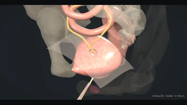
Midline slice from the patient's right, the bladder inside a fractured pelvis. Schematic, not to scale; relationships only.
Stand at the patient's right and look at a midline slice.
Behind is the sacrum, in front the sprung pubic symphysis with its fracture fragments, and between them the bladder, with the prostate and urethra descending in front of the pelvis to the perineum.
Above the bladder the peritoneal reflection runs across the dome, and above that sit bowel loops: everything on the peritoneal side of that reflection is the intraperitoneal world, and a dome tear leaks into it.
Below and in front, the bladder neck lies naked against the pelvic bones and the fracture haematoma: an extraperitoneal tear leaks into that space instead.
The cystogram reads the two leaks by where the contrast goes, and the whole decision, catheter versus laparotomy, hangs on which side of the peritoneal reflection the tear sits.
›Peritonealised dome - the part of the bladder under peritoneum; a tear here leaks urine straight into the peritoneal cavity.
›Bladder neck against the ring - the extraperitoneal bladder wall, lying directly on the pubic bones and their fractures; the site of the leak into the haematoma.
›Pelvic haematoma - the fracture haematoma surrounding the extraperitoneal bladder; it absorbs the leak, and it is the tissue that heals the tear.
›Peritoneal reflection + bowel - the plane that separates the two rupture patterns and the reason intraperitoneal urine outlines bowel loops on imaging.
›The catheter corridor - the urethral route for drainage and the suprapubic route as the fallback, both of which must be reconciled with orthopaedic metalwork.
The mechanismBlunt deceleration or crush breaks the pelvic ring: the symphysis springs, the rami fracture, and the bony cage around the bladder changes shape. Two injuries follow from one force. The bladder neck, fixed against the bones, is torn where the ring displaces, and that leak drains into the pelvic haematoma. The dome, free and distended, bursts when compression squeezes it against a closed outlet, and that leak drains into the peritoneal cavity. One force, two leaks, in two different spaces. where it bites: the same pelvic fracture threatens both the bladder and the urethra: blood at the meatus changes the whole pathway.
Assessment firstThe trauma survey comes before the urology: the sources of shock are found and addressed, and the bladder is studied once the patient is resuscitating rather than instead of resuscitation. Gross haematuria after pelvic trauma names the urinary tract for imaging, and the finding is pursued with the same seriousness as the fracture itself. The meatus is inspected and the prostate assessed on gentle rectal examination before anything is passed. The bladder diagnosis is made on imaging, never by catheterisation against resistance. where it bites: never force a catheter through blood at the meatus: the urethral injury must be excluded first.
CT cystogram, properly filledOnce the urethra is cleared, contrast is run retrograde into the bladder and the catheter is clamped so the organ fills to genuine distension before scanning. The filling is active and retrograde, not the passive arrival of excreted contrast, and the clamp stays on throughout. The reason is mechanical: an underfilled bladder collapses, its wall apposes and seals the tear, and the study reads falsely normal. A cystogram is only as good as the distension behind it. where it bites: the underfilled cystogram is a false-negative machine: an empty bladder wall apposes and seals the tear during scanning.
Read the leakThe pattern of extravasation names the leak. Contrast striping into the pelvic haematoma around the bladder neck and symphysis, contained below the peritoneal reflection, is extraperitoneal rupture. Contrast running free in the peritoneum, outlining bowel loops and pooling in the paracolic gutters, is intraperitoneal rupture from the dome. Both patterns answer one question: which side of the peritoneal reflection does the contrast sit? The answer decides catheter versus theatre. where it bites: contrast around bowel loops means intraperitoneal rupture and a laparotomy, not a catheter.
Extraperitoneal: catheter drainageExtraperitoneal rupture is treated with a freely draining urethral catheter and nothing else in the great majority of patients. The haematoma around the tear both contains the leak and heals it, and the catheter keeps the bladder empty so the edges stay apposed. Drainage is checked constantly, because a catheter blocked by clot turns a healing tear into a leaking one. The catheter stays until a repeat study, again filled properly, shows a sealed bladder. Most of these patients never see a urological operation. where it bites: a blocked catheter converts conservative management into urgent surgery: check drainage constantly.
Intraperitoneal: repair at laparotomyIntraperitoneal rupture goes to laparotomy, where the peritoneal cavity is lavaged and the dome tear is débrided of ragged edges before closure in two layers of absorbable suture. The interior of the bladder is inspected through the tear: the bladder neck is examined for an associated injury and the ureteric orifices are identified so that no injury is closed over unseen. The repair is drained by catheter, urethral or suprapubic as the injuries dictate, while it heals. A dome tear closed without inspecting the neck risks leaving the more dangerous injury behind. where it bites: a neck or ureteric injury found at laparotomy changes the operation: look before closing.
Coordination with orthopaedicsExternal fixator pins, bars and the planned orthopaedic approaches occupy the same few centimetres of lower abdominal wall as the suprapubic corridor, and the timing of packing or fixation changes every catheter plan. The urology plan, its route and its expected duration, is stated in the joint conversation before the metalwork goes on. If suprapubic drainage is needed later, the track is chosen clear of pin sites, under imaging where the anatomy is distorted, because a track driven past a pin seeds the metalwork. One joint plan, written into both sets of notes, made early. where it bites: a suprapubic track driven past fixator pin sites seeds the metalwork; plan the two incisions together.
The spatial relationships the animation is built to make automatic. Before you read each one, ask yourself: at this point in the operation, what is about to go wrong, and what is my first move? The save for each sits in the Complications tab.
RiskThe underfilled cystogram - an empty or half-filled bladder seals its own tears during scanning; fill to genuine distension and clamp before scanning.
RiskThe dome under peritoneum - a leak here is urine into the peritoneal cavity: peritonitis by mechanism, and a laparotomy decision.
RiskThe neck against the fracture - tears here track into the pelvic haematoma; they also travel with urethral injuries and orthopaedic hardware.
When the case turns: the problem, then the next move.
ActionCystogram negative but the patient is peritonitic - the study may be underfilled: repeat with genuine distension; a peritonitic abdomen after pelvic trauma is explored rather than explained away.
ActionCatheter blocked by clot while draining a rupture - hand irrigation with a bladder syringe; if clot cannot be cleared and drainage stays obstructed, change the catheter over a guide or take the patient for washout.
ActionBladder neck injury found at laparotomy for a dome tear - convert the repair to include the neck: débride, close with absorbable sutures, and divert with catheter drainage; inspect the ureteric orifices before closing.
ActionFixator bar sits over the planned suprapubic site - agree a new track with orthopaedics, image-guided if needed; urethral drainage is the cleaner answer when the urethra is safe.
The examiner stems this scene prepares you for. Try to answer out loud first, then reveal, and learn the discriminator, the detail that turns a pass into a strong pass.
QHow do you perform and interpret a CT cystogram in suspected bladder rupture?reveal answer
The cystogram is only as good as the filling.
After the urethra has been cleared, or with a catheter already safely placed, contrast is instilled retrograde into the bladder under gravity, and the catheter is clamped so the bladder distends fully before scanning.
The reason is mechanical: a tear in an empty or barely filled bladder is apposed by the collapsing wall, and contrast can fail to extravasate, which is how underfilled studies produce false negatives.
The interpretation follows the contrast.
Extraperitoneal rupture shows contrast striping into the pelvic soft tissues around the bladder neck and symphysis, within the haematoma planes: it is managed conservatively with a freely draining catheter.
Intraperitoneal rupture shows contrast beyond the bladder outlining bowel loops and pooling in the dependent peritoneal spaces: the dome has burst under peritoneum, urine is in the peritoneal cavity, and the patient goes to laparotomy for repair.
Two pitfalls deserve naming.
First, a passive filling technique without clamping under-distends the bladder and mimics a negative study.
Second, clot or a catheter balloon tamponading a neck tear can mask extravasation, so a high-pressure injury pattern with a negative study still earns careful review.
The cystogram therefore answers one question properly, which side of the peritoneal reflection is the leak, and only when the bladder is genuinely full.
Strong pass
Say both
Naming the underfilled bladder as the mechanism of the false negative
Stating that contrast around bowel loops is the intraperitoneal signature mandating laparotomy
Examiner note
Naming the underfilled bladder as the mechanism of the false negative and stating that contrast around bowel loops is the intraperitoneal signature mandating laparotomy.
QWhen does extraperitoneal bladder rupture still need an operation?reveal answer
Extraperitoneal rupture heals on a draining catheter, with a small minority needing surgery, and the exceptions are worth enumerating because each one changes the indication.
The first is the retained catheter failure: if clot cannot be kept clear and drainage obstructs, or if urine continues to extravasate on follow-up imaging despite drainage, the bladder needs formal repair and washout.
The second is the bony problem: a bone fragment or spike driven into the bladder neck may need to be dealt with at orthopaedic fixation, with the bladder repaired at the same anaesthetic, which is one reason urology stands by during pelvic fixation.
The third is the associated injury: a bladder neck injury extending into the urethra, or a concomitant rectal or vaginal injury, converts a conservative plan into a repair, because a shared wall injury leaking into another organ space will not heal on drainage alone.
The fourth is the planned orthopaedic exposure: when internal fixation hardware will cross a contaminated extraperitoneal tear, repair reduces the infection risk to the metalwork.
The principle through all four: catheter drainage is the treatment of the tear, but it does not treat retained clot, bone, associated organ injuries or hardware, and each of those is an operative reason.
Strong pass
Say all four
Listing bone spike
Associated rectal or vaginal injury
Hardware contamination as the specific conversions
Not just failed drainage
Examiner note
Listing bone spike, associated rectal or vaginal injury, and hardware contamination as the specific conversions, not just failed drainage.
QHow do you coordinate bladder drainage with pelvic fracture management?reveal answer
The coordination problem is spatial: the external fixator or pelvic binder, the suprapubic corridor, the pubic surgical approaches and the bladder all occupy the same few centimetres of lower abdomen and pelvis.
The first decision is the catheter route.
A urethral catheter is the answer whenever the urethra is safe: blood at the meatus, a high-riding prostate or an inability to pass a catheter gently trigger a urethral study, and only then, if the urethra is unsafe, does suprapubic access become the plan.
The second decision is timing: suprapubic catheter insertion is cleanest done before fixator pins are placed, or under imaging once hardware is in the way, because a track driven past pin sites seeds the metalwork.
The third is the shared anaesthetic: when the orthopaedic team is fixing the pelvis, that is the moment to reassess the bladder, clear clot, repair a neck tear if indicated, and site any suprapubic catheter under direct vision rather than blind.
Communication is the intervention: the urology plan, catheter route, expected duration and who removes what, written into the joint notes, prevents the classic failures, a catheter nobody owns, a pin site made infected by a track, or a suprapubic catheter that orthopaedics did not know existed.
Strong pass
Say all four
Treating coordination as a spatial
Timing problem: urethral route first
Suprapubic before pins
Bladder reassessment on the orthopaedic table
Examiner note
Treating coordination as a spatial and timing problem: urethral route first, suprapubic before pins, and bladder reassessment on the orthopaedic table.
QA patient with a pelvic fracture develops peritonitis two days after a negative cystogram. What is happening and what do you do?reveal answer
The likeliest explanation is a missed intraperitoneal bladder rupture: the index cystogram under-filled the bladder, the wall apposed the dome tear, the study read negative, and urine has been leaking into the peritoneal cavity since.
Chemical peritonitis then progresses: increasing pain, distension, absent bowel sounds, tachycardia and then sepsis.
The differential that must be actively excluded is a missed bowel injury from the same blunt force, which presents on a similar trajectory and is the more dangerous partner.
Management is in sequence.
Resuscitate, take cultures, start broad-spectrum antibiotics and analgesia.
Re-image, but do not delay for a study in a deteriorating patient: a properly distended retrograde CT cystogram in a stable patient confirms contrast around bowel loops, while a peritonitic, unstable patient goes to theatre, where the diagnosis is made directly.
At laparotomy: lavage the peritoneal cavity, débride and close the dome tear in two absorbable layers with a general surgical colleague standing by for the bowel, inspect the bladder neck, and leave the bladder drained.
The missed diagnosis carries the lesson forward: fill the bladder properly the first time, and treat a peritonitic abdomen after pelvic trauma as a laparotomy problem until proven otherwise.
Strong pass
Say both
Naming the underfilled index study as the mechanism
Keeping missed bowel injury in the differential while proceeding to laparotomy in the unwell patient
Examiner note
Naming the underfilled index study as the mechanism and keeping missed bowel injury in the differential while proceeding to laparotomy in the unwell patient.
Say it aloud, then reveal. Reading is not remembering.
01The relationshipSay the critical spatial relationship out loud, then reveal.reveal
Memory frame
Stand at the patient's right and look at a midline slice.
Why?
Behind is the sacrum, in front the sprung pubic symphysis with its fracture fragments, and between them the bladder, with the prostate and urethra descending in front of the pelvis to the perineum. Above the bladder the peritoneal reflection runs across the dome, and above that sit bowel loops: everything on the peritoneal side of that reflection is the intraperitoneal world, and a dome tear leaks into it. Below and in front, the bladder neck lies naked against the pelvic bones and the fracture haematoma: an extraperitoneal tear leaks into that space instead. The cystogram reads the two leaks by where the contrast goes, and the whole decision, catheter versus laparotomy, hangs on which side of the peritoneal reflection the tear sits.
02The first moveMissed intraperitoneal leak. Your first move?reveal
First move
Recognise the missed leak: increasing abdominal pain, distension, ileus and systemic upset after a supposedly negative study.
03The escalation triggerMissed intraperitoneal leak. What makes you call for help?reveal
Escalate when
Peritonitis with deterioration, or a missed associated bowel injury → urgent laparotomy with general surgical support.
04The discriminatorWhat turns a pass into a strong pass?reveal
Strong pass
Say both
Naming the underfilled bladder as the mechanism of the false negative
Stating that contrast around bowel loops is the intraperitoneal signature mandating laparotomy
Examiner note
Naming the underfilled bladder as the mechanism of the false negative and stating that contrast around bowel loops is the intraperitoneal signature mandating laparotomy.
05The pitfallWhere does this operation bite first?reveal
Pitfall
The underfilled cystogram
Why?
an empty or half-filled bladder seals its own tears during scanning; fill to genuine distension and clamp before scanning.
Revision practice with self-marked answers, not an assessment of clinical competence. Kept in
this browser only; it does not sync between devices.