The commonest urological procedure and the one most often done badly: a catheter passed blind through two curves and a sphincter into a bladder that must prove it is there before the balloon goes up.
This page is the feel of it, curve by curve, and the ladder to climb when it will not pass.
Core tierFRCS core urology, emergency and accessInteractive 3D
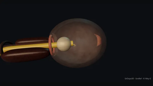
Along the male urethra from the meatus, as the catheter passes. Schematic, not to scale; relationships only.
From the meatus the urethra runs straight along the penis, turns upward at the penoscrotal angle through the bulb, passes the external sphincter as a ring, and then curves up and over the median lobe through the prostate to the bladder neck.
Holding the penis up straightens the first curve; a coudé tip at twelve o'clock manages the second.
The balloon inflates only when the tip is at the hub and urine has proved the bladder.
›Meatus and navicular fossa - the entry and the widest part of the penile urethra; meatal stenosis is met here.
›Penile urethra - straight while the penis is held up at ninety degrees; the first place a stricture sits in a young man.
›Bulbar urethra and the penoscrotal angle - the first curve, the widest and most dependent part; where a forced catheter leaves the lumen.
›External sphincter and membranous urethra - the definite resistance two centimetres beyond the bulb; opens with breathing, injured by pushing.
›Prostatic urethra - the second curve, up over the median lobe to the neck; the tightest part in a big gland.
›Bladder neck - the last ring; the balloon is drawn back to sit on it, never inflated before it.
›The catheter - 14 or 16 French Foley, coudé tip as the second choice in an older man, 10 ml of water in the balloon.
Before the catheter: indication, safety, consentEvery catheter has a reason written down: retention, output monitoring, a wound to keep dry, a bladder to rest. Then the question that stops a urethral catheter: is the urethra safe? Blood at the meatus, a pelvic fracture, a high-riding prostate or recent urethral surgery means stop and ask; the same for a known stricture or an artificial sphincter that has not been deactivated. The patient is told what will happen and that it will be uncomfortable but not painful. On the trolley: sterile gloves, cleaning solution, a drape, a 10 ml syringe of sterile water, 11 ml of 2 per cent lidocaine gel, the catheter, a bag and a specimen pot.
The catheter: size and tipA 14 or 16 French two-way Foley for a man, 12 or 14 for a woman; a three-way 22 French only for haematuria that needs irrigation. Silicone for anyone likely to keep it more than a few weeks. The coudé tip has a fixed upward bend that rides over a large median lobe and a high bladder neck; it is the second catheter to try in an older man, not the fifth. The balloon takes 10 ml of sterile water, never saline, which crystallises. A smaller catheter is not a gentler catheter: it buckles at the first resistance and makes a false passage.
Prepare: gel and positionClean the glans with the foreskin retracted, drape, and put the whole syringe of gel into the urethra, not onto the catheter. Squeeze the glans gently for two minutes to hold it there; the anaesthetic needs the time and the lubrication needs to be ahead of the tip. The penis is held up at ninety degrees to the body with the non-dominant hand, gently stretched. This straightens the penoscrotal angle and takes the first curve out of the urethra before the catheter reaches it. where it bites: a foreskin left retracted at the end of the procedure becomes a paraphimosis by the morning.
The penile urethraThe catheter goes in by hand, held near the tip, a few centimetres at a time, and never with forceps. The navicular fossa just inside the meatus is the widest part; beyond it the penile urethra runs straight while the penis is held up. Resistance here in a young man is a meatal stenosis or a stricture, and it is met with gel and a smaller step down the ladder to a wire under vision, not with force.
The bulbar curveAt the penoscrotal angle the tip enters the bulbar urethra, the widest and most dependent part, and meets the first curve. The hand holding the penis brings it down toward the feet as the catheter passes the curve, so that the tip follows the lumen round rather than driving into the roof of the bulb. A catheter that stops here has met the curve, not a stricture: lower the penis and advance again. This is where a forced catheter leaves the urethra and enters the spongiosum, and the bleeding that follows is the sign of it. where it bites: the bulbar curve is where a false passage is made; the fix is the angle of the penis, not more push.
The external sphincterTwo centimetres beyond the bulb the tip meets a definite resistance: the external urethral sphincter, a ring of striated muscle that closes reflexly on the foreign body. This is the moment most catheters are pushed too hard. The move is steady gentle pressure held on the catheter while the patient is asked to take slow deep breaths, or to try to pass urine; both relax the sphincter and the catheter goes through, often with a small give. Once it has passed, the veru sits on the floor just beyond and the prostatic urethra begins. where it bites: pushing through a closed sphincter injures it; the sphincter opens for a patient who is breathing, not for a house officer who is pushing.
The prostatic urethraThe prostatic urethra curves upward and forward over the median lobe to the bladder neck: the second curve, and in a big gland the longest and tightest part of the whole passage. A standard catheter tends to dig into the median lobe or the floor of the neck; a coudé tip with its bend kept at twelve o'clock rides over the lobe. The bladder neck is a final ring of resistance in a long prostatic urethra, and beyond it the catheter runs free.
In the bladder: urine before balloonThe catheter is advanced to the hub before anything else is done, so that the tip and the balloon are well inside the bladder and not sitting in the prostatic urethra. Then wait for urine. If nothing comes, press suprapubically, wait a minute, flush 20 ml of saline down the catheter and aspirate. Urine back means the bladder; nothing back means either an empty bladder or a tip that is not in it, and a bladder scan settles which. Only when the catheter has proved its position does the balloon go up: 10 ml of water, slowly, stopping at any pain. where it bites: a balloon inflated in the prostatic urethra splits it; urine first, balloon second, every time.
Seat, secure and writeDraw the catheter back until the balloon sits on the bladder neck and the catheter stops. Replace the foreskin over the glans. Connect the bag and hang it below the level of the bladder so it drains; secure the catheter to the thigh so the balloon is not dragged against the neck. The note records the indication, the size and type, the balloon volume, the residual volume drained, the colour of the urine, whether it was easy or difficult and why, and the plan: the date of the trial without catheter or the reason it stays.
The difficult catheter: the ladderTwo gentle failed attempts is the limit for one pair of hands. The ladder, in order: more gel and more time at the sphincter with the patient breathing; a coudé tip; a size up, 18 French, because a stiffer catheter follows the lumen where a floppy one buckles. Then urology. The registrar passes a flexible cystoscope, sees the true lumen, places a guidewire into the bladder and railroads a catheter over it; or, if the urethra will not pass at all and the bladder is palpable, puts in a suprapubic catheter under ultrasound. Nobody should have the urethra instrumented five times in one night.
The spatial relationships the animation is built to make automatic. Before you read each one, ask yourself: at this point in the operation, what is about to go wrong, and what is my first move? The save for each sits in the Complications tab.
RiskThe bulbar curve - a catheter forced here enters the spongiosum: a false passage with bleeding and no urine.
RiskThe external sphincter - closes reflexly on the catheter; opened with breathing and steady pressure, injured by force.
RiskThe prostatic urethra - the balloon inflated here splits the urethra; advance to the hub and wait for urine first.
RiskThe retracted foreskin - left back after the procedure it becomes a paraphimosis; always replaced.
Trigger each of these live in the Animation tab, then read the save: the first move, the escalation trigger, and the line that goes in the op note.
False passage
the catheter pushed out of the lumen, usually at the bulbar curve: it stops short, bleeds, and drains nothing.
The save
Stop. Withdraw the catheter; do not inflate a balloon that has not drained urine.first move
One further gentle attempt with more gel, the penis lowered at the curve, and a larger coudé catheter.
If that fails, urology: a flexible cystoscope to find the true lumen, a guidewire into the bladder and a catheter railroaded over it.
A urethra that cannot be passed under vision, or a patient in retention with a palpable bladder: suprapubic catheter under ultrasound.escalation trigger
Note: the number of attempts, where it stopped, the bleeding, the route finally used and the plan for the trial without catheter.document
Balloon inflated in the urethra
the balloon filled before the tip reached the bladder: sudden pain, blood at the meatus, no drainage.
The save
Deflate the balloon immediately.first move
Advance the catheter to the hub, wait for urine, then re-inflate slowly in the bladder.
Haematuria afterwards is expected and usually settles; keep the catheter in and the bag under observation.
Heavy bleeding, clot retention or a catheter that will not drain: urology review and a three-way catheter with irrigation.escalation trigger
Note: what happened, the volume and where, the bleeding, and that the patient has been told; stricture follow-up is planned.document
Urethral bleeding after a difficult pass
fresh blood at the meatus or in the bag: urethral trauma, and a false passage until proved otherwise.
The save
If the catheter is draining, it stays in: it is splinting the injury.first move
If it is not draining, withdraw it; the next attempt is under vision, not blind.
Bleeding that continues or a patient who cannot pass urine after the catheter comes out needs urology the same day.escalation trigger
Leave the catheter in for longer than planned and warn the patient that a stricture may follow months later.
Note: the attempts, the bleeding, whether the catheter drains, and the plan.document
No urine after insertion
a catheter to the hub with nothing in the bag: not in the bladder, an empty bladder, or a blocked lumen.
The save
Leave the balloon down. Suprapubic pressure, a minute of patience.first move
Flush 20 ml of saline and aspirate: urine back confirms the bladder.
Nothing back: bladder scan. An empty bladder means the indication was wrong; a full one means the tip is not in it and the catheter comes out.
A full bladder that cannot be reached urethrally after two attempts: urology for vision-guided access or a suprapubic catheter.escalation trigger
Note: the scan volume, the flush result and the decision.document
Paraphimosis
the foreskin left retracted swells behind the glans and cannot be replaced.
The save
Firm circumferential compression of the glans for several minutes to squeeze out the oedema, then pull the foreskin forward over it.first move
Gel, ice in a glove, or dextrose-soaked gauze helps the swelling down first.
A foreskin that will not reduce: urology for a dorsal slit under local anaesthetic, with circumcision later.escalation trigger
Note: how long it was retracted, the reduction, and that the foreskin is now forward; teach the ward.document
The examiner stems this scene prepares you for. Try to answer out loud first, then reveal, and learn the discriminator, the detail that turns a pass into a strong pass.
QDescribe how you would catheterise a man in acute retention on the ward.reveal answer
I would first confirm the indication and that the urethra is safe: no blood at the meatus, no pelvic fracture and no recent urethral surgery, and I would ask about strictures and an artificial sphincter.
With consent, aseptic preparation and the foreskin retracted, I would instil 11 ml of lidocaine gel into the urethra and hold it for two minutes.
With the penis held up at ninety degrees to straighten the penoscrotal angle, I would pass a 14 or 16 French Foley by hand, lowering the penis as the tip passes the bulbar curve.
At the external sphincter I would hold steady gentle pressure and ask him to breathe slowly or try to void, never pushing against the resistance.
I would advance to the hub, wait for urine, and only then inflate the balloon with 10 ml of water, stopping at pain.
I would draw it back onto the neck, replace the foreskin, secure it, record the residual, and plan the trial without catheter.
Strong pass
Say all four
Urine before balloon
The penis angle at the bulbar curve
Breathing at the sphincter
Rather than a generic aseptic checklist
Examiner note
Urine before balloon, the penis angle at the bulbar curve, and breathing at the sphincter, rather than a generic aseptic checklist.
QThe catheter will not pass. What do you do?reveal answer
I would stop after two gentle failed attempts and think about where it stopped.
Early resistance in a young man suggests a stricture and I would not persist blind.
Resistance at the bulb is the curve: I would lower the penis and use more gel.
Resistance two centimetres later is the sphincter, and I would hold steady pressure with the patient breathing.
If a standard catheter fails I would try a coudé tip, and then a larger, stiffer catheter such as an 18 French, because a small catheter buckles and creates a false passage.
After that I would call urology.
The registrar can pass a flexible cystoscope to see the true lumen, place a guidewire into the bladder and railroad a catheter over it, or place a suprapubic catheter under ultrasound if the bladder is palpable and the urethra will not pass.
I would document every attempt and the bleeding, and would never inflate a balloon that had not drained urine.
Strong pass
Bigger not smaller, a coudé second, and knowing that the escalation is vision-guided access or a suprapubic catheter, not a fifth blind attempt.
QWhat are the complications of urethral catheterisation and how are they avoided?reveal answer
The intra-procedural complications are false passage and urethral trauma from force, usually at the bulbar curve or the sphincter, and balloon inflation in the urethra from filling it before the tip has reached the bladder;
both are prevented by technique: gel and time, the penis angle, breathing at the sphincter, advancing to the hub and waiting for urine.
Paraphimosis follows a foreskin left retracted and is prevented by replacing it every time.
Early complications are catheter-associated infection, minimised by asepsis, a closed system and removing the catheter as soon as it is not needed;
haematuria;
bypassing and blockage;
and after chronic retention, decompression haematuria and a post-obstructive diuresis that needs fluid balance monitoring.
The late complication is stricture, most likely after a traumatic insertion or a prolonged catheter, so a difficult insertion is documented and followed up.
Strong pass
Say all three
Grouping the complications by timing
Pairing each with its specific prevention
Including decompression haematuria and diuresis after chronic retention
Examiner note
Grouping the complications by timing and pairing each with its specific prevention, including decompression haematuria and diuresis after chronic retention.
QA patient has a catheter in and blood in the bag after a difficult insertion by the night team. What do you do?reveal answer
I would assume urethral trauma with a possible false passage.
If the catheter is draining urine, it is in the bladder and is splinting the injury, so it stays in and I would leave it longer than originally planned;
I would monitor the haematuria, which usually settles, and check the haemoglobin if the bleeding is heavy.
If it is not draining, or if the patient has pain and a distended bladder, I would suspect that the catheter or its balloon is in the urethra: I would deflate the balloon, remove it, and arrange catheterisation under vision with a flexible cystoscope and a guidewire, or a suprapubic catheter.
Clot retention would need a three-way catheter and irrigation.
I would document the events and warn the patient about a possible stricture, with follow-up arranged.
Strong pass
Say both
The draining catheter stays and splints
The non-draining one comes out and the next attempt is under vision
Examiner note
The draining catheter stays and splints; the non-draining one comes out and the next attempt is under vision.
Say it aloud, then reveal. Reading is not remembering.
01The relationshipSay the critical spatial relationship out loud, then reveal.reveal
Memory frame
From the meatus the urethra runs straight along the penis, turns upward at the penoscrotal angle through the bulb, passes the external sphincter as a ring, and then curves up and over the median lobe through the prostate to the bladder neck.
Why?
Holding the penis up straightens the first curve; a coudé tip at twelve o'clock manages the second. The balloon inflates only when the tip is at the hub and urine has proved the bladder.
02The first moveFalse passage. Your first move?reveal
First move
Stop.
More detail
Withdraw the catheter; do not inflate a balloon that has not drained urine.
03The escalation triggerFalse passage. What makes you call for help?reveal
Escalate when
A urethra that cannot be passed under vision, or a patient in retention with a palpable bladder: suprapubic catheter under ultrasound.
04The discriminatorWhat turns a pass into a strong pass?reveal
Strong pass
Say all four
Urine before balloon
The penis angle at the bulbar curve
Breathing at the sphincter
Rather than a generic aseptic checklist
Examiner note
Urine before balloon, the penis angle at the bulbar curve, and breathing at the sphincter, rather than a generic aseptic checklist.
05The pitfallWhere does this operation bite first?reveal
Pitfall
The bulbar curve
Why?
a catheter forced here enters the spongiosum: a false passage with bleeding and no urine.
Revision practice with self-marked answers, not an assessment of clinical competence. Kept in
this browser only; it does not sync between devices.