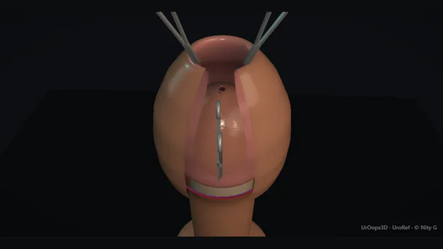
A sleeve circumcision in eleven moves: the outer skin line marked with the foreskin in its natural position, a layered dorsal slit with the meatus in view, eversion, a 0.5 cm mucosal collar, excision of the sleeve, and bipolar with a box stitch at the frenulum before the ring is closed.
Core tierFRCS andrology, genital surgeryInteractive 3D
The foreskin as a ring, dorsal at 12 and ventral at 6 o'clock. Schematic, not to scale; relationships only.
The operation is two circles and a sleeve.
The outer circle is the skin line, marked with the foreskin in its natural position; the inner circle is the 0.5 cm mucosal collar on the sulcus.
At 12 o'clock the slit joins them, the outer layer back to the skin incision and the inner layer stopping 0.5 cm short, with the meatus in view below both blades.
At 6 o'clock the frenular artery sits over the urethra, which is why it is controlled with bipolar and a box stitch rather than blind diathermy.
›Foreskin sleeve - outer skin and inner mucosa folded at the preputial orifice; the sleeve between the external line and the collar is what is removed.
›Coronal sulcus - the level of the external mark with the foreskin in its natural position, and the landmark the 0.5 cm collar is measured from.
›Mucosal collar - the 0.5 cm of inner layer left on the sulcus; the deep dorsal slit stops at it and the closure is made to it.
›Urethral meatus - at the tip of the glans under a tight foreskin; kept in view through the dorsal slit so no blade enters it.
›Frenulum + frenular artery - the ventral tether and its named artery; the classic bleeder, controlled with bipolar and a box stitch.
›Dorsal neurovascular bundle - deep dorsal vein midline, paired arteries and nerves under Buck's fascia at 12 o'clock; the reason the external incision and the slit stay superficial.
›Urethra - runs just under the ventral skin; the reason the ventral cut and any energy there stay superficial.
External markingWith the foreskin in its natural position and the penis flaccid and unstretched, the outer skin line is drawn circumferentially with a marker pen at the level of the corona. This line decides how much shaft skin is kept, so it is checked for symmetry from all four compass points before anything is cut. A line drawn on stretched or retracted skin moves, and that is the usual route to taking too much shaft skin. where it bites: a line marked on stretched skin sits too far back and removes shaft skin that cannot be put back.
Two clips on the distal foreskinTwo artery forceps are placed on the distal edge of the foreskin, one either side of the dorsal midline. They hold the foreskin, let it be retracted in any direction, and frame the line of the dorsal slit between them. The jaws take the free edge only, clear of the glans.
External incisionThe marked outer line is incised with the knife all the way round, skin only. The cut stays in the skin: the superficial vessels lie in the dartos immediately beneath, and the dorsal neurovascular bundle lies deeper under Buck's fascia at 12 o'clock. where it bites: a deep outer incision at 12 o'clock is where the dorsal bundle is put at risk.
Layered dorsal slitThe foreskin is opened at 12 o'clock with scissors, in two layers, with the urethral meatus kept in view throughout so that neither blade is ever inside it. The superficial (outer) layer is divided first, in the midline, all the way back to the external incision. The deep (inner) layer is then divided, stopping 0.5 cm short of the external incision, so that the future mucosal collar is left intact. where it bites: a blade passed blind under a tight foreskin can enter the meatus and cut the glans or urethra; the meatus is seen before every cut.
Evert the foreskinWith the slit made, the clips draw the foreskin back and it is everted over the corona, turning the inner layer outwards. The glans, the coronal sulcus and the frenulum are now in view, and the everted inner layer is the surface the collar will be marked on.
Mark the collarA 0.5 cm mucosal collar is marked with the marker pen on the everted inner layer, measured from the coronal sulcus, around the whole circumference. The mark meets the apex of the deep dorsal slit, which was stopped 0.5 cm short for this reason. An even collar is what gives an even suture line. where it bites: too long a collar leaves redundant, swollen mucosa; too short puts the suture line against the corona.
Collar incisionThe collar mark is incised with the knife through the mucosa all the way round, joining the apex of the dorsal slit. At 6 o'clock the cut is kept shallow over the frenulum, where the frenular artery runs.
Excise the sleeveScissors join the internal (collar) incision to the external incision around the circumference, dividing the tissue between them in the superficial plane. The foreskin sleeve comes away as one piece and is sent for histology. Staying superficial keeps the dorsal bundle and the urethra out of the specimen. where it bites: a deep plane here strays into the dorsal neurovascular bundle at 12 or the urethra at 6.
Frenulum: bipolar and box stitchThe frenulum is inspected under direct vision, and a short frenulum that still tethers the glans is released in a shallow plane. The frenular artery, the classic bleeder of the operation, is controlled precisely at the vessel with bipolar diathermy rather than swept with broad energy. A box stitch at the frenulum then brings the ventral skin to the collar and secures the frenular bed. where it bites: blind or deep energy under the frenulum risks the urethra, which lies immediately beneath.
ClosureThe shaft skin is closed to the mucosal collar all the way round with interrupted Vicryl Rapide. With the frenulum held by the box stitch, a suture at 12 o'clock divides the ring into halves, and each gap is then halved again, which is what keeps the spacing even and avoids dog ears. Knots sit clear of the meatus, and the line is checked for gaps before the dressing goes on.
DressingJelonet (paraffin gauze) is laid on the suture line, dry gauze goes over it, and a crepe bandage is added if wanted; the crepe is optional. The dressing is snug enough to control ooze without constricting the glans, and the tip is checked for colour before the patient leaves theatre. A catheter is rarely needed and is reserved for a defined indication such as a documented urethral leak or retention. where it bites: a catheter placed for reassurance alone adds urethral irritation to a healing suture line.
The spatial relationships the animation is built to make automatic. Before you read each one, ask yourself: at this point in the operation, what is about to go wrong, and what is my first move? The save for each sits in the Complications tab.
RiskUrethral meatus at the dorsal slit - under a tight foreskin the deep blade can enter the meatus instead of the preputial space; the meatus is seen before each cut.
RiskShaft skin and collar length - the outer line is marked on unstretched skin and the collar at 0.5 cm; either misjudged changes how much skin is left to close.
RiskFrenular artery - divides in the ventral frenular bed and retracts beneath the cut edge; controlled with bipolar at the vessel and a box stitch.
RiskDorsal neurovascular bundle at 12 - vein, arteries and nerves under Buck's fascia; a deep external incision or careless diathermy does real damage.
RiskVentral urethra - no more than skin-deep at 6 o'clock; the ventral cut and blind diathermy both respect it.
Trigger each of these live in the Animation tab, then read the save: the first move, the escalation trigger, and the line that goes in the op note.
Glans or meatal injury at the dorsal slit
with a tight foreskin the deep blade of the scissors is passed into the meatus instead of the preputial space, and the slit cuts the glans or the distal urethra.
The save
Stop before the blades close; withdraw the blade and look.first move
Re-site the blade under direct vision of the meatus, between the inner layer and the glans, before any further cut.
If the glans or meatus has been cut, inspect the extent with the field dry and do not extend the slit to see better.
A recognised clean injury is repaired primarily with fine absorbable sutures by a competent operator; consider a catheter when the urethra is involved.
Urethral involvement, a meatal defect or any doubt about the repair → senior assessment in theatre before closure.escalation trigger
Op note: how the injury occurred, its site and extent, the repair, catheter decision and the follow-up plan.document
Bleeding: frenular artery
the artery divides in the ventral frenular bed and retracts beneath the cut edge; bleeding is brisk and positional.
The save
Direct, sustained pressure to the ventral cut edge; do not lift and re-look repeatedly.first move
Elevate and inspect once the field is controlled: the frenular artery usually sits just beneath the cut edge at 6 o'clock.
Control it precisely at the vessel with bipolar, then secure the frenular bed with the box stitch or under-run it with a fine absorbable suture; blind deep diathermy is not acceptable.
Keep all energy superficial ventrally: the urethra lies immediately beneath.
Expanding haematoma, uncontrolled bleeding or an unstable patient → return to theatre for formal exploration.escalation trigger
Op note: bleeding point identified, method of control, dressing applied, follow-up plan.document
Glans / urethral injury
a deep ventral cut, a deep frenular stitch or blind diathermy breaches the thin ventral tissue over the urethra.
The save
Recognise it: a deep ventral bite, urine leak from the cut edge, or a spraying, deviated stream on first void.first move
Stop dissecting; do not enlarge the wound to chase the injury blind.
Catheterise with a suitable catheter to decompress and divert; most small ventral injuries heal on a catheter.
Inspect the meatus for involvement and document the site and depth.
Persistent leak, a large defect or meatal involvement → formal repair planning and specialist review.escalation trigger
Op note: site, size, catheter size and the plan for removal and review.document
Meatal stenosis
a late narrowing at the meatus from ischaemia or over-cauterisation; presents with a spraying, deviated stream weeks to months later.
The save
Recognise the presentation: spraying, straining or a thin deviated stream after an initially normal void.first move
Examine the meatus directly; a flow rate settles it when the history is unclear.
Ventral meatotomy under local anaesthesia is the standard correction.
Prevent the next one: minimal diathermy at the meatus and no tight ventral sutures at primary surgery.
Recurrent stenosis or lichen sclerosus involvement → meatoplasty and specialist review.escalation trigger
Note: timing, technique, and review of the primary operation for cause.document
When the case turns: the problem, then the next move.
ActionTight foreskin, meatus not visible for the slit - lift the distal edge on the two clips and pass the blade only under direct vision of the meatus; never cut blind.
ActionOuter line drawn with the skin stretched or retracted - wipe it and re-mark with the foreskin in its natural position before the knife touches it.
ActionCollar uneven after eversion - re-mark the 0.5 cm collar around the whole circumference from the sulcus before incising.
ActionFrenular bleeding obscures the field - direct pressure, then bipolar precisely at the artery and the box stitch; charred blind diathermy is not a substitute.
ActionDog ears at the join - space the sutures evenly before excising anything more; conservative management first, revision rare and delayed.
ActionMeatus looks ragged at the end - leave it alone; minimal handling, and catheterise only for a defined leak.
The examiner stems this scene prepares you for. Try to answer out loud first, then reveal, and learn the discriminator, the detail that turns a pass into a strong pass.
QWhat do you tell an adult man consenting for circumcision?reveal answer
I consent against the specific anatomy the operation crosses.
Bleeding is the commonest significant risk and I name the frenular artery as its usual source, including the small proportion who return to theatre.
Infection is usually superficial wound infection.
Meatal stenosis is the important late risk, more so with over-cauterisation or lichen sclerosus, and I would mention it because it presents after the patient has stopped thinking about the operation.
I discuss appearance honestly: the result will look circumcised, with a suture line and a mucosal collar, and symmetry cannot be guaranteed on the day.
I also discuss altered sensation of the glans, acknowledging the evidence is limited but that the concern is real to patients.
Rarer but mentionable risks are urethral or meatal injury and dissatisfaction with the cosmetic outcome.
Finally I check the indication, because consent for pathological phimosis or lichen sclerosus is a different conversation from consent for preference.
Strong pass
Say both
Naming meatal stenosis and the frenular artery specifically
Setting expectations about appearance and sensitivity rather than reciting a generic list
Examiner note
Naming meatal stenosis and the frenular artery specifically, and setting expectations about appearance and sensitivity rather than reciting a generic list.
QA man returns four hours after circumcision with bleeding through the dressing. How do you manage him?reveal answer
First, physiology: is he stable, is the bleeding active or ooze, is there a haematoma.
Most early bleeding is edge ooze that settles with direct pressure and a firm dressing, and a stable man with minimal soiling can be observed.
Active bleeding or an expanding haematoma goes to theatre.
Under anaesthesia I open the wound, evacuate clot and inspect systematically, because venous ooze from the dorsal join and arterial bleeding from the frenular bed look identical through a dressing.
The frenular artery is the named target: it retracts just beneath the ventral cut edge, so I pick it up precisely, control it with bipolar at the vessel and secure the frenular bed with a box stitch or an under-running absorbable suture.
The dorsal veins are controlled with bipolar or suture at the dorsal join.
The one thing I will not do is blind deep diathermy at the ventrum, because the urethra sits immediately beneath the skin and a bleeder becomes a fistula.
Haemoglobin and a group and save if the loss is meaningful, and a documented op note of the vessel found and the method used.
Strong pass
Naming the frenular artery as the source and the urethra as the reason blind diathermy is forbidden at the ventrum.
QA man presents with a painful, swollen glans and a constricting ring proximal to it. Walk me through your reduction.reveal answer
That is paraphimosis, and it is a time-critical compression problem: the retracted foreskin ring is obstructing venous and then arterial flow, so the glans swells and the ring tightens.
Analgesia first, usually a dorsal penile block or ring block with or without procedural sedation.
Then reduce the oedema before attempting reduction: ice, firm manual compression of the glans for several minutes, or multiple puncture techniques to express the oedema fluid.
With the oedema down, reduction is bimanual: thumbs on the glans, fingers behind the ring, steady pressure to push the glans back through while the ring is brought forward.
Once reduced, the glans is inspected for viability and the patient is scheduled for definitive elective circumcision, because recurrence is otherwise likely.
If manual reduction fails, the constricting ring is released with a dorsal incision under anaesthesia.
Delay risks necrosis, so this is not a wait-and-see problem.
Strong pass
Say both
Reducing the oedema before attempting reduction
Planning elective circumcision afterwards rather than treating it as a one-off event
Examiner note
Reducing the oedema before attempting reduction, and planning elective circumcision afterwards rather than treating it as a one-off event.
QWhen does circumcision need a different plan?reveal answer
Several situations change the operation rather than cancel it.
Acute balanoposthitis is treated and settled first; operating through infected tissue invites wound breakdown and a worse cosmetic result, though an abscess still needs drainage.
Lichen sclerosus, formerly balanitis xerotica obliterans, is often the indication for circumcision, but it changes the plan: the meatus must be assessed because involvement there means meatal stenosis is already present or imminent, so meatotomy or meatoplasty may be needed alongside, and the excision may need to be wider to clear diseased skin, with histology of what is removed.
Suspicion of penile cancer is a different operation altogether: no local circumcision, but imaging, staging and oncological excision with MDT discussion.
Finally, a man whose main motive is appearance or perceived benefit gets a careful conversation about realistic outcomes, because dissatisfaction is the complication no dressing fixes.
Strong pass
Say both
Distinguishing lichen sclerosus managed by circumcision with meatal assessment from penile cancer requiring oncological referral
Saying that lichen sclerosus changes the plan rather than contraindicating it
Examiner note
Distinguishing lichen sclerosus managed by circumcision with meatal assessment from penile cancer requiring oncological referral, and saying that lichen sclerosus changes the plan rather than contraindicating it.
Say it aloud, then reveal. Reading is not remembering.
01The relationshipSay the critical spatial relationship out loud, then reveal.reveal
Memory frame
The operation is two circles and a sleeve.
Why?
The outer circle is the skin line, marked with the foreskin in its natural position; the inner circle is the 0.5 cm mucosal collar on the sulcus. At 12 o'clock the slit joins them, the outer layer back to the skin incision and the inner layer stopping 0.5 cm short, with the meatus in view below both blades. At 6 o'clock the frenular artery sits over the urethra, which is why it is controlled with bipolar and a box stitch rather than blind diathermy.
02The first moveGlans or meatal injury at the dorsal slit. Your first move?reveal
First move
Stop before the blades close; withdraw the blade and look.
03The escalation triggerGlans or meatal injury at the dorsal slit. What makes you call for help?reveal
Escalate when
Urethral involvement, a meatal defect or any doubt about the repair → senior assessment in theatre before closure.
04The discriminatorWhat turns a pass into a strong pass?reveal
Strong pass
Say both
Naming meatal stenosis and the frenular artery specifically
Setting expectations about appearance and sensitivity rather than reciting a generic list
Examiner note
Naming meatal stenosis and the frenular artery specifically, and setting expectations about appearance and sensitivity rather than reciting a generic list.
05The pitfallWhere does this operation bite first?reveal
Pitfall
Urethral meatus at the dorsal slit
Why?
under a tight foreskin the deep blade can enter the meatus instead of the preputial space; the meatus is seen before each cut.
Revision practice with self-marked answers, not an assessment of clinical competence. Kept in
this browser only; it does not sync between devices.