
UrOops3D, danger-zone atlas
Operative animation, Clot evacuation and haemostasis

Clot retention: evacuation and endoscopic haemostasis

The bleeding bladder that has blocked its own catheter: day three after a prostate resection, a bladder tumour on anticoagulants, radiation cystitis. The patient comes first, then the bedside washout, then theatre with the widest sheath there is.

This page is the order of it: clear the clot, find the source, burn the point, leave a catheter that works, and ask why it bled.

Core tier FRCS core urology, emergency and haematuria Interactive 3D

Operative visualisation

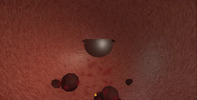
Schematic scene preview: Clot retention: evacuation and haemostasis
Preparing the 3D anatomy…

UrOops3D · UroRef · © Nity G

Click me next Section 2 of 8 Anatomy The structures this operation works between, and the one it must not cross.

The structures in the scene

Clot evacuation and haemostasis: how the structures relateThrough the resectoscope sheath, the bladder and the prostatic cavity. Clot on the bladder floor: drawn toward the neck and out 26 French sheath and the Ellik. Clot on the bladder floor: over-filled, the thin dome splits The dome (danger). Clot on the bladder floor: cleared first; diathermy in a red field burns Bladder neck at five and seven (danger). Bladder neck at five and seven: a centimetre apart; coagulate the point Trigone and orifices (danger). Clot on the bladder floor: venous ooze; coagulated, the sinus opens Prostatic bed and its sinuses (danger).Clot on the bladderfloor26 French sheath andthe Ellik!The domeBladder neck at fiveand seven!Trigone and orifices!Prostatic bed andits sinusesdrawn toward theneck and outover-filled, thethin dome splitscleared first;diathermy in a redfield burnsa centimetre apart;coagulate the pointvenous ooze;coagulated, thesinus opensTargetDangerContext
Through the resectoscope sheath, the bladder and the prostatic cavity. Schematic, not to scale; relationships only.
  • Through the sheath the bladder is a red chamber and the clot lies on its floor; the Ellik draws it toward the neck and out.
  • Once clear, the survey runs wall by wall and then back through the neck into the prostatic cavity, where the arterial points sit at five and seven o'clock just inside the neck and the venous ooze wells from the bed beyond.
  • The orifices are a centimetre from those arteries.
  • ›The bladder neck at five and seven o'clock - where the arteries enter; the commonest arterial bleeding point after a resection, and close to the orifices.
  • ›The prostatic bed and its sinuses - venous ooze that the catheter controls and the button opens.
  • ›The dome - the thinnest wall, thinner still three days after a resection; where an over-filled bladder splits.
  • ›Trigone and orifices - seen before and after any coagulation at the neck.
  • ›The 26 French sheath - the widest channel there is for clot; the Ellik and the Toomey work through it.
  • ›The button electrode - coagulation at the point, short bursts, the bed not painted.
  • ›The three-way catheter - 22 French, 30 ml balloon, irrigation until rosé, traction for hours only.
Click me next Section 3 of 8 Steps The operation in order, with the landmark that guards each move.

Operative steps

  1. The patient before the catheter Clot retention is painful and frightening and can hide a big bleed. Before anything is done to the catheter: pulse, blood pressure, a large-bore line, haemoglobin, clotting, group and save or crossmatch, and the drug chart for the anticoagulant or antiplatelet that is held and, if the bleeding is serious, reversed with the haematologist. The abdomen is examined for the bladder and for peritonism. The history is taken: what was done to the bladder or prostate and when, what has bled before, the tumour, the radiotherapy, the infection.
  2. The bedside washout A large catheter, at least 20 French and three-way if one is being changed, and a 50 ml bladder syringe: saline in, drawn back, and the clot comes with it, repeated until the return is clear. The washout is gentle and the bladder is never filled hard; the clot is broken up by the syringe, not forced through. Most clot retention clears this way and continuous irrigation through a three-way catheter keeps it clear. A catheter that blocks again within the hour, a return that stays red, or clot that will not come is the failure that goes to theatre.
    where it bites: a bladder syringe pushed hard into a bladder that cannot drain perforates it; the syringe aspirates, it does not pump.
  3. To theatre Consent names cystoscopy, evacuation of clot, diathermy of bleeding points, a possible return to the prostatic bed or a tumour resection, a catheter with irrigation, and the chance of a return to theatre. Blood is available. A general or spinal anaesthetic, lithotomy, the diathermy plate on if monopolar. On the trolley: a 26 French resectoscope sheath with its visual obturator, the widest channel there is for clot; the Ellik evacuator and a Toomey syringe; the loop, for breaking up old clot and for a resection if a tumour is the cause; the button or roller electrode; a 22 French three-way catheter; and the irrigation set for continuous flow.
  4. The red field The sheath goes in under vision on the visual obturator; the prostatic bed is passed with care because it is where the bleeding may be. In the bladder the view is red and the field is full of clot, some fresh and jelly-like, some old and rubbery. The irrigation is set to run but not high, and nothing is coagulated yet: diathermy in a red field burns whatever the electrode lands on, which may be the orifice or the wrong wall, and does not find the point that matters.
  5. The Ellik The telescope and bridge come out of the sheath and the Ellik evacuator goes onto it, its chamber full of saline and free of air. Squeeze and release: the clot is drawn into the chamber and settles, and the process is repeated until the return is clear. Old clot that will not come through the sheath is broken up with the loop, without current, and drawn out in pieces; a Toomey syringe finishes the last of it. Each squeeze fills the bladder, so it is emptied between cycles and never over-distended.
    where it bites: the Ellik fills the bladder with every squeeze; three days after a resection the wall is thin and a distended bladder splits at the dome.
  6. The survey With the telescope back in and the irrigation slowed, the bladder is surveyed first, wall by wall, because the cause may be there: a tumour, a perforation, a radiation-damaged wall, the ulcer of a catheter balloon. Both orifices are found and their jets seen; the trigone is inspected. Only then the neck and the prostatic bed: the cavity is looked at sector by sector with the irrigation slowed until the point that is bleeding declares itself. A bleeding point is watched until it is certain, not guessed.
  7. The source An arterial jet spurts across the field in a pulse; it is coagulated at its point with the button electrode on coagulation, a short burst until it whitens, and the bed around it is not painted. The commonest place after a TURP is the bladder neck at five and seven o'clock where the arteries enter, and the coagulation there is done with the orifices in mind. Venous ooze from the bed is dark and welling and is not chased, because coagulating a sinus opens it: the catheter and the irrigation control it. A bleeding tumour is resected down to a clean base and the base coagulated.
    where it bites: the arteries at five and seven o'clock sit close to the orifices; the point is coagulated, never the sector.
  8. The check The irrigation is turned off for a moment and the field watched: what fills it now is what the catheter has to control, and a jet that appears when the pressure drops is coagulated. Both orifices are looked at again with their jets, because coagulation at the neck was near them. The bladder is emptied through the sheath and the sheath withdrawn under vision.
  9. The catheter and the note A 22 French three-way catheter goes in over the bed with 30 ml in the balloon and continuous irrigation running until the effluent is rosé. For venous ooze the balloon is pulled down onto the bladder neck and the catheter taped to the thigh under traction, which is released within a few hours because a balloon left on the neck erodes it. The note records what was found, the clot burden, what was coagulated and where, the orifices seen with efflux, the catheter and the irrigation plan, and the question still open: why it bled, with the imaging, the cystoscopy or the anticoagulation review that answers it.
Click me next Section 4 of 8 Danger zones Where this operation bites, and why it bites there.
Critical relationship

Danger zones

The spatial relationships the animation is built to make automatic. Before you read each one, ask yourself: at this point in the operation, what is about to go wrong, and what is my first move? The save for each sits in the Complications tab.

  • RiskThe distended bladder - the Ellik and the bladder syringe both fill it; a thin post-operative wall splits at the dome.
  • RiskThe red field - diathermy before the clot is out burns whatever the electrode lands on.
  • RiskThe neck arteries and the orifices - a centimetre apart; the point is coagulated, not the sector.
  • RiskThe venous sinus - coagulating it opens it and absorbs irrigant; it is left to the catheter.
Practise it Take me into the scene Opens the animation at a danger moment and asks you to predict the mechanism and your first move before it reveals.
Click me next Section 5 of 8 Complications The save for each one: the first move, the escalation trigger, the op-note line.

Complications and immediate saves

Trigger each of these live in the Animation tab, then read the save: the first move, the escalation trigger, and the line that goes in the op note.

Bladder perforation by over-distension

  • the Ellik or the syringe used on a full bladder: the dome splits, the irrigant does not return, the abdomen fills.
The save
  1. Stop irrigating. Empty the bladder through the sheath. first move
  2. Look for the perforation at the dome; fat or bowel means intraperitoneal.
  3. Extraperitoneal: a catheter for a week and a cystogram before it comes out.
  4. Intraperitoneal, peritonism or a large deficit: laparotomy, repair in two layers, washout, senior help. escalation trigger
  5. Note: the site, the type, the deficit, the repair or drainage and the imaging plan. document

Bleeding that will not stop

  • the field will not clear whatever is coagulated: the whole bed, a clotting problem, or an artery the button cannot reach.
The save
  1. Three-way catheter, 30 ml balloon, traction on the neck, fast irrigation; blood and clotting products with the anaesthetist. first move
  2. Correct the clotting: reverse the anticoagulant, platelets, tranexamic acid where appropriate.
  3. Interventional radiology for embolisation of the prostatic or vesical arteries if the bleeding continues. escalation trigger
  4. Open packing of the prostatic bed or the bladder is the last resort, with a consultant.
  5. Note: the blood loss, the products given, the traction, and the radiology plan. document

Orifice burned

  • the coagulation at five or seven o'clock has taken an orifice: no jet, a white scar where the slit was.
The save
  1. Stop coagulating that corner. first move
  2. Cannulate the orifice with a wire or ureteric catheter if it can be found and place a stent for four to six weeks.
  3. If it cannot be found: ultrasound at two weeks and three months for hydronephrosis.
  4. Loin pain, fever or a rising creatinine: image and drain by nephrostomy. escalation trigger
  5. Note: which orifice, stented or not, and the imaging plan. document

The missed cause

  • the bleeding was never the prostate: a bladder tumour, a radiation-damaged wall, a balloon ulcer.
The save
  1. Every clot retention has its bladder surveyed wall by wall once the clot is out, before the prostate is blamed. first move
  2. A tumour found is resected to a clean base and coagulated, and staged as any tumour.
  3. Radiation cystitis that will not stop bleeding: fulguration, then formalin, alum or hyperbaric oxygen with the specialist team. escalation trigger
  4. Note: the cause found or excluded, and the investigation of the upper tracts if none was seen. document

Fluid absorption

  • an hour of irrigation through an open bed: hyponatraemia with glycine, fluid overload with saline.
The save
  1. Stop resecting and stop the inflow; tell the anaesthetist. first move
  2. Send a sodium; with glycine and confusion or bradycardia treat as TUR syndrome.
  3. Saline irrigation avoids the hyponatraemia but not the volume: a poor heart goes into failure. Furosemide and oxygen. escalation trigger
  4. Note: the irrigant, the time, the deficit, and the sodium. document
Click me next Section 6 of 8 Troubleshooting When the case turns: the problem, then the next move.

Intra-operative troubleshooting

When the case turns: the problem, then the next move.

  • ActionThe catheter blocks again within the hour - theatre, not another washout; the clot is forming faster than a syringe can clear it.
  • ActionCannot see anything in the bladder - clot, not blood: Ellik until the return is clear before looking for a point.
  • ActionThe clot will not come through the sheath - break it with the loop without current, then Ellik; a Toomey for the last pieces.
  • ActionThe bladder keeps distending - the outflow is blocked with clot: stop, empty through the sheath, clear the outflow.
  • ActionEverything oozes and nothing spurts - venous: stop coagulating, catheter with traction and fast irrigation, correct the clotting.
  • ActionThe effluent is still red at two hours - the catheter is blocking or the bleeding is arterial: syringe washout, then back to theatre or radiology.
Click me next Section 7 of 8 FRCS viva Say it out loud, then mark yourself against the model answer.

FRCS viva questions

The examiner stems this scene prepares you for. Try to answer out loud first, then reveal, and learn the discriminator, the detail that turns a pass into a strong pass.

QA patient three days after TURP develops clot retention on the ward. Manage them.reveal answer
  • I would see the patient rather than the catheter first: pulse, blood pressure, a large-bore cannula, haemoglobin, clotting, group and save, and I would stop and consider reversing any anticoagulant.
  • Then a bedside washout with a 50 ml bladder syringe through a large-bore catheter, exchanged for a 22 French three-way if not already in, aspirating clot gently until the return is clear, and then continuous irrigation.
  • If the washout fails, the catheter blocks again quickly or the patient is unstable, I would take him to theatre with consent for cystoscopy, evacuation and diathermy and with blood available.
  • There I would use a 26 French resectoscope sheath, evacuate the clot with the Ellik without over-distending the bladder, survey the bladder wall by wall for another cause, check both orifices, and then inspect the prostatic bed sector by sector to find the arterial point, usually at the neck at five or seven o'clock, and coagulate it with the button.
  • Venous ooze I would leave to a three-way catheter with traction and irrigation.
  • I would document the findings and plan the investigation of why it bled.
Strong pass

Say all three

  • The patient resuscitated before the catheter is touched
  • The bladder surveyed before the prostate is blamed
  • The arterial point coagulated while ooze is left to the catheter
Examiner note
The patient resuscitated before the catheter is touched, the bladder surveyed before the prostate is blamed, and the arterial point coagulated while ooze is left to the catheter.
QWhat is the difference between arterial and venous bleeding in the prostatic bed and how does it change what you do?reveal answer
  • Arterial bleeding spurts in a pulsatile jet across the field and comes from a named point, most often at the bladder neck at five and seven o'clock where the prostatic arteries enter, or from a vessel at the capsule.
  • It does not stop with a catheter and it is found and coagulated at its point with the button, a short burst until it blanches, with the orifices in mind.
  • Venous bleeding is dark, welling ooze from the sinuses of the bed; it gets worse when the irrigation pressure falls and it is controlled by pressure, not by current.
  • Coagulating a sinus opens it further and absorbs irrigant, so venous ooze is left to a three-way catheter with the balloon on traction against the neck and fast irrigation until the effluent is rosé, with traction released within hours.
  • Turning the irrigation off for a moment at the end shows which kind of bleeding is left and whether the catheter alone will manage it.
Strong pass

Say all three

  • Arteries coagulated at the point
  • Sinuses left to traction and irrigation
  • The irrigation-off test to decide which is left
Examiner note
Arteries coagulated at the point, sinuses left to traction and irrigation, and the irrigation-off test to decide which is left.
QWhat causes fluid absorption during a prolonged washout and how do you manage it?reveal answer
  • Irrigant is absorbed through the open venous sinuses of a resected prostatic bed and through a perforation, in proportion to the pressure of the irrigation and the time.
  • With glycine, which is hypotonic, the result is dilutional hyponatraemia, TUR syndrome, presenting under regional anaesthesia with confusion, restlessness and visual disturbance, and under general anaesthesia with bradycardia and hypertension from raised intracranial pressure, then hypotension.
  • With normal saline and bipolar equipment there is no hyponatraemia but the volume load remains and a patient with a poor heart goes into pulmonary oedema.
  • Management is to stop the resection and the inflow, secure haemostasis and catheterise, inform the anaesthetist, and send an urgent sodium.
  • Acute symptomatic hyponatraemia is treated with hypertonic saline in a monitored setting because the danger is cerebral oedema; volume overload with oxygen and furosemide.
  • Prevention is a low irrigation bag, a short procedure, saline where possible, and stopping to reassess when the time passes an hour.
Strong pass

Say both

  • Separating the hyponatraemia of glycine from the volume load of saline
  • Hypertonic saline for the acute symptomatic case
Examiner note
Separating the hyponatraemia of glycine from the volume load of saline, and hypertonic saline for the acute symptomatic case.
QThe bleeding will not stop despite coagulation and a catheter on traction. What next?reveal answer
  • I would ensure resuscitation is ahead of the bleeding: blood, fresh frozen plasma or platelets guided by the clotting and the haematologist, reversal of any anticoagulant, and tranexamic acid where there is no contraindication.
  • I would check the catheter is draining and not blocked with clot, that the balloon is large and on traction against the neck, and that the irrigation is fast.
  • If bleeding continues I would call the consultant and involve interventional radiology for angiography and selective embolisation of the prostatic or inferior vesical arteries, which controls most arterial bleeding without a further operation.
  • If radiology is unavailable or fails, the surgical options are a return to theatre to re-inspect and coagulate under vision with senior help, and as a last resort open packing of the prostatic bed or the bladder, or suture of the bleeding vessels through a cystotomy.
  • Throughout I would document the blood loss and the products, and keep the patient in a monitored bed.
Strong pass

Say all three

  • Naming embolisation as the next step after traction
  • Clotting correction
  • With open packing as the last resort
Examiner note
Naming embolisation as the next step after traction and clotting correction, with open packing as the last resort.
Click me next Section 8 of 8 Evidence The guidance each of these answers is built on.

Evidence and guidance

Two minutes

Close the lap: recall it before you leave.

Say it aloud, then reveal. Reading is not remembering.

  1. 01 The relationship Say the critical spatial relationship out loud, then reveal. reveal
    Memory frame

    Through the sheath the bladder is a red chamber and the clot lies on its floor; the Ellik draws it toward the neck and out.

    Why?
    Once clear, the survey runs wall by wall and then back through the neck into the prostatic cavity, where the arterial points sit at five and seven o'clock just inside the neck and the venous ooze wells from the bed beyond. The orifices are a centimetre from those arteries.
  2. 02 The first move Bladder perforation by over-distension. Your first move? reveal
    First move

    Stop irrigating.

    More detail
    Empty the bladder through the sheath.
  3. 03 The escalation trigger Bladder perforation by over-distension. What makes you call for help? reveal
    Escalate when

    Intraperitoneal, peritonism or a large deficit: laparotomy, repair in two layers, washout, senior help.

  4. 04 The discriminator What turns a pass into a strong pass? reveal
    Strong pass

    Say all three

    • The patient resuscitated before the catheter is touched
    • The bladder surveyed before the prostate is blamed
    • The arterial point coagulated while ooze is left to the catheter
    Examiner note
    The patient resuscitated before the catheter is touched, the bladder surveyed before the prostate is blamed, and the arterial point coagulated while ooze is left to the catheter.
  5. 05 The pitfall Where does this operation bite first? reveal
    Pitfall

    The distended bladder

    Why?
    the Ellik and the bladder syringe both fill it; a thin post-operative wall splits at the dome.

Revision practice with self-marked answers, not an assessment of clinical competence. Kept in this browser only; it does not sync between devices.