
Revision sheet
Clot retention: evacuation and haemostasis
8 steps, each one the authored title and caption from the interactive animation, with the frame captured from that step. Read it before the case, or print it and work through the steps in order.
Teaching only, not for diagnosis or treatment decisions. The text on this sheet is the animation's own step copy, reproduced verbatim.
UrOops3D · UroRef · © Nity G
- 01
The patient before the catheter
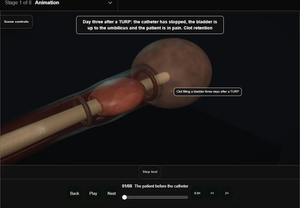
- Clot retention on day three: pain, a bladder to the umbilicus, a catheter that has stopped.
- Pulse, pressure, haemoglobin, clotting, group and save, anticoagulant held.
- A 50 ml bladder syringe washout at the bedside clears most.
- 02
To theatre
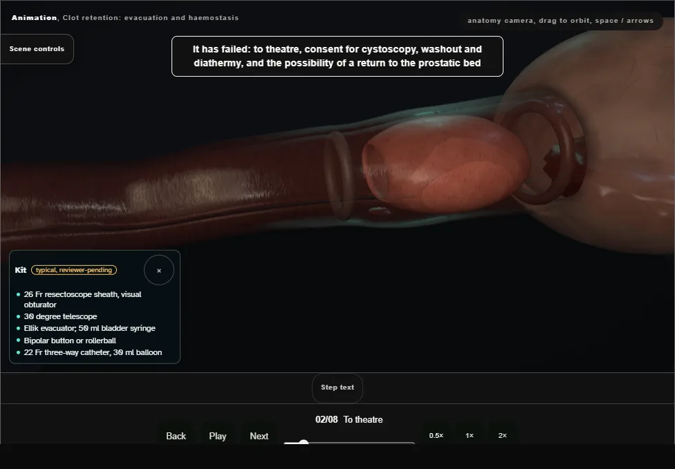
- Washout failed: consent for cystoscopy, evacuation and diathermy, and for a return to the bed.
- A 26 French resectoscope sheath under vision, the widest channel there is for clot, the Ellik on the trolley.
- 03
The red field
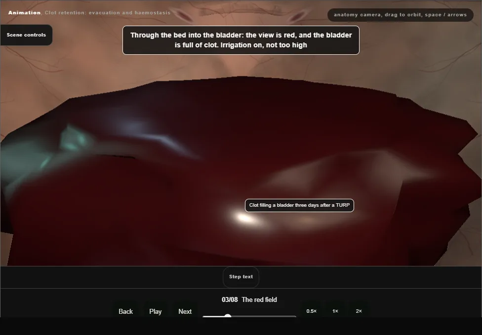
- Through the bed into a bladder full of clot.
- Irrigation on, not high.
- No blind diathermy: in a red field it burns the wrong thing.
- 04
The Ellik
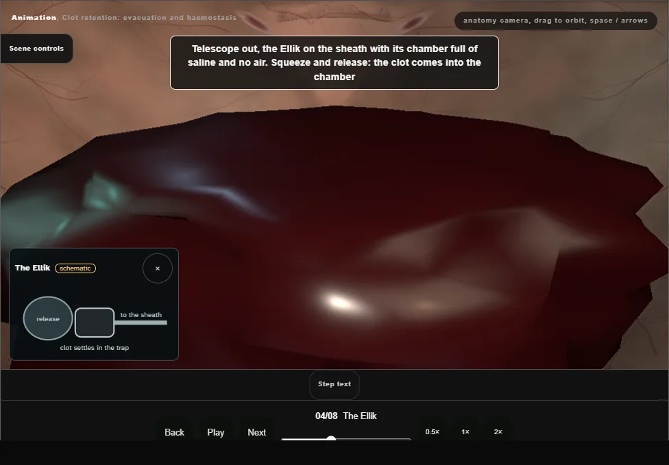
- Telescope out, the Ellik on the sheath, chamber full and no air.
- Squeeze and release; old clot broken up with the loop without current.
- The bladder is never over-filled: three days after a resection it is thin.
- 05
The survey
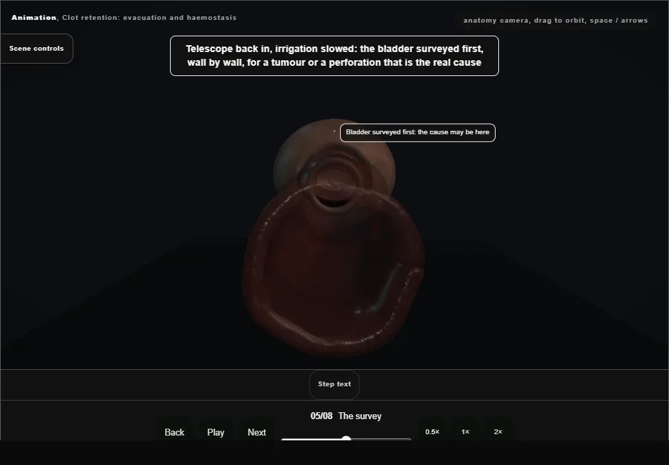
- Bladder first, wall by wall, for the tumour or the perforation that is the real cause; both orifices with their jets; then the neck and the prostatic bed, sector by sector.
- 06
The source
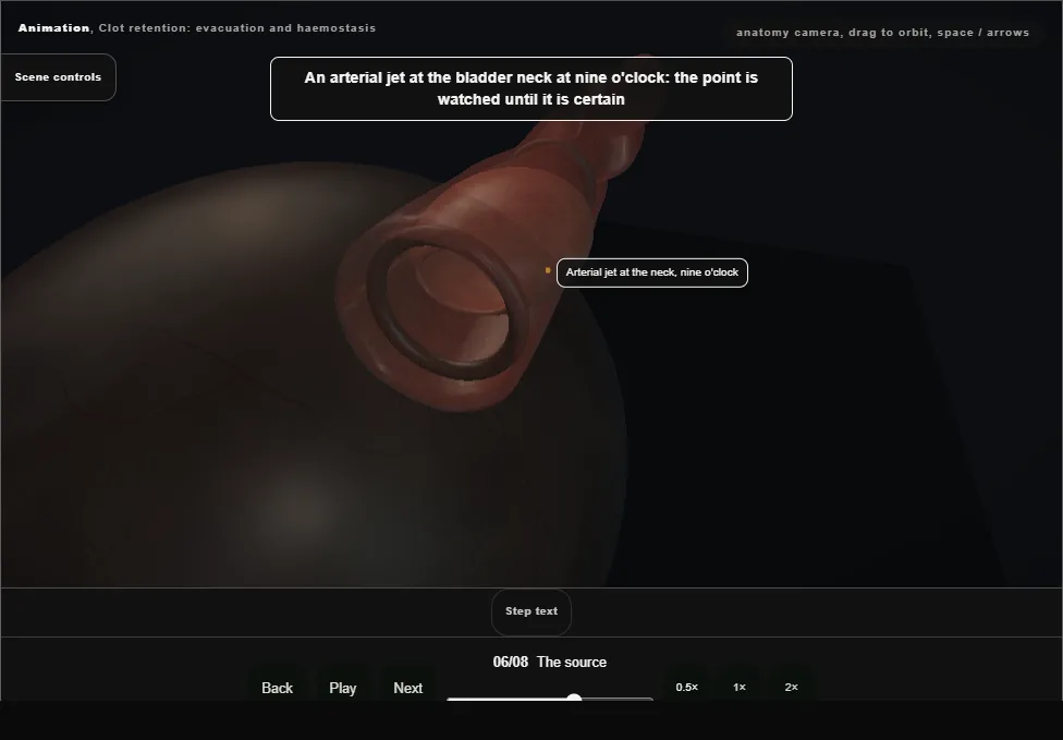
- An arterial jet at the neck at nine o'clock.
- The button on the point, a short burst until it whitens, the bed around it untouched.
- Venous ooze is for the catheter.
- 07
The check
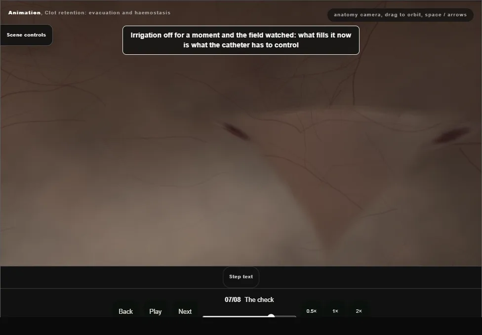
- Irrigation off for a moment: what fills the field now is what the catheter must control.
- Both orifices seen again, because the coagulation at nine o'clock was near the right one.
- 08
The catheter and the note
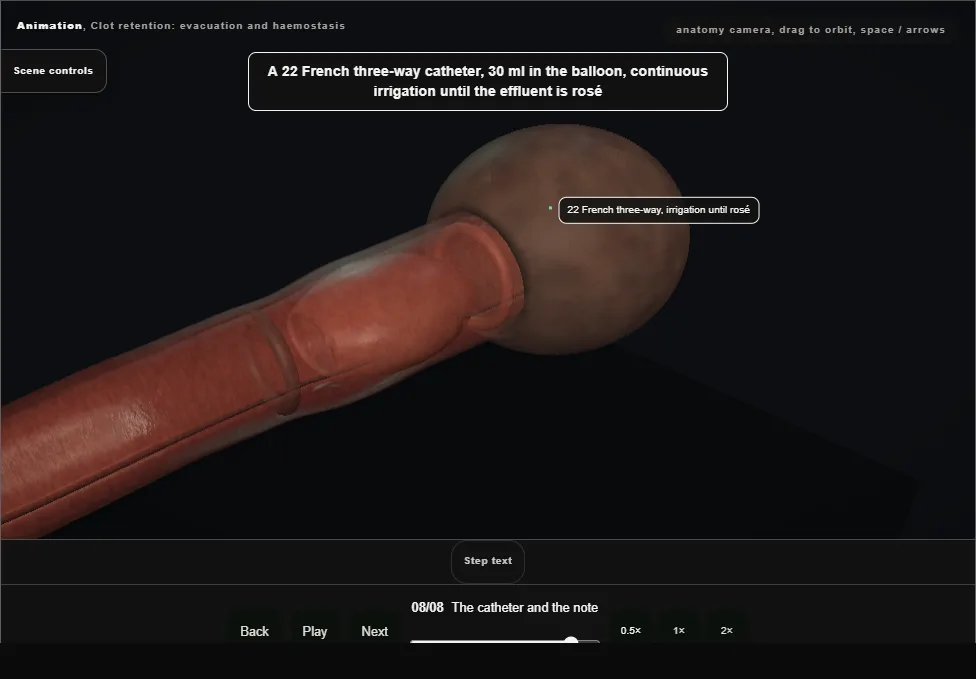
- A 22 French three-way, 30 ml in the balloon, irrigation until rosé, traction only for ooze and released within hours.
- The note names the source, the burn, the orifices seen, the plan, and the question still open: why did it bleed?
Derived from endoscopy/ClotEvacuationScene.tsx at build time: every step title and caption above is the authored
ProcedureDef text, reproduced verbatim, and this page shares its readable-points renderer with the animation
itself. 8 of 8 steps have a captured frame.
Text fingerprint 3f7c6c7f03807e77. A caption edit without a re-capture fails the production
check, so the sheet and the animation cannot drift apart silently.