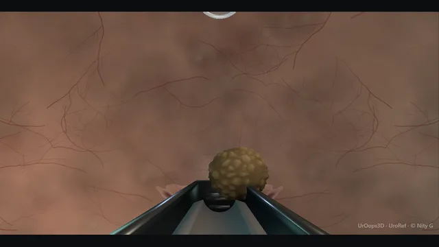
A rigid scope climbs the urethra from the meatus to a stone sitting at the bladder base, breaks it with the laser or the stone punch under vision, and washes the chips out through the sheath.
The thin dome under sustained irrigation is the structure that perforates when the bladder is over-distended, and the obstructing outlet below is the reason the stone exists in the first place.
One road: meatus up through the outlet to the stone and the dome. Schematic, not to scale; relationships only.
The operation is a vertical stack with one road.
The scope enters at the meatus, runs the bulbar and prostatic urethra where the outlet resists it, and arrives at a stone lying dependant at the base behind that outlet.
Fragmentation happens with the wall always in view beyond the stone;
evacuation happens by washing the dependent base up to the scope tip and out along the same road;
and the survey happens because the dome above and the base below are where tumour, diverticulum and retained chips hide.
The two danger structures sit at the ends of the road: the urethra at the bottom, which is traumatised on entry, and the dome at the top, which is perforated by pressure.
The stone punch adds one rule to the same map: the stone is lifted off the base into the centre of the chamber, and the crunch happens there, away from floor, walls and dome.
›Bladder dome - the thinnest wall and the one under most pressure during irrigation; the perforation site of over-distension.
›Stone at the base - sits dependant, behind the outlet that failed to clear it; fragmented from the edge so fragments are born evacuable.
›Prostatic outlet - the usual cause in men; the same obstruction that grew the stone also resists the scope on the way in.
›Urethra - the single road in and out; every passage pays a toll, and repeated forced passages buy a stricture.
›Bladder base + mucosa - inspected fully at the end; stones and chronic irritation coexist with tumour and with the diverticulum that retains chips.
Urethral access under visionThe whole operation lives or dies on the road in, so the scope passes with lubricant, irrigation running and vision on from the meatus forward, watching the lumen accept the instrument rather than feeling for it. The calibre of the urethra is judged at the external meatus and the bulbar urethra, and the scope size is chosen for that urethra rather than for the stone. Where the outlet fights the scope, the decision is made early and deliberately: a smaller instrument, a visual dilatation or a staged approach, never a harder push. where it bites: the first passage sets the tone for the whole list: forced insertion through a tight outlet is the classic source of a false passage and the delayed stricture.
Cystoscopy + stone burdenWith the scope in the bladder, fill is kept to a comfortable working volume and the whole mucosa is surveyed before anything is broken. Burden is judged honestly: size, number, apparent hardness and position, and the state of the outlet the stone grew behind, because a stone secondary to obstruction behaves differently from a stone in a drained, catheterised bladder. The fill matters as much as the findings: a working bladder is moderately full, leaving wall thickness and compliance in reserve. where it bites: the tumour, diverticulum or fistula found at survey changes the operation: the stone is the finding, not the diagnosis.
Fragmentation: punch or laserThe stone is broken from one edge, not hammered at its centre. With the laser the fibre is worked along one edge so fragments are born small enough to evacuate; with the stone punch the jaws open and take the stone by an edge under direct vision, and the stone is lifted to the centre of the bladder before any crushing force is applied (the punch sequence is shown at the end of the animation). The bladder is the one place in the urinary tract that tolerates high fragmentation energy: the wall is thick compared with a ureter, irrigation dilutes the dust and the outflow carries it away, so the stone can be broken hard and fast under vision where the same energy in the ureter would be reckless. The fibre is the larger core, 365, 500 or 550 micron, because a larger core carries more energy per pulse and nothing in the bladder has to deflect; the holmium setting in the bladder is 1.5 to 2.0 J at 20 to 50 Hz (owner-confirmed values). where it bites: firing towards the wall with the stone between fibre and mucosa, or crushing blind inside a crater, is how the bladder gets perforated: the wall behind the stone stays in view for every shot.
Evacuation under visionIrrigation washes the chips pooling at the dependent base up to the scope and out along the sheath, the Ellik principle applied under direct vision rather than blind. The base is swept systematically, because chips settle in the same dependant spot and hide in the same trabeculations. Evacuation counts as finished when the base is visibly and palpably clear, not when the field looks mostly empty. where it bites: over-distension: repeated filling with a thin dome under pressure is the setup for perforation, and the temptation rises exactly when vision is cloudy and the operator wants one more wash.
Final surveyWith the burden cleared and the bladder refilled to a moderate working volume, the full mucosa is inspected systematically, base to dome and every wall, including the dome where the pressure sat all case. Chronic irritation, infection and outlet obstruction are the company a bladder stone keeps, and a tumour can bleed with it and hide behind it. The survey also finds the trabeculation and diverticula that will harbour the next generation of fragments. where it bites: skipping the survey because the stone is gone: the stone was the presentation, the bladder behind it is the diagnosis.
Treat the causeA bladder stone is a symptom of drainage failure: the obstructing prostate, a long-term catheter, a neurogenic bladder or a foreign body. The cause gets a plan the same day, whether that is a bladder-neck incision, a resection, a catheter change regimen or a referral, because fragmentation alone is a lease, not a cure. where it bites: fragmentation without a cause plan guarantees the patient returns, with a bigger burden and a tighter outlet.
Exit + catheter decisionThe scope comes out under vision and the urethra is left as gently as it was entered. A catheter rests the bladder briefly when the mucosa is raw, the outlet was tight or bleeding is expected, and drainage otherwise is a decision, not a reflex. The metabolic workup and the cause treatment are booked before discharge, because the stone that leaves behind an untreated cause books its own return. where it bites: discharging the stone without the follow-up plan attached: the operation ends when the cause has an appointment.
Alternative: the stone punchThe stone punch is chosen over the fibre when the stone is hard, large or several, when the laser is slow or unavailable, or when the bladder needs clearing in minutes rather than lasering for an hour: on a big hard stone it is faster and needs no laser. Its costs are calibre and force: a wider sheath through the urethra, and a mechanical bite that crushes whatever sits between the jaws. So the sequence is fixed. The jaws open and take the stone by an edge under direct vision, the wall behind it in view. The scope then looks up, towards the anterior wall and the dome, and lifts the stone off the base into the middle of the bladder, where the lens is clear, the whole stone is in view and nothing but water is within reach of the jaws. where it bites: the jaws must be seen closing on stone alone. A fold of wall drawn into the window with the stone is the classic punch perforation, and a lens that cannot see the jaws has no business closing them.
Crunch, then evacuateOnly then does the punch close. The crunch happens in the centre of the bladder at a moderate fill, never against the mucosa and never blind in a cloudy field. The pieces fall to the base; anything still too large to evacuate is taken again the same way, edge first, lifted, then crushed, until the fragments are small enough to be washed out through the sheath under vision. Evacuation, the base check and the survey then run exactly as after the laser, and the fragments go for stone analysis. where it bites: closing on a stone resting on the wall crushes the wall with it; closing in a cloudy field crushes what cannot be seen. Grasp, look up, then crunch.
The spatial relationships the animation is built to make automatic. Before you read each one, ask yourself: at this point in the operation, what is about to go wrong, and what is my first move? The save for each sits in the Complications tab.
RiskThin dome under irrigation - over-distension puts it under the highest stress in the whole system; perforation risk tracks filling discipline, not laser power.
RiskProstatic urethra + meatus - the narrowest point of the road; repeated forced passage here makes the false passage and the delayed stricture.
RiskDependent base behind the stone - where chips pool, where evacuation is judged, and where the retained nidus of the next stone is left behind.
RiskThe jaws of the stone punch - they crush whatever is between them; the wall drawn in with the stone, or a stone crushed while resting on the wall, is the mechanical perforation. Grasp under vision, lift to the centre, then crunch.
Trigger each of these live in the Animation tab, then read the save: the first move, the escalation trigger, and the line that goes in the op note.
Bladder perforation
over-distension, a fibre fired towards the wall or a crater dusted blind splits the thin dome; return stops matching inflow, vision clouds and the abdominal contour changes.
The save
Stop inflow immediately and let the bladder decompress; do not refill to see better.first move
Assess the patient, not just the image: haemodynamics, abdominal signs and whether the contour is draining.
Leave a catheter to drain the bladder; most extraperitoneal tears settle on free drainage.
Image with contrast if the pattern is unclear or intraperitoneal injury is plausible.
Intraperitoneal perforation, bowel injury or a failing patient → theatre for repair, with a low threshold for a general surgical opinion.escalation trigger
Op note: mechanism, site suspected, drainage strategy, imaging plan and the catheter removal plan.document
Urethral trauma
repeated forced passage through a tight outlet makes a false passage or a raw urethra; blood at the meatus and a scope that stops running freely are the clues.
The save
Stop advancing. Withdraw and re-assess under vision rather than pushing through resistance.first move
Use the smallest adequate scope, plenty of lubricant and, for a tight outlet, a visual urethrotomy or a catheter-first strategy rather than force.
If a false passage is suspected, pass a catheter only under vision or over a wire; never blind down a track that may be extra-luminal.
Leave a catheter for a defined period to splint a clean mucosal injury, and plan a flow study or flexible check at review.
Retention, persistent bleeding or an unable-to-catheterise urethra → senior help and suprapubic access rather than more attempts.escalation trigger
Op note: passages attempted, injury recognised, catheter plan, stricture-risk counselling at review.document
Retained fragments
chips left at the dependent base become the nidus for the next stone and the next infection; evacuation is judged by a visibly and palpably clear base, not a field that looks mostly empty.
The save
Finish the survey at the base specifically: look behind the prostate, in any diverticulum and at the dome after tilting.first move
Wash with the scope tip at the lowest point of the bladder and the patient tilted so the base drains to the tip.
Fragment anything left to dust rather than leaving chips sized to re-aggregate.
Book imaging before discharge if clearance is in doubt, and treat the cause in the same admission.
A large retained burden or an infected residue → staged clearance, not optimistic discharge.escalation trigger
Op note: clearance achieved or residual declared, imaging plan, metabolic workup and cause treatment plan.document
Bladder wall in the punch jaws
a fold of mucosa is drawn into the window with the stone, or the stone is crushed while resting on the wall; the wall tears with the stone and the field bleeds and clouds.
The save
Stop. Do not close the jaws; open them and let the stone drop.first move
Stop inflow, let the bladder settle, and look at the wall where the jaws were: a mucosal tear, a bleeding point or a full-thickness defect.
A superficial tear with a clear view: continue, with the stone lifted to the centre before every crunch, and drain the bladder afterwards.
A full-thickness defect or a distending abdomen: manage as a perforation; stop, decompress, catheter, image.
Intraperitoneal injury, bowel involvement or a failing patient → theatre for repair.escalation trigger
Op note: the mechanism, the site, the depth judged, the catheter and imaging plan.document
When the case turns: the problem, then the next move.
ActionStone rolls away from the fibre - Trap it against the bladder base or dome with the sheath before firing, or fragment in a partially empty bladder where it cannot tumble.
ActionVision goes cloudy mid-fragmentation - Stop lasering, empty and refill once rather than repeatedly, and check for bleeding from the mucosa or prostate before continuing.
ActionChips will not wash out - Reposition: scope tip to the lowest point, patient tilted, irrigation in bursts; persist with positioning before persisting with pressure.
ActionThe punch jaws will not take the stone - Reduce the fill a little so the stone cannot roll, approach an edge rather than the widest diameter, and if it still will not sit in the window, break it first with the laser and punch the pieces.
ActionThe bladder wall comes into the window with the stone - Do not close. Open the jaws, let the stone go, refill slightly, and grasp again only when the whole stone and the wall behind it are seen; lift to the centre before any force.
ActionCloudy view with the stone in the jaws - Hold still and do not crunch. Empty and refill once; if the view does not clear, open the jaws and let the stone drop before changing anything else.
ActionThe burden is too large or too hard for transurethral clearance - Change the plan deliberately: open cystolithotomy for a truly giant or hard stone, or fragmentation plus cause treatment staged, rather than a long traumatic transurethral session.
The examiner stems this scene prepares you for. Try to answer out loud first, then reveal, and learn the discriminator, the detail that turns a pass into a strong pass.
QA man of retiring age is found to have a bladder stone on a scan for poor flow. What matters more than the fragmentation?reveal answer
The drainage failure that grew it.
A bladder stone in an adult is nearly always a symptom: of an obstructing prostate, a neuropathic or underactive bladder, a long-term catheter, a retained foreign body or, less often, a stone migrated from above with a bladder that could not clear it, and sometimes a tumour or diverticulum shares the same field.
So the assessment before fragmentation is a full history of voiding, catheters, neurological disease and previous stones;
a proper examination including a neurological screen where the story suggests it;
urine testing including culture, because infected residual fragments restart the whole cycle;
and flexible or rigid cystoscopy at the time of treatment to see the mucosa, the outlet and any diverticulum.
Fragmentation then takes its proper place as the symptomatic step, planned around the cause: stone composition and hardness, outlet size for scope passage, and whether the bladder has the capacity and sensation to tolerate a long irrigation procedure.
An operation that clears the stone perfectly and leaves the cause untreated has treated the X-ray and abandoned the patient, who returns with a bigger stone, a tighter outlet and a harder problem.
Strong pass
Say both
Saying that the stone is the symptom and the outlet or the bladder is the diagnosis
Linking culture and cystoscopic survey to the recurrence risk before discussing lasers at all
Examiner note
Saying that the stone is the symptom and the outlet or the bladder is the diagnosis, and linking culture and cystoscopic survey to the recurrence risk before discussing lasers at all.
QWhen is open cystolithotomy the right operation for a bladder stone?reveal answer
When the transurethural road costs more than the abdominal one.
The classic indications are size and hardness: a very large or a very hard stone, where the hours of laser time and irrigation volume needed to fragment and evacuate it carry more risk of over-distension, perforation, bleeding and infection than a short open retrieval.
The second group is anatomy: a urethra that will not admit an adequate scope, a narrowed meatus or a strictured road where forcing passage buys the stricture that plagues the patient for years;
here open retrieval with a concurrent urethral procedure planned properly is kinder.
The third group is the combined operation: the patient who needs open or large-instrument bladder surgery anyway, most often a bladder outlet procedure or repair of a large diverticulum, where adding the stone through the same incision costs minutes.
The fourth is burden with sepsis or heavy infection, where a quick open retrieval in a drained bladder may be safer than a long irrigated endoscopic session.
What open surgery is not, is a failure: it is a deliberate route choice, consented as such, with its own honest risks including wound infection, leak from the cystotomy and longer recovery.
The wrong answer is the purist one, either direction.
Strong pass
Say all three
Framing the decision around the urethra
The physical behaviour of the stone and the concurrent operation
Openly naming the risks of the open route rather than defending endoscopy on principle
Examiner note
Framing the decision around the urethra, the physical behaviour of the stone and the concurrent operation, and openly naming the risks of the open route rather than defending endoscopy on principle.
QDuring evacuation the return stops matching inflow, the abdomen is distending and the patient, under spinal, complains of abdominal pain. What has happened and what do you do?reveal answer
The bladder has perforated, and under spinal anaesthesia the patient is doing the monitoring: new abdominal pain with a distending abdomen and return that no longer matches inflow is perforation until proven otherwise.
The sequence is fixed.
Stop irrigation at once and let the bladder drain; the instinct to refill for a better look is the instinct that enlarges the hole.
Examine the abdomen with the bladder empty: generalised peritonism or shifting signs point intraperitoneal; localised suprapubic findings point extraperitoneal.
Check the whole patient: observations, abdominal stiffness over time, and urine output from whatever drain is running.
Then decide with imaging: a cystogram or CT cystogram defines the tear and whether contrast escapes freely into the peritoneal cavity.
Most extraperitoneal perforations are managed without surgery: catheter drainage, antibiotics where indicated, and observation with a repeat study before removal.
Intraperitoneal perforation, bowel injury suspected from the mechanism, a deteriorating abdomen or sepsis goes to theatre for repair and washout, with general surgical input where the mechanism involves bowel.
The bladder is closed, drained properly, and the cause plan, which the stone was announcing all along, is written in the same notes.
Strong pass
Say both
Naming the spinal patient as the monitor
Dividing management on the extraperitoneal versus intraperitoneal line with imaging before surgery rather than exploration by default
Examiner note
Naming the spinal patient as the monitor, and dividing management on the extraperitoneal versus intraperitoneal line with imaging before surgery rather than exploration by default.
QHow do you stop this patient growing another bladder stone?reveal answer
By closing every loop the first stone opened.
The cause first: an obstructing prostate is treated or at least formally assessed and scheduled;
a neurogenic bladder has its drainage optimised, which may mean intermittent self-catheterisation taught properly rather than tolerated badly;
a catheter history prompts review of what is indwelling and why, with material and technique addressed;
a foreign body or suture nidus is removed.
The chemistry second: stone analysis when fragments are retrieved, a metabolic screen for the recurrent former, and dietary advice that is specific to composition rather than generic water advice, although fluid intake generous enough to keep the urine pale is the backbone for everyone.
The infection loop third: culture before and after treatment, eradication of treated infection, and a low threshold to look for chronic retention or diverticulum when infection recurs.
The surveillance loop last: imaging arranged at a defined interval so that recurrence is caught small, and the patient taught the feeling of incomplete emptying as a symptom to report rather than endure.
None of this is sophisticated; all of it is skippable, and skipping it is why the same patient reappears with the same stone and less bladder.
Strong pass
Say all three
Treating stone analysis
Composition-specific advice and cause surgery as one linked plan
Naming intermittent self-catheterisation and surveillance imaging as recurrence prevention rather than afterthoughts
Examiner note
Treating stone analysis, composition-specific advice and cause surgery as one linked plan, and naming intermittent self-catheterisation and surveillance imaging as recurrence prevention rather than afterthoughts.
Say it aloud, then reveal. Reading is not remembering.
01The relationshipSay the critical spatial relationship out loud, then reveal.reveal
Memory frame
The operation is a vertical stack with one road.
Why?
The scope enters at the meatus, runs the bulbar and prostatic urethra where the outlet resists it, and arrives at a stone lying dependant at the base behind that outlet. Fragmentation happens with the wall always in view beyond the stone; evacuation happens by washing the dependent base up to the scope tip and out along the same road; and the survey happens because the dome above and the base below are where tumour, diverticulum and retained chips hide. The two danger structures sit at the ends of the road: the urethra at the bottom, which is traumatised on entry, and the dome at the top, which is perforated by pressure. The stone punch adds one rule to the same map: the stone is lifted off the base into the centre of the chamber, and the crunch happens there, away from floor, walls and dome.
02The first moveBladder perforation. Your first move?reveal
First move
Stop inflow immediately and let the bladder decompress; do not refill to see better.
03The escalation triggerBladder perforation. What makes you call for help?reveal
Escalate when
Intraperitoneal perforation, bowel injury or a failing patient → theatre for repair, with a low threshold for a general surgical opinion.
04The discriminatorWhat turns a pass into a strong pass?reveal
Strong pass
Say both
Saying that the stone is the symptom and the outlet or the bladder is the diagnosis
Linking culture and cystoscopic survey to the recurrence risk before discussing lasers at all
Examiner note
Saying that the stone is the symptom and the outlet or the bladder is the diagnosis, and linking culture and cystoscopic survey to the recurrence risk before discussing lasers at all.
05The pitfallWhere does this operation bite first?reveal
Pitfall
Thin dome under irrigation
Why?
over-distension puts it under the highest stress in the whole system; perforation risk tracks filling discipline, not laser power.
Revision practice with self-marked answers, not an assessment of clinical competence. Kept in
this browser only; it does not sync between devices.