Bladder exstrophy: primary closure in the modern staged repair
Indications
Classic bladder exstrophy in a newborn: the bladder open on the lower abdominal wall, the pubic bones apart and an epispadiac penis or bifid clitoris.
A failed earlier closure, with the bladder or the wall come apart.
Alternatives
Complete primary repair: the bladder closure and the epispadias repair in one operation.
Radical soft-tissue mobilisation after Kelly, as the second stage after a neonatal closure or combined with a delayed closure.
Delayed closure in a few months, with or without osteotomy, where the plate is small or the baby unwell.
Risks
Dehiscence of the wall or the bladder, and bladder prolapse.
Fistula or breakdown of the urethral repair.
Ureteric obstruction, reflux and damage to the upper tracts.
Persistent incontinence or an unsafe, small, high-pressure bladder.
Injury or curvature of the penis or clitoris, and later sexual problems.
Morbidity from the osteotomy and the immobilisation.
Stones, infection and a raised risk of cancer later in life.
Repeated staged and revision operations.
Primary closure of classic bladder exstrophy in a newborn boy, the first operation of the modern staged repair.
The plate and the urethral plate are freed from the abdominal wall, the posterior urethra and bladder neck are tubularised, and the bladder is closed and dropped into the pelvis behind a pubis brought together after osteotomy.
The epispadias repair follows in the first years and the bladder neck reconstruction once the bladder has grown. The lesson is the blood supply of the plate, the ureters in its trigone, and the tension that a wide pelvis puts on every closure.
Advanced tierFRCS paediatric urology, congenital anomalies of the bladder and urethraInteractive 3D
How to use this module. Play the nine steps, or use Back and Next. Watch the plate lift off the wall, the outlet roll into a tube, the bladder close and sink behind the pubis, and the pelvis turn as the bones meet. The triggers show a failed closure, an obstructed ureter, a fistula and continence that fails.
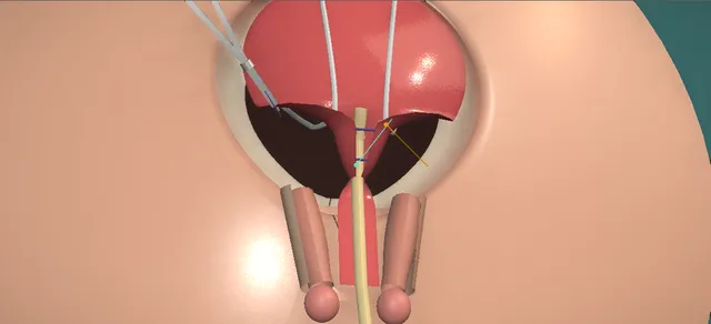
A newborn with classic bladder exstrophy, seen from the front, head at the top. Schematic, not to scale; relationships only.
Seen from the front with the head at the top, the bladder plate lies open on the lower abdominal wall with the umbilicus at its upper edge and both ureteric orifices low on it.
Below it, the urethral plate runs on along the dorsum of a short penis between two splayed corpora.
Under the plate the pubic bones are apart, so the pelvis is open in front.
The closure rolls the plate into a bladder, drops it behind the pubis and brings the pubis together in front of the bladder neck.
›Bladder plate - the open bladder on the lower abdominal wall; its mucosa becomes the lining of the closed bladder.
›Ureteric orifices and trigone - low on the plate, close to the bladder neck; stented through the closure.
›Urethral plate - runs from the bladder neck along the dorsum of the penis between the corpora; tubularised at the bladder neck now, and along the penis at the epispadias repair.
›Corpora cavernosa - splayed apart with the pubic bones, short and curved upward; brought together over the urethra at the epispadias repair.
›Pubic bones - apart in front; brought together with a stitch after the osteotomies.
›Ilium - cut by the iliac osteotomy above the hip joint so that each half of the pelvis can turn forward.
›Rectus fascia and paraexstrophy tissue - lateral to the plate; the plate is freed from it and it is closed in the midline over the bladder.
›Umbilicus - low, at the upper edge of the plate; kept above the incision.
›Peritoneum and bowel - just above and behind the dome of the plate.
›Pelvic vessels and nerves - near the osteotomy and deep in the pelvis beside the bladder neck.
Newborn assessmentA newborn boy with classic bladder exstrophy is seen by the exstrophy team. The umbilical cord is tied rather than clamped, and the plate is covered with plastic film so that it stays moist and is not rubbed. The size and quality of the plate, the width of the pubic gap, the penis and the hips are assessed, the kidneys are scanned, and the family meets the team that will look after the child for life. where it bites: a plate that dries, is rubbed by dressings or is crushed by a cord clamp becomes inflamed and polypoid, and a poor plate closes badly.
Circumferential incisionThe line is marked round the bladder plate, below the umbilicus, and carried down both edges of the urethral plate on the dorsum of the penis. It is incised where the mucosa meets the skin. The paraexstrophy skin lateral to the plate is kept, with its own supply. where it bites: the line keeps to the junction: an incision into the plate narrows it, and one wide of it takes skin needed later.
Free the plateThe plate is lifted and freed from the abdominal wall and the paraexstrophy tissue, with the dissection kept on the side of the wall so that the plate keeps its blood supply. The freeing is carried deep to the bladder neck and the urethral plate, toward the pelvic floor, so that both can be brought back into the pelvis without tension. The ureters enter the plate from behind, low down. where it bites: the blood supply of the plate and of the genitalia runs in this tissue; the ureters lie just behind the trigone, and the peritoneum and bowel just above the dome.
Tubularise the outletA fine stent is passed up each ureter. A catheter is laid along the urethral plate into the bladder neck, and the posterior urethra and the bladder neck are rolled into a tube round it. No formal continence procedure is done at this stage: tightening the outlet now would obstruct a bladder that has yet to grow. where it bites: the orifices sit close to the neck in an abnormal trigone; a stitch placed low in the tube can take a ureter.
Pelvic osteotomyThe orthopaedic surgeon makes the pelvic osteotomies, under the same anaesthetic and usually before the urological closure begins. An iliac osteotomy on each side lets the two halves of the pelvis turn forward, so that the pubic bones can be brought together without tension. Many centres use it when the closure is not done in the first days of life or when the pubic gap is wide. where it bites: the pelvic vessels and nerves lie close behind the cut; a closure under tension without osteotomy is a common reason for dehiscence.
Close the bladder and drop itThe plate is rolled into a bladder and closed in layers in the midline, from the neck up to the dome, with the two stents brought out through the dome. A suprapubic tube is placed through the dome to drain it. The bladder and the posterior urethra are then dropped back into the pelvis, behind the pubis. where it bites: a bladder closed under tension or with its dome left too high is the bladder that prolapses.
Bring the pubis togetherA stitch is passed round the two pubic bones, in front of the bladder neck, and tied as the halves of the pelvis turn forward and the bones meet. The bladder neck and posterior urethra now lie deep in the pelvis, behind the symphysis. where it bites: the bladder neck lies directly behind the stitch; a knot on the urethra causes erosion and fistula.
Close the abdominal wallThe rectus fascia is closed in the midline over the bladder, then the skin. The two ureteric stents and the suprapubic tube leave through the closed wall. The pelvis is held still afterwards, in traction or with an external fixator, while the bones and the wound heal. where it bites: movement of the pelvis in the weeks after closure pulls on the repair; immobilisation and the drains are kept until healing allows.
The later stagesIn the first years of life the epispadias is repaired: the urethral plate is rolled into a tube and the corpora are brought together over it. Once the bladder has grown enough, usually at about school age, the bladder neck reconstruction is done for continence, often with the ureters reimplanted. Follow-up of the kidneys, the bladder, continence, sexual function and cancer risk goes on for life. where it bites: a bladder that does not grow, or a neck reconstruction that fails, leads to augmentation, a catheterisable channel and their own long-term risks.
The spatial relationships the animation is built to make automatic. Before you read each one, ask yourself: at this point in the operation, what is about to go wrong, and what is my first move? The save for each sits in the Complications tab.
RiskBlood supply of the plate and the genitalia - the dissection keeps to the wall side; a plate or a urethral plate stripped of its supply fails.
RiskUreters in an abnormal trigone - the orifices lie close to the neck; they are stented, and kept out of the stitches that tubularise the outlet.
RiskPelvic vessels and nerves - close behind the osteotomy and deep beside the bladder neck during the pubic closure.
RiskBowel and peritoneum - just above the dome; the peritoneum is pushed up off the bladder, not opened.
RiskTension on the closure - a wide pelvis closed without osteotomy or immobilisation pulls the repair apart.
Trigger each of these live in the Animation tab, then read the save: the first move, the escalation trigger, and the line that goes in the op note.
Wound dehiscence and bladder prolapse
the wall and the bladder come apart in the first days or weeks; tension, infection and a closure without osteotomy are the usual causes, and a bladder prolapse through the wound is the same failure.
The save
Cover the exposed plate with plastic film at once so that it stays moist, and keep the drains and the stents running.first move
Look for infection and treat it; record what has come apart: skin, fascia, bladder, pubis.
Tell the exstrophy team the same day: whether to close again now or later, and with which osteotomy, is their decision.escalation trigger
Protect the plate until it is closed again: every failed closure leaves a smaller, more scarred plate.
Record the day it failed, what was exposed, the cultures and the plan agreed.document
Ureteric obstruction after closure
a ureter kinked or narrowed where it enters the closed bladder, or caught in a stitch at the neck; the kidney swells once the stents are out.
The save
Scan the kidneys after the stents come out, and again if the baby is unwell or has a urinary infection.first move
A dilated system with a falling or poor function is drained: a stent or a nephrostomy.
A ureter that stays obstructed after drainage is reimplanted; the exstrophy team decides when.escalation trigger
Record the scan findings, the drainage and the function on the follow-up scan.document
Urethrocutaneous fistula after the epispadias repair
urine leaks from a hole in the repaired urethra, most often at the base of the penis where the repair meets the bladder neck.
The save
Keep the urine draining and let the tissue settle; a small fistula may close by itself.first move
Look beyond it: a stricture or a narrow meatus downstream keeps a fistula open.
A fistula that persists is repaired electively by the reconstructive team, with a layer between the urethra and the skin.escalation trigger
Record where it is, its size, and the calibre of the urethra beyond it.document
Long-term continence failure
the bladder stays small or the neck does not hold, despite the staged repair; the next steps are augmentation of the bladder and a catheterisable channel.
The save
Measure what there is: bladder capacity, pressure, the upper tracts and the pattern of wetting.first move
A small bladder may still grow; a failed neck reconstruction may be revised or bulked.
Where neither will do, the team plans augmentation with bowel and a catheterisable channel, often with closure of the bladder neck.escalation trigger
Teach catheterisation, and plan lifelong follow-up for stones, infection, metabolic change and cancer.
Record the urodynamic findings, the options discussed with the family and the plan.document
When the case turns: the problem, then the next move.
ActionThe plate is small, polypoid or inflamed at birth - closure can be delayed for a few months, the plate kept under film; the team decides whether the plate will make a bladder at all.
ActionThe baby is more than a few days old, or the pubic gap is wide - many centres add a pelvic osteotomy, so that the pubis can be closed without tension.
ActionThe pubis will not come together without tension - do not force the stitch: tension on the closure is what makes it fail; the osteotomy, the turn of the pelvis and immobilisation are what relieve it.
ActionThe penis is very short after the closure - that is expected; length is gained at the epispadias repair, when the corpora are freed and brought together.
ActionA girl with exstrophy - the same closure; the bifid clitoris and the labia are brought together at the closure, and the vagina is assessed again at puberty.
The examiner stems this scene prepares you for. Try to answer out loud first, then reveal, and learn the discriminator, the detail that turns a pass into a strong pass.
QWhat is bladder exstrophy, and where does it sit in the exstrophy-epispadias complex?reveal answer
It is a defect of the lower abdominal wall and the pelvis in which the bladder lies open on the abdomen.
The accepted explanation is that the cloacal membrane is abnormally large and is not reinforced by the ingrowth of mesoderm, so when it breaks down the lower abdominal wall, the anterior bladder, the urethra and the pubic symphysis fail to form in front.
The complex runs from isolated epispadias, through classic bladder exstrophy, to cloacal exstrophy, in which the bowel is involved as well, with an omphalocele, an imperforate anus and spinal anomalies.
In classic exstrophy I would expect an open bladder plate with both ureteric orifices on it, a low umbilicus, a wide pubic diastasis with the hips externally rotated, and an epispadiac penis that is short and curved upward, or a bifid clitoris in a girl.
The anus lies further forward than usual.
It is rare, more common in boys, and in the United Kingdom it is managed in nationally commissioned centres.
Strong pass
Say all three
Describing the bony defect as part of the anomaly
Not just the bladder
Placing classic exstrophy correctly between epispadias and cloacal exstrophy
Examiner note
Describing the bony defect as part of the anomaly, not just the bladder, and placing classic exstrophy correctly between epispadias and cloacal exstrophy.
QA baby with exstrophy has just been born in your hospital. What do you do?reveal answer
I would make the baby safe and protect the plate, and I would call the exstrophy centre straight away.
The cord should be tied rather than clamped, because a plastic clamp lying on the plate injures it, and the plate is covered with plastic film, not gauze, so that it stays moist and is not rubbed.
I would examine the baby fully: the size and quality of the plate, the width of the pubic gap, the penis or the clitoris, the anus, the hips and the spine, and I would look for features of cloacal exstrophy.
A renal ultrasound shows the upper tracts.
Many cases are now suspected before birth: the bladder is not seen on the scan, the cord inserts low, the pubic rami are wide and there is a mass on the lower abdomen.
When that happens the parents should meet the team before delivery, and the baby should be born where the closure will be done.
Transfer is planned with the centre, and the parents are told this is a lifelong condition with a lifelong team.
Strong pass
Say all three
Tying rather than clamping the cord
Covering the plate with film
Handing the child to a specialist centre rather than attempting a closure locally
Examiner note
Tying rather than clamping the cord and covering the plate with film, and handing the child to a specialist centre rather than attempting a closure locally.
QCompare the modern staged repair with the complete primary repair. What results would you quote?reveal answer
The modern staged repair separates the work.
The bladder, the posterior urethra and the abdominal wall are closed in the newborn, with an osteotomy where it is needed.
The epispadias is repaired in the first years, and the bladder neck is reconstructed once the bladder has grown, often with reimplantation of the ureters.
In the Johns Hopkins series of Baird and colleagues, 47 of 67 boys and girls closed primarily at that centre, 70 per cent, were dry day and night and voided per urethra without augmentation or catheterisation, and children whose capacity before the neck repair was larger did better.
The complete primary repair, after Mitchell, closes the bladder and repairs the epispadias in one newborn operation, with disassembly of the penis.
In the long-term Seattle review of Ellison and colleagues, 12 of 29 eligible children were continent, voiding with dry intervals of at least three hours, at a mean follow-up of more than twelve years;
42 per cent of those needed no further continence surgery, and some became continent only in adolescence.
These are single-centre retrospective series with different definitions of continence, so I would not set one figure against the other directly.
A third approach is the radical soft-tissue mobilisation after Kelly; in the Melbourne series of Jarzebowski and colleagues, 21 of 30 patients without diversion were completely or partially dry.
Strong pass
Say both
Quoting the figures with their definitions and their source
Saying why they cannot be compared head to head
Examiner note
Quoting the figures with their definitions and their source, and saying why they cannot be compared head to head.
QWhy would you do a pelvic osteotomy, and what does it cost the child?reveal answer
I would use it to take tension off the closure.
The pubic gap is part of the anomaly.
Bringing the pubis together in front of the bladder neck takes tension off the abdominal closure and puts the bladder neck and the posterior urethra deep in the pelvis, where the pelvic floor can support them.
In a newborn closed in the first days the pelvis is malleable and some centres close without an osteotomy, but many add one when the closure is later or the gap is wide, because a closure under tension is a common reason for dehiscence.
The osteotomy is made by the orthopaedic surgeon, and an iliac cut on each side lets each half of the pelvis turn forward.
It costs a longer anaesthetic, a period of immobilisation in traction or with an external fixator, pin-site and wound problems, and a risk to the vessels and nerves near the cut.
In the female series of Purves and colleagues, the six girls who were dry after the closure alone had all had a transverse innominate and vertical iliac osteotomy.
The Kelly approach goes the other way: Leclair and colleagues closed 27 bladders combined with the soft-tissue mobilisation without osteotomy and reported no dehiscence at a median of a year.
Strong pass
Say both
Explaining the osteotomy as a way to remove tension and to place the bladder neck deep in the pelvis
Knowing that some schools do without it
Examiner note
Explaining the osteotomy as a way to remove tension and to place the bladder neck deep in the pelvis, and knowing that some schools do without it.
QThe child is now eight, wet and has had a bladder neck reconstruction. What next, and what follows them into adult life?reveal answer
I would find out why the child is wet: the capacity and pressure of the bladder on urodynamics, the upper tracts on ultrasound, and the pattern of wetting.
A small bladder may still grow, and a neck that leaks may be revised.
When neither will give a safe, continent bladder, the next steps are augmentation of the bladder with bowel and a continent catheterisable channel, often with closure of the bladder neck.
The EAU guideline notes that these children may need augmentation or diversion with a channel and are then at risk of the long-term complications of bowel in the urinary tract.
Into adult life I would follow the kidneys, stones, infection, metabolic change and sexual function: in men the penis is short and curved upward and ejaculation may be affected, and in women vaginal stenosis and pelvic organ prolapse occur and pregnancy carries a higher risk of preterm labour.
The guideline also records a raised and earlier risk of cancer, mostly adenocarcinoma, with no agreed surveillance protocol, and it recommends a planned transition to adult care.
Strong pass
Say both
Seeing continence surgery as one stage of a lifelong plan
Naming the late risks of bowel in the urinary tract and of cancer
Examiner note
Seeing continence surgery as one stage of a lifelong plan, and naming the late risks of bowel in the urinary tract and of cancer.
Curriculum. Supports learning relevant to the ISCP Urology Curriculum syllabus topic Paediatric urology. Not a workplace-based assessment and not evidence of competence.
Two minutes
Close the lap: recall it before you leave.
Say it aloud, then reveal. Reading is not remembering.
01The relationshipSay the critical spatial relationship out loud, then reveal.reveal
Memory frame
Seen from the front with the head at the top, the bladder plate lies open on the lower abdominal wall with the umbilicus at its upper edge and both ureteric orifices low on it.
Why?
Below it, the urethral plate runs on along the dorsum of a short penis between two splayed corpora. Under the plate the pubic bones are apart, so the pelvis is open in front. The closure rolls the plate into a bladder, drops it behind the pubis and brings the pubis together in front of the bladder neck.
02The first moveWound dehiscence and bladder prolapse. Your first move?reveal
First move
Cover the exposed plate with plastic film at once so that it stays moist, and keep the drains and the stents running.
03The escalation triggerWound dehiscence and bladder prolapse. What makes you call for help?reveal
Escalate when
Tell the exstrophy team the same day: whether to close again now or later, and with which osteotomy, is their decision.
04The discriminatorWhat turns a pass into a strong pass?reveal
Strong pass
Say all three
Describing the bony defect as part of the anomaly
Not just the bladder
Placing classic exstrophy correctly between epispadias and cloacal exstrophy
Examiner note
Describing the bony defect as part of the anomaly, not just the bladder, and placing classic exstrophy correctly between epispadias and cloacal exstrophy.
05The pitfallWhere does this operation bite first?reveal
Pitfall
Blood supply of the plate and the genitalia
Why?
the dissection keeps to the wall side; a plate or a urethral plate stripped of its supply fails.
Revision practice with self-marked answers, not an assessment of clinical competence. Kept in
this browser only; it does not sync between devices.