
UrOops3D, danger-zone atlas
Operative animation, Focal ablation of the prostate

Focal ablation for localised prostate cancer

The tumour and a margin destroyed with heat, cold or electrical pulses, and the rest of the prostate, the sphincter and the nerves left alone, in a man whose cancer is confined to one part of a gland mapped by MRI and biopsy.

Less incontinence and less impotence than removing or irradiating the whole gland; the price is a gland that stays, must be watched, and may need treating again.

This page is the selection, the planning of the zone, the energy and its monitoring, the rectum and the urethra, and the follow-up that never ends.

Advanced tier FRCS uro-oncology, localised prostate cancer Interactive 3D

Operative visualisation

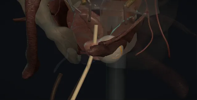
Schematic scene preview: Focal ablation of prostate cancer
Preparing the 3D anatomy…

UrOops3D · UroRef · © Nity G

Click me next Section 2 of 8 Anatomy The structures this operation works between, and the one it must not cross.

The structures in the scene

Focal ablation of the prostate: how the structures relateProstate in section, the treatment zone and what must be spared. Index lesion and its margin: heat or ice past the capsule makes a fistula Rectal wall (danger). Index lesion and its margin: runs through the middle; warmed during a freeze Urethra and the warming catheter (danger). Index lesion and its margin: the zone stops short of the apex Apex and external sphincter (danger). Index lesion and its margin: posterolateral; the untreated side kept whole Neurovascular bundles (danger). Index lesion and its margin: from the rectum, heat stacked apex to base HIFU probe with cooled balloon. Index lesion and its margin: from the perineum; temperatures at the spared Cryoprobes and thermocouples.Index lesion and itsmargin!Rectal wall!Urethra and thewarming catheter!Apex and externalsphincter!NeurovascularbundlesHIFU probe withcooled balloonCryoprobes andthermocouplesheat or ice pastthe capsule makes afistularuns through themiddle; warmedduring a freezethe zone stopsshort of the apexposterolateral; theuntreated side keptwholefrom the rectum,heat stacked apexto basefrom the perineum;temperatures at thesparedTargetDangerContext
Prostate in section, the treatment zone and what must be spared. Schematic, not to scale; relationships only.
  • In the scene the prostate sits under the bladder with the rectum behind it and the treatment zone shaded on one side, a quadrant or a hemigland with its margin, ending short of the apex and the bladder neck.
  • The HIFU probe lies in the rectum against the posterior surface of the gland with its cooled balloon between them, or the cryoprobes enter from the perineum below and pass upward through the zone with the urethral warmer running through the middle.
  • The neurovascular bundle on the untreated side runs posterolateral and untouched; the bundle on the treated side lies at the edge of the shaded zone.
  • ›The index lesion and its margin - the MRI contour plus the positive cores, with five to ten millimetres around it; the treatment volume is the smallest unit that contains it.
  • ›The rectal wall - millimetres behind the posterior capsule; cooled by the HIFU balloon, watched by the cryotherapy thermocouple, pushed away by saline.
  • ›The urethra and the warming catheter - runs through the middle of the gland; warmed saline keeps its mucosa alive during a freeze beside it.
  • ›The apex and the external sphincter - the zone stops a few millimetres short of it, giving up margin that cannot be regrown.
  • ›The neurovascular bundles - posterolateral against the capsule; the untreated side keeps its bundle, the treated side loses it if the tumour lies against it.
  • ›The HIFU probe - in the rectum with a cooled balloon; rows of small heat lesions stacked from apex to base within the planned zone.
  • ›The cryoprobes and thermocouples - 17 gauge needles through a perineal template; argon freeze, the iceball watched on ultrasound, temperatures read at the structures to be spared.
Click me next Section 3 of 8 Steps The operation in order, with the landmark that guards each move.

Operative steps

  1. Who is a focal candidate A man with localised prostate cancer confined to one region of the gland on MRI and on a biopsy that has sampled the whole gland, targeted and systematic, so that the map is trusted: intermediate risk on the whole, grade group 2 and selected grade group 3, PSA below about 15 to 20, stage T1 or T2, with a life expectancy that makes treatment worthwhile and a wish to keep erections and continence that outweighs the chance of a second treatment. Low-risk disease is surveillance, not ablation; high-risk or multifocal significant disease is a whole-gland treatment. He hears what NICE says about the evidence, that the procedure is offered under special arrangements or within a trial or registry, and that surveillance after it is not optional.
  2. HIFU, cryotherapy or electroporation High-intensity focused ultrasound is delivered from a probe in the rectum, the sound focused into a lesion of a few millimetres, stacked to fill the planned volume with heat: suited to posterior and mid-gland disease in a gland the focus can reach without calcification in the beam path. Cryotherapy is needles through the perineum on a template, argon freezing to an iceball watched on ultrasound: suited to anterior and apical disease, the larger gland, and the gland after radiotherapy. Irreversible electroporation uses needle electrodes through the perineum and high-voltage pulses to open cell membranes without heat, sparing the collagen of urethra and rectum, under general anaesthesia with relaxation, and in the UK a research procedure. The modality is chosen for the lesion, not the machine.
  3. Planning the zone: the lesion and its margin The MRI lesion is contoured, and the biopsy cores that were positive are placed on the same map, since the tumour is usually larger than its MRI footprint. Around that a margin of about five to ten millimetres is drawn, and the treatment volume becomes the smallest anatomical unit that contains it: a quadrant, a hemiablation of one side to the midline, or a hockey stick that takes the posterior gland across the midline for a lesion near it. The volume stops short of the sphincter at the apex, the rectal wall behind, the bladder neck above, and the neurovascular bundle on the untreated side. The plan is agreed with the radiologist and drawn on the treatment software, and it is the plan that is delivered.
    where it bites: a margin drawn to the MRI lesion alone undertreats; the positive systematic cores extend the zone beyond what the MRI shows.
  4. Anaesthesia, probe and the urethral warmer General or spinal anaesthesia, lithotomy, an enema beforehand and antibiotic prophylaxis. For HIFU the treatment probe goes into the rectum with a cooled water-filled balloon around it, the prostate is imaged and registered to the MRI plan, and a suprapubic or urethral catheter drains the bladder during and after. For cryotherapy a transrectal ultrasound probe on a stepper images the gland, a brachytherapy template against the perineum guides 17 gauge cryoprobes into the planned zone, thermocouples are placed at the sphincter, the rectal wall and the neurovascular bundle, and a urethral warming catheter circulates saline at about 40 degrees through the urethra so that the mucosa survives the freeze beside it. The warmer is in and running before any freezing starts.
    where it bites: a cryotherapy freeze without the urethral warmer running sloughs the urethra along its length; the warmer is checked before the first probe is activated.
  5. Protecting the rectum The rectal wall lies a few millimetres behind the posterior capsule, and every modality reaches it if it is not watched. In HIFU the probe balloon circulates cooled water against the anterior rectal wall, the lesions are planned with a safety margin from the wall, and the distance from probe to capsule is checked on the live image before each row. In cryotherapy the iceball edge is watched on the sagittal ultrasound as it advances toward the rectum, the freeze on the posterior probes is reduced as it approaches, the rectal thermocouple is kept above freezing, and saline can be injected between prostate and rectum before the freeze to push the wall away. After radiotherapy the rectal wall heals badly, and the margin behind is wider still.
    where it bites: the rectal wall is millimetres from the posterior capsule; a lesion or an iceball that reaches it is a rectourethral fistula weeks later.
  6. The ablation, watched in real time HIFU is delivered as rows of lesions from apex to base across the planned zone, each a few seconds, the tissue change watched as hyperechoic clouds and the probe repositioned row by row; the software records what has been treated, and the operator, not the software, decides whether the zone is covered. Cryotherapy is two freeze-thaw cycles: argon on to bring the zone below about minus 40 degrees, seen as the iceball with its shadow, a passive or helium thaw, then a second freeze, since a double freeze kills what a single one leaves; the thermocouples give the temperatures at the structures to be spared. Electroporation delivers its pulses between paired electrodes synchronised to the heart. The endpoint is the zone covered and nothing outside it.
  7. The untreated side and the neurovascular bundle The point of the operation is what is not treated. The contralateral gland is left with its neurovascular bundle intact and the ipsilateral bundle is spared where the lesion allows, which is why erections are usually preserved; where the tumour lies against the bundle, the bundle is included in the zone and the man has been told that erections on that side will suffer, because an untreated margin against the tumour is a recurrence. The thermocouple or the planned margin marks the limit. The bladder neck and the sphincter are outside the zone in a focal treatment, and the zone at the apex ends a few millimetres short of the sphincter, sacrificing a little margin there because the sphincter cannot be regrown.
    where it bites: the neurovascular bundle sits posterolateral against the capsule; the margin on the treated side takes it if the tumour touches it, and the untreated side keeps it whole.
  8. After: catheter, retention, slough The catheter stays for a few days after HIFU and about a week after cryotherapy, because the treated gland swells and then sheds. He goes home the same or next day with an alpha blocker, analgesia and antibiotics by protocol. For some weeks he passes necrotic tissue in the urine, with dysuria and sometimes a plug that stops the stream. Retention after the catheter comes out is a catheter again and, if it recurs, a cystoscopy to clear the fossa. Infection is treated on culture. Blood in the urine and semen settles. He is told which symptoms are expected and which are not: rectal bleeding, faeces or air in the urine, or urine from the rectum are a fistula until proved otherwise and are seen the same day.
  9. Follow-up: PSA, MRI, biopsy The gland remains and so does the risk, and the man agreed to be watched. PSA every three months in the first year and six-monthly after, read as a trend from the nadir, because untreated gland still makes PSA. MRI at six to twelve months to show the ablation scar and the untreated gland. Biopsy of the treated zone and the rest of the gland at about a year in most protocols, and for a rising PSA or a new lesion. In-field recurrence can be ablated again; out-of-field disease is a decision at the meeting between another focal and a whole-gland treatment. Prostatectomy or radiotherapy remain possible after ablation, with more scar and a higher complication rate. He stays on the registry.
Click me next Section 4 of 8 Danger zones Where this operation bites, and why it bites there.
Critical relationship

Danger zones

The spatial relationships the animation is built to make automatic. Before you read each one, ask yourself: at this point in the operation, what is about to go wrong, and what is my first move? The save for each sits in the Complications tab.

  • RiskThe rectal wall - reached by heat or ice that passes the posterior capsule; a rectourethral fistula that declares itself weeks later, worst after radiotherapy.
  • RiskThe external sphincter and bladder neck - outside the zone in a focal treatment; incontinence or bladder neck stenosis if the apex or base is overtreated.
  • RiskThe urethra - in the middle of every hemiablation; sloughs if it is frozen without the warmer, strictures if it is heated.
  • RiskThe neurovascular bundles - the reason for the operation; the untreated bundle is kept whole and the treated bundle is sacrificed only where the tumour demands it.
Practise it Take me into the scene Opens the animation at a danger moment and asks you to predict the mechanism and your first move before it reveals.
Click me next Section 5 of 8 Complications The save for each one: the first move, the escalation trigger, the op-note line.

Complications and immediate saves

Trigger each of these live in the Animation tab, then read the save: the first move, the escalation trigger, and the line that goes in the op note.

Retention, slough and outlet obstruction

  • a swollen then sloughing gland: the stream that stops when the catheter comes out, the plug of necrotic tissue, and the man who keeps blocking for weeks.
The save
  1. A catheter for the expected period and an alpha blocker before it is removed; a bladder scan at the trial without catheter. first move
  2. Retention after removal: catheter again and a second trial at a week; the slough is explained and expected for several weeks.
  3. Recurrent retention or a stream that never recovers: flexible cystoscopy, and clearance of slough or a bladder neck incision in theatre.
  4. Retention with fever or a gland that is exquisitely tender: a prostatic abscess in necrotic tissue; image, drain and admit. escalation trigger
  5. Note: the modality and zone, the catheter type and removal date, the alpha blocker, and the warning given about slough. document

Infection and sepsis

  • necrotic tissue in the urinary tract with a catheter in it: urinary infection, epididymitis, or sepsis, and after HIFU a rectal probe that adds the rectal flora.
The save
  1. Antibiotic prophylaxis by protocol and a catheter that is not left longer than it needs to be. first move
  2. Fever after the treatment: urine and blood cultures and antibiotics started before the result; the catheter changed if it is in.
  3. Examine the perineum and the rectum; a tender fluctuant gland or a perineal collection is imaged.
  4. Hypotension, rigors or no response within a day: the sepsis pathway, critical care review, and drainage of any abscess. escalation trigger
  5. Note: the prophylaxis, the cultures sent, the antibiotic and the time, and the review plan. document

Rectourethral fistula and rectal injury

  • the rare complication that ends the argument for the treatment: faeces or air in the urine, urine from the rectum, or pelvic sepsis weeks after energy reached the rectal wall.
The save
  1. Prevent it: the cooled balloon, the posterior margin, the rectal thermocouple, the saline injection behind the gland, and a wider margin still after radiotherapy. first move
  2. Any faeces, air or rectal leakage of urine: seen the same day; examination, cystoscopy, flexible sigmoidoscopy and MRI or CT to define the tract.
  3. Divert the urine with a suprapubic catheter and the faeces with a colostomy where the fistula is large or infected; small fistulae occasionally close with diversion alone.
  4. A fistula that does not close, or one in an irradiated pelvis: staged repair with a reconstructive urologist and colorectal surgeon, often with a gracilis or omental interposition. escalation trigger
  5. Note: the rectal protection used and the temperatures or distances recorded at the time; later, the fistula size, the diversion and the repair plan. document

Incontinence, stricture and bladder neck stenosis

  • energy carried to the apex, the bladder neck or the urethra: stress leakage, a stream that slows over months, or a neck that closes to a pinhole.
The save
  1. Keep the zone off the sphincter and the neck at the planning stage; run the urethral warmer through the whole freeze. first move
  2. Leakage after the catheter comes out: pelvic floor physiotherapy and time; most is urgency and slough and settles.
  3. A falling flow rate at review is a cystoscopy; a urethral stricture is dilated or incised, a bladder neck stenosis is incised.
  4. Stress incontinence persisting beyond a year, or a stricture that recurs after two treatments: urodynamics and a continence or reconstructive surgeon. escalation trigger
  5. Note: the distance from the zone to the apex and the neck on the plan, and the flow and pad count at each visit. document

Residual, recurrent or out-of-field cancer

  • the gland is still there: a rising PSA from the nadir, a new lesion on the follow-up MRI, or positive cores at the protocol biopsy, in or outside the treated zone.
The save
  1. PSA three-monthly in the first year, read as a trend from the nadir, with MRI at six to twelve months and a biopsy at about a year or for cause. first move
  2. A rise or a new lesion: targeted and systematic biopsy of the treated zone and the untreated gland, so that in-field and out-of-field are distinguished.
  3. In-field significant recurrence: repeat ablation of that zone is reasonable once; out-of-field disease is discussed at the meeting.
  4. Multifocal or high-grade recurrence, or a second failure: whole-gland treatment, radical prostatectomy or radiotherapy, with the man told the complication rate is higher after ablation. escalation trigger
  5. Note: the nadir PSA, each subsequent value, the MRI dates, the biopsy map, and the registry entry. document
Click me next Section 6 of 8 Troubleshooting When the case turns: the problem, then the next move.

Intra-operative troubleshooting

When the case turns: the problem, then the next move.

  • ActionCalcification shadows the beam path on HIFU - the lesion behind it will not be treated; replan around it or choose cryotherapy, which does not care about calcification.
  • ActionThe gland is too deep for the HIFU focus at the anterior lesion - the anterior-posterior distance exceeds the focal range; this is a cryotherapy case through the perineum, not a compromise with the HIFU probe.
  • ActionThe rectal thermocouple is falling toward zero during the freeze - reduce or stop the argon on the posterior probes, let the iceball edge retreat on the sagittal view, and inject saline behind the gland before the second cycle.
  • ActionThe urethral warmer alarms or the flow stops - stop the freeze; check the circuit and the catheter position, restart the warmer and confirm the flow before any probe is reactivated.
  • ActionThe registration between MRI and ultrasound does not match the gland - do not treat to a misregistered plan; re-register on the capsule and the urethra, and fall back to cognitive placement of the zone by the landmarks.
  • ActionThe man returns with a plug of tissue and a full bladder - catheterise, and if the catheter will not pass, a suprapubic catheter and a cystoscopy to clear the slough; explain that this is the gland shedding, not the cancer.
Click me next Section 7 of 8 FRCS viva Say it out loud, then mark yourself against the model answer.

FRCS viva questions

The examiner stems this scene prepares you for. Try to answer out loud first, then reveal, and learn the discriminator, the detail that turns a pass into a strong pass.

QWho is a candidate for focal therapy, and how does it sit against the other options for localised prostate cancer?reveal answer
  • I would consider focal therapy for a man with localised prostate cancer whose disease has been mapped with multiparametric MRI and a biopsy that sampled the whole gland, targeted and systematic, and is confined to one region: predominantly grade group 2, selected grade group 3, PSA below about 15 to 20 and stage T1 to T2, with a life expectancy that justifies treatment and a strong wish to preserve erections and continence.
  • Low-risk disease is managed by active surveillance, where ablation adds harm without benefit; high-risk, multifocal or extraprostatic disease needs whole-gland treatment by prostatectomy or radiotherapy.
  • The advantages are lower rates of incontinence and erectile dysfunction, a day-case procedure and a preserved gland;
  • the disadvantages are the need for a second treatment in a proportion of men, lifelong surveillance with PSA, MRI and biopsy, and a weaker evidence base, so that NICE guidance places it under special arrangements or within a trial or registry rather than as a standard alternative.
  • I would present it at the multidisciplinary meeting and to the man as a trade of oncological certainty for function, and I would not offer it to a man who would not accept the surveillance.
Strong pass

Say both

  • The candidate defined by a whole-gland biopsy map
  • The evidence position stated as NICE states it rather than as an established equal
Examiner note
The candidate defined by a whole-gland biopsy map, and the evidence position stated as NICE states it rather than as an established equal.
QDescribe focal HIFU and focal cryotherapy, and how you protect the rectum, urethra and sphincter.reveal answer
  • Both begin with a plan: the MRI lesion and the positive cores contoured with a margin of five to ten millimetres, forming a quadrant, hemigland or hockey-stick zone that stops short of the sphincter, the bladder neck and the rectal wall.
  • For HIFU, under general or spinal anaesthesia in lithotomy with a catheter draining the bladder, the transrectal probe is placed with a cooled water balloon against the anterior rectal wall, the gland is registered to the plan, and rows of focused ultrasound lesions are delivered from apex to base within the zone while the tissue change is watched on the live image and the probe-to-capsule distance checked before each row;
  • the cooled balloon and the posterior margin protect the rectum, and the zone ends short of the apex to protect the sphincter.
  • For cryotherapy, a transrectal probe images the gland, cryoprobes are placed through a perineal template into the zone, thermocouples at the sphincter, the rectal wall and the neurovascular bundle, and a urethral warming catheter runs throughout;
  • two freeze-thaw cycles are delivered, the iceball watched on the sagittal image as it approaches the rectum, the posterior probes reduced as it does, and saline injected behind the gland to lift the rectal wall away.
  • In both, the operator decides when the zone is covered.
Strong pass

Say both

  • The urethral warmer running before the first freeze and the rectal thermocouple or balloon as active monitoring
  • Not as passive safety features
Examiner note
The urethral warmer running before the first freeze and the rectal thermocouple or balloon as active monitoring, not as passive safety features.
QWhat are the complications of focal ablation?reveal answer
  • I would group them as early and functional, structural, and oncological.
  • Early: retention and slough in the weeks after treatment as the ablated gland swells and sheds, managed with a catheter, an alpha blocker and occasionally cystoscopic clearance;
  • urinary infection, epididymitis and rarely a prostatic abscess in necrotic tissue;
  • haematuria and haematospermia.
  • Structural: urethral stricture or bladder neck stenosis from energy at the neck or along the urethra, more after whole-gland treatment;
  • and the rare rectourethral fistula from energy reaching the rectal wall, presenting weeks later with faeces or air in the urine, needing diversion and staged repair, and far more likely in the irradiated pelvis, which is why salvage ablation carries a different consent.
  • Functional: stress incontinence is uncommon in focal treatment because the sphincter is outside the zone, in the order of a few per cent, and erectile function is preserved in most men, with the ipsilateral bundle lost where the tumour lay against it;
  • ejaculation is reduced or absent in many.
  • Oncological: residual or recurrent disease in the treated zone, and new disease in the untreated gland, found by the PSA trend from the nadir, MRI and the protocol biopsy, with a proportion of men needing a second focal or a whole-gland treatment over the following years.
  • The figures are current teaching.
Strong pass

Say both

  • The fistula tied to the rectal wall and to salvage after radiotherapy
  • Recurrence divided into in-field and out-of-field with the surveillance that finds each
Examiner note
The fistula tied to the rectal wall and to salvage after radiotherapy, and recurrence divided into in-field and out-of-field with the surveillance that finds each.
QHow do you follow a man after focal therapy, and what do you do when the PSA rises?reveal answer
  • The gland remains, so follow-up is oncological surveillance, not a discharge.
  • I would measure PSA every three months in the first year and six-monthly thereafter, and interpret it as a trend from the post-treatment nadir rather than against the thresholds used after prostatectomy or radiotherapy, since untreated gland continues to produce PSA.
  • I would arrange multiparametric MRI at six to twelve months to confirm the ablation zone and examine the untreated gland, and a biopsy of the treated zone and the remaining gland at about a year in most protocols, and at any time for a rising PSA, a new lesion or a suspicious examination.
  • If the PSA rises I would exclude infection and slough as causes, repeat it, obtain an MRI and then a targeted and systematic biopsy, because the management depends on where the disease is.
  • Significant in-field recurrence can be ablated again once;
  • out-of-field or multifocal disease is discussed at the meeting and usually means whole-gland treatment, radical prostatectomy or radiotherapy, with the man told that scarring from the ablation raises the complication rate of both.
  • Low-volume grade group 1 disease found at the protocol biopsy can be watched.
  • Throughout he remains on the registry, and the record shows the nadir, every PSA, the imaging and the biopsy maps in one place.
Strong pass

Say both

  • PSA interpreted from the nadir with the reason
  • The biopsy that separates in-field from out-of-field before any second treatment is chosen
Examiner note
PSA interpreted from the nadir with the reason, and the biopsy that separates in-field from out-of-field before any second treatment is chosen.
Click me next Section 8 of 8 Evidence The guidance each of these answers is built on.

Evidence and guidance

Two minutes

Close the lap: recall it before you leave.

Say it aloud, then reveal. Reading is not remembering.

  1. 01 The relationship Say the critical spatial relationship out loud, then reveal. reveal
    Memory frame

    In the scene the prostate sits under the bladder with the rectum behind it and the treatment zone shaded on one side, a quadrant or a hemigland with its margin, ending short of the apex and the bladder neck.

    Why?
    The HIFU probe lies in the rectum against the posterior surface of the gland with its cooled balloon between them, or the cryoprobes enter from the perineum below and pass upward through the zone with the urethral warmer running through the middle. The neurovascular bundle on the untreated side runs posterolateral and untouched; the bundle on the treated side lies at the edge of the shaded zone.
  2. 02 The first move Retention, slough and outlet obstruction. Your first move? reveal
    First move

    A catheter for the expected period and an alpha blocker before it is removed; a bladder scan at the trial without catheter.

  3. 03 The escalation trigger Retention, slough and outlet obstruction. What makes you call for help? reveal
    Escalate when

    Retention with fever or a gland that is exquisitely tender: a prostatic abscess in necrotic tissue; image, drain and admit.

  4. 04 The discriminator What turns a pass into a strong pass? reveal
    Strong pass

    Say both

    • The candidate defined by a whole-gland biopsy map
    • The evidence position stated as NICE states it rather than as an established equal
    Examiner note
    The candidate defined by a whole-gland biopsy map, and the evidence position stated as NICE states it rather than as an established equal.
  5. 05 The pitfall Where does this operation bite first? reveal
    Pitfall

    The rectal wall

    Why?
    reached by heat or ice that passes the posterior capsule; a rectourethral fistula that declares itself weeks later, worst after radiotherapy.

Revision practice with self-marked answers, not an assessment of clinical competence. Kept in this browser only; it does not sync between devices.