
Revision sheet
Focal ablation of prostate cancer (HIFU)
9 steps, each one the authored title and caption from the interactive animation, with the frame captured from that step. Read it before the case, or print it and work through the steps in order.
Teaching only, not for diagnosis or treatment decisions. The text on this sheet is the animation's own step copy, reproduced verbatim.
UrOops3D · UroRef · © Nity G
- 01
Who is a focal candidate
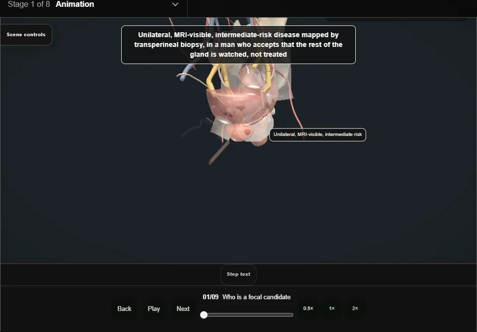
- Unilateral, MRI-visible, intermediate-risk disease mapped by transperineal biopsy, in a man who accepts that the rest of the gland is watched, not treated.
- Not for bilateral disease, high-grade cores, or a lesion the imaging cannot see.
- 02
HIFU, cryotherapy or electroporation
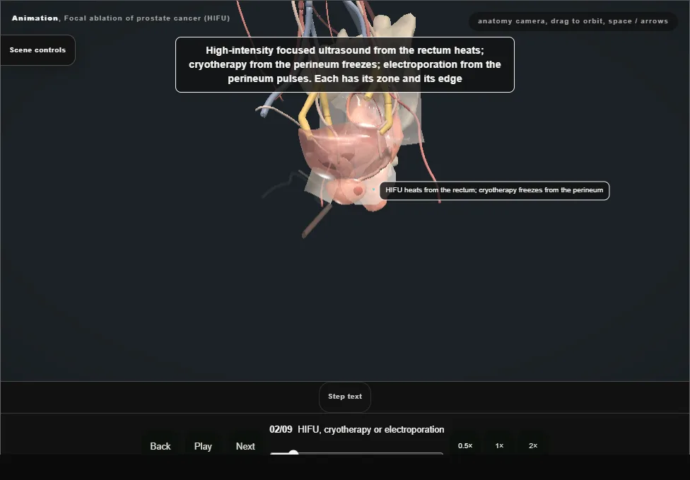
- Focused ultrasound from the rectum heats; cryotherapy from the perineum freezes; electroporation pulses.
- HIFU suits the posterior gland the probe reaches, cryotherapy the anterior and apical gland it cannot.
- 03
Planning the zone
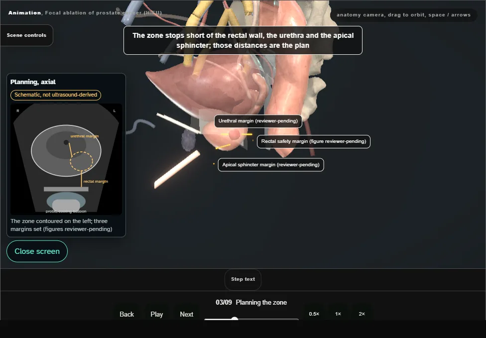
- The lesion and a margin, or the whole half of the gland.
- The zone stops short of the rectal wall, the urethra and the apical sphincter; those distances are the plan.
- 04
Anaesthesia and the treatment probe
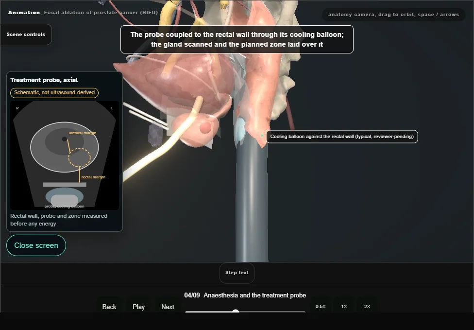
- General anaesthetic, lithotomy, a catheter or suprapubic; the treatment probe in the rectum on its stand, coupled to the rectal wall through its cooling balloon.
- (For cryotherapy, a urethral warmer instead: see the branch.)
- 05
Protecting the rectum
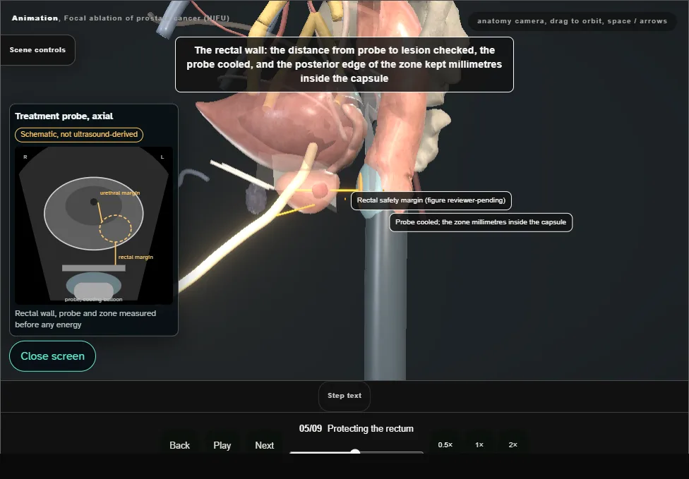
- The distance from probe to lesion checked, the probe cooled, the posterior edge of the zone kept millimetres inside the capsule; where the gap is thin the plan changes before any energy is delivered.
- 06
The ablation, watched in real time
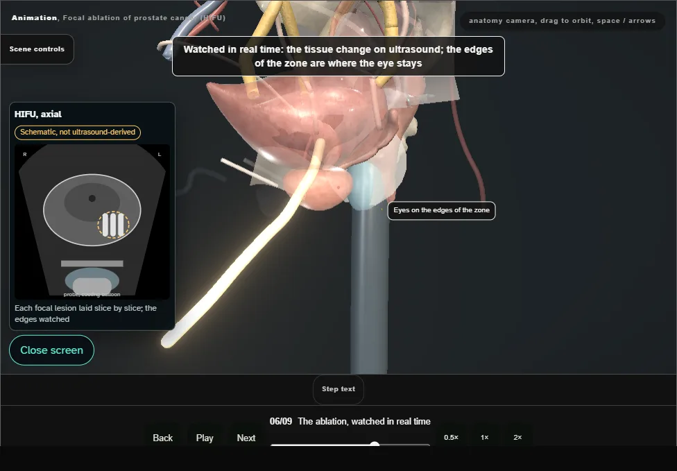
- Slice by slice, focal lesion by focal lesion, the zone filling on the screen.
- The tissue change on ultrasound: the edges of the zone are where the eye stays.
- 07
The untreated side and the bundle
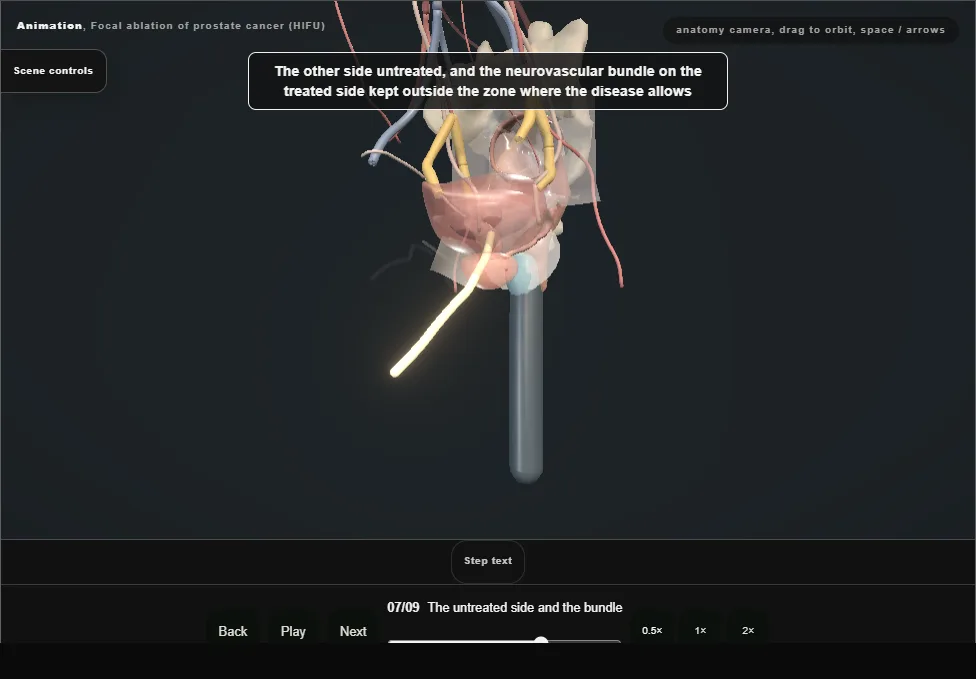
- The other side untreated, and the neurovascular bundle on the treated side kept outside the zone where the disease allows.
- Erections, continence, and a gland that can be treated again are why the man chose this.
- 08
After
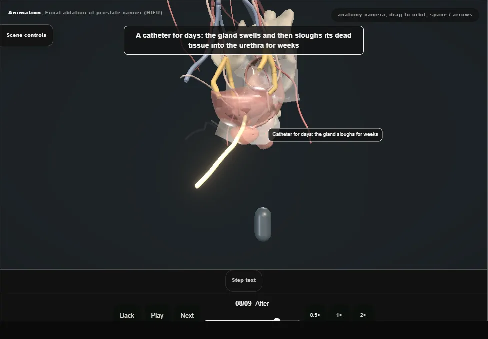
- A catheter for days: the gland swells then sloughs dead tissue into the urethra for weeks.
- Retention, dysuria, debris and infection are the first month; a fistula is the rare disaster.
- 09
Follow-up
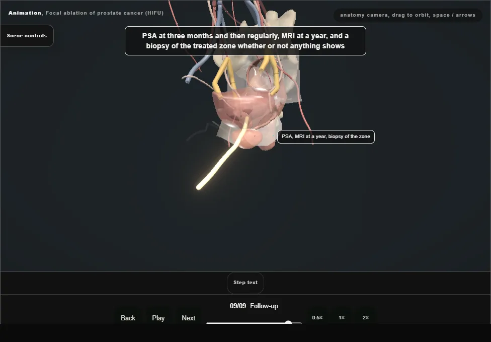
- PSA at three months and then regularly, MRI at a year, and a biopsy of the treated zone.
- Recurrence in the zone is retreated or moved to whole-gland treatment; disease on the other side is a new decision.
Derived from pelvis/FocalAblationScene.tsx at build time: every step title and caption above is the authored
ProcedureDef text, reproduced verbatim, and this page shares its readable-points renderer with the animation
itself. 9 of 9 steps have a captured frame.
Text fingerprint e205c4e8ad7e5721. A caption edit without a re-capture fails the production
check, so the sheet and the animation cannot drift apart silently.