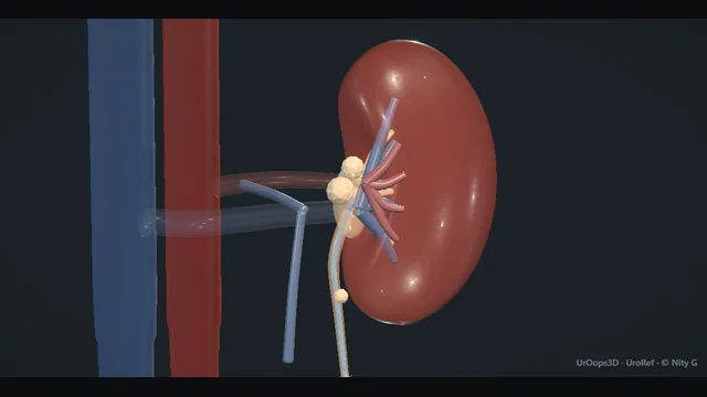
A JJ stent left long enough becomes a stone cast of itself: crusted at both coils, along the shaft, and welded to the ureter and bladder it was sent to protect.
Removal is planned backwards from the imaging: free the bladder coil with the laser from below, test traction gently, and finish a heavy burden with combined percutaneous and cystoscopic clearance in one anaesthetic rather than by force.
Along the encrusted stent, kidney to bladder. Schematic, not to scale; relationships only.
The stent is a cast of the urinary tract, and its two ends are two different problems.
The bladder coil sits in the widest, most accessible space: it can be seen, lasered and basketed from below, so it is freed first.
The renal coil sits in the least accessible space, buried in a narrow pelvis encrusted to the wall, and it is the coil that refuses traction;
it is reached percutaneously, in line with the collecting system, exactly as any other stone.
Between them the shaft runs the length of the ureter, and encrustation along it is what snaps when the two ends are pulled against each other.
The whole strategy reads off that geometry: soft access from below where the tract is wide, direct access from above where the tract is narrow, and no force anywhere the ureter is the weaker partner.
›Renal coil - the upper pigtail, the commonest welded point; freed percutaneously rather than by force from below.
›Bladder coil - the visible, graspable end; decrustated cystoscopically until it moves, then taken with the basket.
›Encrusted shaft - calcification strung along the ureteric course; the segment that fractures under traction.
›Ureter - the structure that avulses when force meets a welded coil; traction is tested, never imposed.
›Kidney behind the stent - silently obstructing or infected in a proportion of cases; the reason the culture comes first.
Encrustation burden on imagingA plain film with a cross-sectional map locates every crusted segment of the stent before any instrument is picked up, and the two are read together rather than either alone. The plain film shows the whole stent in one view; the cross-section shows which coils are welded into tissue and which move free. The strategy follows the map: where the stent is free it will come, where it is welded it must be decrustated first. A heavy burden at both ends is planned as a combined case from the outset, not discovered mid-list. The same images show the kidney behind the stent: a dilated or poorly functioning system puts drainage ahead of clearance and changes the order of the day. where it bites: judging the burden from the referral letter rather than fresh imaging: encrustation grows, and last year's film understates today's problem.
Infection settled firstUrine is cultured before instrumentation, every time, and the sample goes before antibiotics so the result has something to guide. An obstructed infected system behind an encrusted stent is drained first: the stent is changed over a wire where it still passes, and a nephrostomy is made where it does not. Antibiotics run before the scope is introduced, and clearance is booked for a later, well list. Instrumenting a closed infected system under pressure showers that infection into the circulation, which is how a stent problem becomes a septic one. where it bites: lasering into a closed infected system converts a stent problem into a septic one; decompression and antibiotics come first, clearance comes when the patient is well.
Cystoscopy + laser decrustationThe cystoscope reaches the bladder coil and the laser peels its calcification from the outside in, the fibre kept in full view until the coil is soft enough to move. Irrigation clears the debris between bursts, because the work proceeds on visible crust and never through a white-out. Decrustation first, traction second: the order is not negotiable, because a coil that still resists the basket has told the operator the crust is still on. A cuff of encrustation at the vesico-ureteric junction is divided under vision as well, or it shreds the shaft as the stent is drawn through it. where it bites: the crust is stone on a plastic core: firing blind into a white-out of debris risks the bladder wall behind it.
Gentle tractionThe basket takes the freed coil and traction is gentle and sustained, never jerky, watched on the screen rather than judged by feel alone. A stent that has been properly freed yields to steady coaxing; one that does not has identified where it is still welded. The stent either yields or it does not, and the ureter must never be the thing that yields instead. A change in resistance to a grating, grinding pull means the shaft is failing, and the pulling stops at that moment rather than after the fracture. where it bites: avulsion is the catastrophic endpoint of force: resistance met by harder pulling, rather than by a change of plan.
Combined clearance for the heavy coilWhen the upper coil stays welded, the same anaesthetic continues rather than repeating. A percutaneous tract is made to the renal coil exactly as for any renal stone, built on the imaging planned before the list. The laser decrustates the renal coil from above while the cystoscope holds the shaft from below, and with both ends controlled the whole stent comes out in one piece. One piece is the outcome that matters: a fractured retained segment is a stone with a plastic core and a harder second act. The combined approach is the planned exit for a heavy coil, chosen on the imaging, not an improvisation after force has failed. where it bites: repeated failed attempts under the same anaesthetic multiply ureteric trauma; the combined approach is the planned exit, not an admission of defeat.
The registerThe operation that ends this case started months ago as an entry: a removal date recorded at the moment of insertion. Around it sit the patient-held diary and the departmental register with an owner and a recall that acts. Any new stent left across a traumatised ureter carries its own registered removal date from the day it is placed. The prevention plan leaves with the patient: stone analysis of the encrustation, a metabolic workup and a means of tracking any future stent. Every forgotten stent is a systems failure, not a patient failure, and the discharge summary is where the system is either repaired or repeated. where it bites: discharging the patient without the recurrence prevention plan: stone analysis, metabolic workup and a means of tracking any new stent.
The spatial relationships the animation is built to make automatic. Before you read each one, ask yourself: at this point in the operation, what is about to go wrong, and what is my first move? The save for each sits in the Complications tab.
RiskThe welded renal coil - encrusted into the pelvis; forced traction here avulses the upper ureter, one of the worst injuries in endourology.
RiskVesico-ureteric junction cuff - a ring of crust where the stent exits the ureter; resists the basket and shreds the shaft when pulled through it.
RiskKidney behind the stent - obstruction and infection hide behind an encrusted stent; instrumentation without a culture and a drainage plan is the catastrophic move.
Trigger each of these live in the Animation tab, then read the save: the first move, the escalation trigger, and the line that goes in the op note.
Stent fracture
traction snaps the crusted shaft and a calcified segment stays welded to the ureteric wall; the stent becomes a retained stone with a plastic core.
The save
Stop pulling the moment resistance changes character: a grating, crunching yield means the shaft, not the encrustation, is giving way.first move
Keep all removed pieces and lay them out: a missing length is a retained fragment, and its position is defined on imaging, not assumption.
Convert the problem to a stone problem: laser decrustation of the fragment and basket retrieval, by the same routes as any ureteric stone.
Stent the ureter at the end of the attempt if the mucosa is traumatised, with a registered removal date from the moment of insertion.
A fragment that will not clear, or a system that will not drain → staged combined clearance, with percutaneous access planned on imaging.escalation trigger
Op note: fracture site, fragment site and size, routes used, drainage plan and register entry for any new stent.document
Failed single-procedure removal
the coil will not come: sustained traction moves the bladder but the upper coil stays welded, and every further pull is ureteric roulette.
The save
Stop pulling. Resistance that survives decrustation and steady traction is a welded coil, and the answer is access, not force.first move
Re-image on the table: define what is still crusted and where before choosing the next instrument.
Escalate within the same anaesthetic where the patient and the expertise allow: percutaneous access to the renal coil, decrustation from above, delivery in one piece.
Where combination is not available or the patient is unstable, leave a drainage plan and stage the clearance deliberately.
Repeated blind attempts, rising resistance or ureteric bleeding → abandon retrieval, drain the kidney and refer for combined clearance.escalation trigger
Op note: what was freed, what remains, imaging plan, and the register entry that schedules the next attempt.document
Obstructed infected system
the system behind the encrusted stent is obstructed and infected; instrumentation under pressure showers that infection into a closed space.
The save
Recognise the triad of fever, flank pain and a failing kidney behind the stent: clearance is cancelled, drainage is the operation.first move
Resuscitate, culture blood and urine, and start antibiotics on the likely organisms without waiting for sensitivities.
Decompress the kidney by the most reliable available route: changing the stent over a wire if it passes, otherwise a nephrostomy.
Watch the response: a draining kidney that stays septic needs imaging for pus under pressure and urgent decompression of any collection.
Deterioration despite drainage → critical care and a drainage re-look; the stone burden waits until the patient is well.escalation trigger
Note: infection at presentation, drainage route and result, antibiotic plan, and the staged date for definitive clearance.document
When the case turns: the problem, then the next move.
ActionA wire will not pass beside the encrusted shaft - Do not drill: decrustate the obstructing cuff under vision until a channel opens, or take the upper route percutaneously and meet the stent from above.
ActionTraction yields nothing after decrustation - Stop and re-image on the table; a welded upper coil is a percutaneous problem, and force is the one tool that makes it worse.
ActionVision lost in a blizzard of lasered debris - Irrigate, wait, empty and re-look rather than firing into cloud; the wall behind a white-out is exactly where the laser should not be pointed blind.
ActionNo one knows when the stent went in - Treat it as encrusted until imaging proves otherwise: map the burden first, culture first, and plan access for the worst case rather than the hopeful one.
The examiner stems this scene prepares you for. Try to answer out loud first, then reveal, and learn the discriminator, the detail that turns a pass into a strong pass.
QYour department keeps producing forgotten stents. What actually fixes that?reveal answer
Systems, not memory.
A forgotten stent is a tracking failure, and the fixes that work are the ones that survive staff turnover.
The foundation is a register with a mandatory removal date entered at the moment of insertion, because a stent without a scheduled exit is a stent being forgotten in real time.
The register must be auditable: a named owner, a recall mechanism that generates an action, not just a list, and a report that reaches the governance meeting so that the unreturned patients are counted and chased.
Around it sit the softer layers: patient-facing diaries and cards in their own language so the patient holds a copy of the plan;
a rule that no stent leaves theatre without its date and its owner;
and honesty about the high-risk groups, the opioid-dependent, the itinerant, the complex-reconstruction patients whose stents are elective and easy to leave.
Vigilance also has a clinical arm: duration is the enemy, so indications for stenting are challenged rather than habitual, and every stone operation asks whether a stent is needed at all.
The exam-ready summary is that registries prevent, recall lists rescue, and audit proves both; a department with a register nobody reads has a list, not a system.
Strong pass
Saying the removal date is entered at insertion, not at follow-up, and naming the audit and recall mechanism as the part that makes a register a system.
QDefend a single-setting combined strategy for the heavily encrusted stent.reveal answer
The defence is anatomical and logistical at once.
A heavily encrusted stent is stone disease at two ends of the urinary tract plus a plastic spine between them, and staged attempts treat those ends in isolation, in separate anaesthetics, through a ureter that has already been traumatised by the last attempt.
The combined strategy solves the geometry in one visit: the cystoscope decrustates and controls the bladder coil from below while a percutaneous tract, built exactly as for any renal stone, reaches the welded renal coil from behind, in line with the collecting system.
Between them the two scopes can deliver the whole stent in one piece, which is the outcome that matters, because a fractured retained segment converts a solvable problem into a permanent one.
The logistical defence is equally real: one anaesthetic instead of several for a patient group that often has comorbidity, one exposure to the risk of sepsis rather than several, and a definitive endpoint that the surgeon controls rather than one that depends on the next list surviving winter pressures.
The honest limits: infection uncontrolled, a sick patient, or the lack of percutaneous expertise in the room are all reasons to drain and stage.
But those are reasons to choose the timing, not arguments for repeated half-clearances by force.
Strong pass
Say both
Framing the whole stent in one piece as the primary outcome
Naming uncontrolled sepsis as the brake on single-setting surgery rather than logistical preference
Examiner note
Framing the whole stent in one piece as the primary outcome, and naming uncontrolled sepsis as the brake on single-setting surgery rather than logistical preference.
QA stent has been in place for years and the patient finally presents. What do you warn about before you touch it?reveal answer
The counselling covers the kidney, the ureter and the future.
The kidney first: a stent left for years may have been silently obstructing, and the patient needs to know that function may already be reduced, that the clearance attempt will define how much drainage is recoverable, and that a proportion of these kidneys end up contributing little.
The ureter second: encrustation can fix the stent to the wall, and the honest warnings are fracture of the stent with a retained fragment, the need for a second or combined procedure, and, at the far end of the scale, ureteric avulsion or stricture, the rare injuries that change a stent exchange into a reconstruction.
The here-and-now risks: bleeding and infection around the procedure, with antibiotics before instrumentation because these systems are frequently colonised.
The future third: this patient is now a high-risk stone former for life, and needs to understand encrustation biology in plain words, that urine chemistry and infection grew this cast, that a metabolic workup and stone analysis are part of the treatment, and that any future stent carries a registered exit date from day one.
The tone matters as much as the content: the patient carried this for years, often after a system failed them, and the consultation should end with a prevention plan they own, not a cautionary tale they leave ashamed of.
Strong pass
Say both
Naming reduced renal function and lifelong stone-risk modification as long-term sequelae alongside the operative risks
Giving the patient ownership of the prevention plan
Examiner note
Naming reduced renal function and lifelong stone-risk modification as long-term sequelae alongside the operative risks, and giving the patient ownership of the prevention plan.
QMid-pull, the traction that was moving suddenly grinds to a crunching halt. What is happening and what are your next three moves?reveal answer
The stent has fractured, or is about to, and the crunch is calcified shaft failing under load.
The first move is to stop: the instinctive harder pull is the reflex that completes the fracture and, worse, that avulses a ureter already welded to the crust.
Everything is put down and the field is assessed.
The second move is to define what remains: the removed material is laid out and its length compared against a full stent, and the table imaging, fluoroscopy with the C-arm available, shows where the retained segment sits and how encrusted it is;
a missing length that imaging cannot account for is searched for until found.
The third move is to change the nature of the operation, not the intensity: the retained segment is now a stone with a plastic core, and it is treated exactly as such, decrustated with the laser under vision from below where it is reachable, delivered percutaneously where it is not, with the combined route chosen early rather than after another failed pull.
Throughout, the ureter is protected over anything else: a stent is placed at the end if the mucosa is traumatised, with its removal date registered at the moment of insertion, because this patient has already demonstrated what happens to stents that outstay their plan.
Strong pass
Say both
Stopping on the change in resistance
Reframing the retained segment as a stone problem with a combined-route plan rather than pursuing with stronger traction
Examiner note
Stopping on the change in resistance, and reframing the retained segment as a stone problem with a combined-route plan rather than pursuing with stronger traction.
Say it aloud, then reveal. Reading is not remembering.
01The relationshipSay the critical spatial relationship out loud, then reveal.reveal
Memory frame
The stent is a cast of the urinary tract, and its two ends are two different problems.
Why?
The bladder coil sits in the widest, most accessible space: it can be seen, lasered and basketed from below, so it is freed first. The renal coil sits in the least accessible space, buried in a narrow pelvis encrusted to the wall, and it is the coil that refuses traction; it is reached percutaneously, in line with the collecting system, exactly as any other stone. Between them the shaft runs the length of the ureter, and encrustation along it is what snaps when the two ends are pulled against each other. The whole strategy reads off that geometry: soft access from below where the tract is wide, direct access from above where the tract is narrow, and no force anywhere the ureter is the weaker partner.
02The first moveStent fracture. Your first move?reveal
First move
Stop pulling the moment resistance changes character: a grating, crunching yield means the shaft, not the encrustation, is giving way.
03The escalation triggerStent fracture. What makes you call for help?reveal
Escalate when
A fragment that will not clear, or a system that will not drain → staged combined clearance, with percutaneous access planned on imaging.
04The discriminatorWhat turns a pass into a strong pass?reveal
Strong pass
Saying the removal date is entered at insertion, not at follow-up, and naming the audit and recall mechanism as the part that makes a register a system.
05The pitfallWhere does this operation bite first?reveal
Pitfall
The welded renal coil
Why?
encrusted into the pelvis; forced traction here avulses the upper ureter, one of the worst injuries in endourology.
Revision practice with self-marked answers, not an assessment of clinical competence. Kept in
this browser only; it does not sync between devices.