
UrOops3D, danger-zone atlas
Operative animation, Hydrocele repair

Hydrocele repair: Jaboulay repair

A hydrocele is fluid in the tunica vaginalis around a testis that must itself be examined before anything is cut, because a hydrocele can be a symptom rather than a diagnosis.

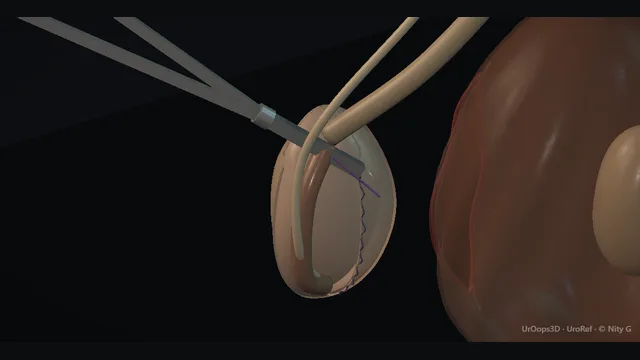
The operation shown is the Jaboulay repair: through an anterior scrotal incision the testis is delivered in its tunica, the fluid is drained, and the tunica is everted and closed with a continuous suture behind the testis and cord, loose enough for a little finger to pass along the cord.

Diathermy and haemostasis follow, in a dependent scrotum where gravity works against haemostasis at every layer.

Core tier FRCS genital surgery, scrotal surgery Interactive 3D

Operative visualisation

Schematic scene preview: Hydrocele repair
Preparing the 3D anatomy…

UrOops3D · UroRef · © Nity G

Click me next Section 2 of 8 Anatomy The structures this operation works between, and the one it must not cross.

The structures in the scene

Hydrocele repair: how the structures relateAnterior approach through the hemiscrotum, skin to testis. Dartos layer: blood drains downhill into it Dependent dead space (danger). Dartos layer: haemostase each layer before the next Tunica vaginalis, fluid bulging anteriorly (danger). Tunica vaginalis, fluid bulging anteriorly: opened, drained, then everted round the testis Everted edges, sutured behind testis and cord. Tunica vaginalis, fluid bulging anteriorly: a careless opening under tension injures it Testis and epididymis, posterior (danger). Testis and epididymis, posterior: closed behind both: a little finger must pass Spermatic cord, entering the top of the tunica (danger). Spermatic cord, entering the top of the tunica: patent in children: repair at the ring Processus vaginalis.Scrotal skinTestis and cord!Dartos layer!Dependent deadspaceTunica vaginalis, fluidbulging anteriorlyEverted edges,sutured behindtestis and cord!Testis and epididymis,posterior!Spermatic cord, enteringthe top of the tunicaProcessusvaginalisblood drainsdownhill into ithaemostase each layer beforethe nextopened, drained,then everted roundthe testisa careless opening undertension injures itclosed behind both: a littlefinger must passpatent in children:repair at the ringTargetDangerLandmarkContext
Anterior approach through the hemiscrotum, skin to testis. Schematic, not to scale; relationships only.
  • The hydrocele occupies the front of the hemiscrotum: testis and epididymis sit posteriorly against the sac wall, fluid bulging anteriorly, which is why the anterior approach reaches the sac through its fullest, safest part and why the testis is at risk only when the opening is careless.
  • Once the testis is delivered in its tunica and the fluid drained, the opened tunica is turned inside out so its cut edges meet behind the testis, and the cord enters at the top of that closure: the continuous suture runs behind the testis and cord, and a little finger passed along the cord proves it is not strangled.
  • Everything sits at the bottom of the patient, which is the whole trouble with haemostasis: blood and lymph drain downhill into exactly the space the operation has just created.
  • ›Tunica vaginalis - the sac: visceral layer on the testis, parietal layer lining the scrotum, and the fluid between them. In the Jaboulay repair the parietal layer is opened and everted.
  • ›Testis + epididymis - lie against the posterior wall of the sac; examined before the operation, protected while it is opened, and inspected once it is drained.
  • ›Spermatic cord - enters the top of the tunica; the closure passes behind it and must leave room for a little finger to pass along it.
  • ›Dartos layer - the vascular layer under the scrotal skin; its vessels are the everyday bleeding of the incision.
  • ›Everted tunica edges - the cut edges, brought together behind the testis and cord with a continuous suture; they bleed and are diathermised before the testis goes back.
  • ›Processus vaginalis - the foetal canal; patent in the communicating paediatric hydrocele, which is why that operation is at the ring, not the sac.
Click me next Section 3 of 8 Steps The operation in order, with the landmark that guards each move.

Operative steps

  1. Examination: transilluminate + examine the testis The swelling is examined standing and lying: it transilluminates brightly and can be got above, which defines it as scrotal and separate from a cord or inguinal swelling. The testis is then palpated against the posterior wall of the sac, because the examination is the one assessment the operation must not replace. Fluid that does not transilluminate, a collection that recurs rapidly after aspiration, or a new hydrocele in an older man is imaged before repair, since a tumour presenting as a primary hydrocele is the classic trap.
    where it bites: the testis inside a hydrocele is the one examination the operation must not replace; a tumour presenting as a hydrocele is the classic trap.
  2. Anterior scrotal incision A vertical incision is made over the fullest, most anterior part of the sac and deepened through skin and dartos, each vessel diathermised as it is met. Taking haemostasis layer by layer from the first cut is the discipline of the operation: the scrotum is dependent, and any vessel left now becomes the haematoma of tomorrow. The incision is carried down until the shiny parietal layer of the tunica vaginalis presents in the wound.
    where it bites: the dartos vessels bleed freely in a dependent sac; haemostasis deferred to the end is the haematoma of tomorrow.
  3. Delivery of the testis in its tunica The parietal tunica is freed from the dartos around it, and the testis is delivered through the wound still inside its unopened tunica, the cord following it. Delivering the sac intact keeps the fluid under control and shows the whole tunica, the testis behind it and the cord at its upper end before anything is cut.
    where it bites: the cord runs into the top of the delivered sac; traction on the testis is traction on the cord.
  4. Opening + draining the tunica vaginalis The tunica vaginalis is picked up between forceps and opened cautiously over its anterior surface, clear of the testis, which lies posteriorly against the sac wall and is injured only by a careless entry. The fluid is drained and the cut edge is seized and controlled as it opens. With the sac empty, the testis and epididymis are inspected before the repair goes any further.
    where it bites: a careless entry into a tense sac is how the testis behind it is cut.
  5. Eversion of the tunica The opened tunica is everted: turned inside out around the testis, so that its secretory lining faces outwards against the dartos and both cut edges come to lie behind the testis and cord. In a very large sac the surplus tunica is trimmed first, with the cord protected, so that the everted cuff is not bulky.
    where it bites: trimming a large sac is done with the cord in view; the cord enters the top of the tunica and is easily caught.
  6. Continuous closure behind the testis and cord The everted edges are closed with a continuous suture behind the testis and cord, working from the lower pole upwards. The closure must not strangle the cord: at the top, a little finger should pass along the cord into the everted tunica. If it will not, the top throws are too tight and are taken out and re-run.
    where it bites: a closure pulled tight round the cord strangles it; the little finger alongside the cord is the check.
  7. Diathermy + haemostasis Diathermy and haemostasis follow while the testis is still delivered and the field can be seen: the everted edges, the tunica and every bleeding point are dealt with until the field is dry, because ooze only declares itself after the wound is closed. The dead space the operation leaves is dependent and fills by gravity alone, which is why the classic complication, the haematoma, is either made in this step or prevented here.
    where it bites: the classic complication of this operation is the haematoma, and it is made in this step or prevented here.
  8. Return, layered closure + support The testis is returned to the scrotum with the cord lying untwisted. The dartos is closed, then the skin, and a scrotal support dressing is applied with the scrotum elevated. Swelling and bruising settle over weeks, and the patient is warned that resolution is slow; persistent or returning fluid means the everted tunica has re-formed a cavity, and the testis is re-examined and imaged before any talk of repeat surgery.
    where it bites: a testis returned with a twisted cord undoes a good repair; check the lie before closing.
Click me next Section 4 of 8 Danger zones Where this operation bites, and why it bites there.
Critical relationship

Danger zones

The spatial relationships the animation is built to make automatic. Before you read each one, ask yourself: at this point in the operation, what is about to go wrong, and what is my first move? The save for each sits in the Complications tab.

  • RiskThe testis beneath the tense sac - posterior in the sac; injured by a careless opening of fluid under tension.
  • RiskThe cord at the top of the closure - the continuous suture runs behind it; pulled tight it strangles the cord, so a little finger must pass alongside.
  • RiskEverted edges and dartos vessels - the everyday bleeders of the operation; diathermised until dry before the testis goes back.
  • RiskThe dependent dead space - the space the fluid came from; gravity fills it with blood unless haemostasis and support close it.
Practise it Take me into the scene Opens the animation at a danger moment and asks you to predict the mechanism and your first move before it reveals.
Click me next Section 5 of 8 Complications The save for each one: the first move, the escalation trigger, the op-note line.

Complications and immediate saves

Trigger each of these live in the Animation tab, then read the save: the first move, the escalation trigger, and the line that goes in the op note.

Haematoma (the classic)

  • a tense, expanding scrotal haematoma in the first day after repair: a dependent scrotum, raw everted edges, and gravity doing the rest.
The save
  1. Assess severity first: a stable man with a static, moderate haematoma is observed with support and elevation. first move
  2. A small haematoma is a dressing problem; firm support, elevation and analgesia, with honest advice that resolution takes weeks.
  3. Examine for extension into the penile base or perineum and for skin threatening tension.
  4. An expanding or tense haematoma, skin compromise or haemodynamic upset → return to theatre for evacuation and haemostasis. escalation trigger
  5. Op note: haemostasis at each layer, support applied, and the post-operative examination findings if the haematoma was managed conservatively. document

Recurrence

  • the fluid returns over months: the everted tunica re-formed a cavity, the closure gave way, or the hydrocele was secondary to underlying testis pathology all along.
The save
  1. Re-examine the testis before anything else; a recurring or atypical hydrocele re-opens the question of underlying disease. first move
  2. Image the scrotum to confirm fluid and assess the testis before offering repeat surgery.
  3. Discuss options honestly: repeat repair, or aspiration with sclerotherapy for the frail patient in whom a second anaesthetic is the greater risk.
  4. Recurrence after technically sound repair, or bilateral new hydroceles → consider underlying causes and refer accordingly. escalation trigger
  5. Note: original technique, interval to recurrence, imaging findings and the chosen option with its rationale. document

Wound infection

  • a red, tender, discharging scrotal wound: skin folds and dependency make the scrotum an unforgiving wound environment.
The save
  1. Assess the wound and the man: spreading erythema, crepitus or systemic upset changes this from a dressing problem to an emergency. first move
  2. Most present as cellulitis or a small discharge and settle with antibiotics and review.
  3. Any collection is opened and drained; the scrotum does not tolerate pus under tension.
  4. Necrotising infection signs, however early they look → immediate surgical exploration, not observation. escalation trigger
  5. Note: wound appearance, organisms if cultured, treatment given and the review interval. document
Click me next Section 6 of 8 Troubleshooting When the case turns: the problem, then the next move.

Intra-operative troubleshooting

When the case turns: the problem, then the next move.

  • ActionClosure tight at the top, round the cord - take the top throws out and re-run them looser; a little finger must pass along the cord into the everted tunica before the testis goes back.
  • ActionVery large or thick-walled sac - trim the surplus tunica, with the cord in view, before eversion, so the everted cuff is not bulky; the edges are still closed behind the testis and cord.
  • ActionThin-walled sac, minimal dissection wanted - Lord's plication is the named alternative, not the operation shown here: radial plication sutures gather the opened sac behind the testis without mobilising it from the dartos.
  • ActionFluid does not transilluminate pre-operatively - image before the operation: blood, chylocele or tumour all present this way and each changes the plan.
  • ActionTestis abnormal on pre-operative imaging - do not simply repair the hydrocele: urological cancer pathways, tumour markers and an oncological plan come first.
  • ActionPaediatric hydrocele in a boy under two - observe, most resolve; beyond that age or with a patent processus confirmed, the operation is at the ring, not the sac.
Click me next Section 7 of 8 FRCS viva Say it out loud, then mark yourself against the model answer.

FRCS viva questions

The examiner stems this scene prepares you for. Try to answer out loud first, then reveal, and learn the discriminator, the detail that turns a pass into a strong pass.

QDescribe the Jaboulay repair of a hydrocele.reveal answer
  • The Jaboulay repair is an eversion operation for a primary hydrocele.
  • Through an anterior scrotal incision over the fullest part of the swelling, taken through skin and dartos with haemostasis at each layer, the parietal tunica is freed from the dartos and the testis is delivered in its unopened tunica.
  • The tunica is opened anteriorly, clear of the testis behind it, the fluid is drained, and the testis and epididymis are inspected.
  • The opened tunica is then everted, turned inside out around the testis, so its secretory lining faces outwards against the dartos, where the fluid it makes is absorbed rather than contained.
  • The cut edges now lie behind the testis and cord and are closed with a continuous suture behind them, from the lower pole upwards.
  • The closure must not strangle the cord: a little finger should pass along the cord into the everted tunica.
  • A very large sac has its surplus trimmed first, with the cord in view.
  • Diathermy and haemostasis follow while the testis is still delivered, and the testis is returned with the cord untwisted, the dartos and skin closed in layers and a scrotal support applied.
  • The named alternative is Lord's plication, which gathers the opened sac with radial plication sutures and no mobilisation from the dartos; it suits the thin-walled sac.
Strong pass

Say both

  • Stating that the closure runs behind the testis and cord and must admit a little finger along the cord
  • Naming Lord's plication as the alternative rather than the default
Examiner note
Stating that the closure runs behind the testis and cord and must admit a little finger along the cord, and naming Lord's plication as the alternative rather than the default.
QIs aspiration or surgical repair the right treatment for a primary hydrocele?reveal answer
  • Repair, for the fit man who wants a definitive answer; aspiration for the defined minority.
  • Aspiration of a primary hydrocele is simple and immediate, but it leaves the sac that made the fluid in place, so recurrence is the rule rather than the exception, often within months, and each aspiration carries a small risk of bleeding and infection into a space that communicates with nothing.
  • Aspiration with sclerotherapy improves durability by sticking the layers together, at the price of pain during and after the injection, and it preserves a role for the frail man in whom anaesthesia is the greater hazard, or the man who declines surgery after honest counselling.
  • Surgical repair, by Jaboulay eversion or, as the alternative, Lord's plication, neutralises the secretory sac and is the definitive operation, at the cost of an anaesthetic, a scrotal wound and the operation's own complications, of which haematoma is the classic.
  • Selection is therefore a conversation about physiology and fitness: the young and fit are offered repair;
  • the frail or refusing are offered aspiration with honest recurrence figures in words, a likely return of the swelling, rather than false permanence.
  • What is never acceptable is aspirating the undiagnosed hydrocele, because the testis beneath it must be assessed before any fluid is removed.
Strong pass

Say both

  • Framing aspiration as a deliberate choice for the unfit with recurrence stated as the expectation
  • Refusing aspiration as a substitute for diagnosis
Examiner note
Framing aspiration as a deliberate choice for the unfit with recurrence stated as the expectation, and refusing aspiration as a substitute for diagnosis.
QHow does the paediatric communicating hydrocele differ as an operation?reveal answer
  • It differs in anatomy, timing and site.
  • The paediatric hydrocele is usually a patent processus vaginalis, the foetal peritoneal canal along the cord, which allows peritoneal fluid to track down into the scrotum; it is a canal problem, not a sac problem.
  • That changes the timing: in infancy a proportion close spontaneously, so observation is legitimate in the first year or two, whereas the adult primary hydrocele never closes itself.
  • It changes the site: the operation is at the ring or the inguinal canal, ligating the patent processus where it leaves the peritoneum, often by a groin crease incision or laparoscopically, and the scrotal sac itself is left alone, because the scrotum did not cause the fluid.
  • Operating on the adult pattern, evacuating and everting the scrotal sac in a child, evacuates today's fluid while leaving the open canal that refills it, so recurrence is near-certain and the child has been given an operation for nothing.
  • The associations matter too: the same patent canal gives inguinal hernia, and the boy presenting with intermittent scrotal swelling that comes and goes with activity has exactly this anatomy.
  • The summary sentence is that the adult operation treats the container and the child's operation treats the connection.
Strong pass

Stating plainly that the paediatric operation is at the ring, not the sac, and that sac surgery in a child guarantees recurrence.

QWhen is a hydrocele a symptom rather than a diagnosis?reveal answer
  • Whenever the fluid is secondary to something else, and the role of the surgeon is to find that something before treating the swelling.
  • The categories run through the testis, the cord and the systemic picture.
  • Within the sac, a testicular tumour presents with a hydrocele in a proportion of cases, classically in the older man with a new, non-transilluminating or rapidly recurring collection;
  • this is why the testis is palpated, and imaged when it cannot be confidently examined, before any repair is offered, and why tumour markers are part of the workup once suspicion exists.
  • Epididymal disease, acute or chronic, can leave reactive fluid.
  • Along the cord, an encysted hydrocele of the cord and, more importantly, a hernia traversing a narrow neck can both masquerade.
  • After the pelvis, renal transplantation, retroperitoneal surgery and lymphatic disruption produce hydroceles by drainage failure.
  • Systemically, heart failure and, in the relevant geography, filariasis cause bilateral collections through lymphatic overload; a symmetrical hydrocele in a cardiac patient is a medical finding first.
  • The practical rule: examine the testis in every hydrocele, image the ones that do not transilluminate, recur after sound repair, appear new in an older man, or sit in a transplant or cardiac context, and treat the cause before the container.
Strong pass

Say both

  • Organising the secondary causes by anatomical compartment
  • Naming the new hydrocele in the older man as the tumour trap
Examiner note
Organising the secondary causes by anatomical compartment and naming the new hydrocele in the older man as the tumour trap.
Click me next Section 8 of 8 Evidence The guidance each of these answers is built on.

Evidence and guidance

Two minutes

Close the lap: recall it before you leave.

Say it aloud, then reveal. Reading is not remembering.

  1. 01 The relationship Say the critical spatial relationship out loud, then reveal. reveal
    Memory frame

    The hydrocele occupies the front of the hemiscrotum

    Why?
    The hydrocele occupies the front of the hemiscrotum: testis and epididymis sit posteriorly against the sac wall, fluid bulging anteriorly, which is why the anterior approach reaches the sac through its fullest, safest part and why the testis is at risk only when the opening is careless. Once the testis is delivered in its tunica and the fluid drained, the opened tunica is turned inside out so its cut edges meet behind the testis, and the cord enters at the top of that closure: the continuous suture runs behind the testis and cord, and a little finger passed along the cord proves it is not strangled. Everything sits at the bottom of the patient, which is the whole trouble with haemostasis: blood and lymph drain downhill into exactly the space the operation has just created.
  2. 02 The first move Haematoma (the classic). Your first move? reveal
    First move

    Assess severity first: a stable man with a static, moderate haematoma is observed with support and elevation.

  3. 03 The escalation trigger Haematoma (the classic). What makes you call for help? reveal
    Escalate when

    An expanding or tense haematoma, skin compromise or haemodynamic upset → return to theatre for evacuation and haemostasis.

  4. 04 The discriminator What turns a pass into a strong pass? reveal
    Strong pass

    Say both

    • Stating that the closure runs behind the testis and cord and must admit a little finger along the cord
    • Naming Lord's plication as the alternative rather than the default
    Examiner note
    Stating that the closure runs behind the testis and cord and must admit a little finger along the cord, and naming Lord's plication as the alternative rather than the default.
  5. 05 The pitfall Where does this operation bite first? reveal
    Pitfall

    The testis beneath the tense sac

    Why?
    posterior in the sac; injured by a careless opening of fluid under tension.

Revision practice with self-marked answers, not an assessment of clinical competence. Kept in this browser only; it does not sync between devices.