
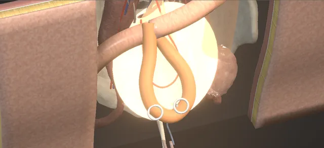
UrOops3D, danger-zone atlas

Schematic model on the open pelvis atlas.

Operative animation, Ileal ureter

Ileal ureter: total replacement of a long ureteric loss

The operation for the ureter that cannot be repaired by anything shorter: a segment of ileum isolated on its pedicle, the bowel rejoined above it, the segment laid isoperistaltically in the right retroperitoneum and joined to the renal pelvis above and the bladder below, both joins tested before the bladder is closed.

The lesson is where the segment comes from, which way round it goes, what keeps it alive, and who should not have one.

Advanced tier FRCS reconstruction, ureter, urinary diversion Interactive 3D

Operative visualisation

Schematic scene preview: Ileal ureter
Preparing the 3D anatomy…

UrOops3D · UroRef · © Nity G

How to use this module. Play through the nine steps, or use Back and Next. Watch the arcades light up against the mesentery, the segment turn to lie with its oral end at the kidney, both joins made, and the saline test. The triggers show a dusky segment, a bowel leak, an upper join leak and the metabolic price.

Click me next Section 2 of 8 Anatomy The structures this operation works between, and the one it must not cross.

The structures in the scene

Ileal ureter: how the structures relateThe right retroperitoneum after the colon is reflected, renal pelvis to bladder. Ileal segment on its pedicle, isoperistaltic: oral end sewn to the pelvis Renal pelvis: oblique pyelotomy, upper join. Ileal segment on its pedicle, isoperistaltic: aboral end drawn through into the bladder Posterolateral bladder window, lower join. Ileal segment on its pedicle, isoperistaltic: lives on it; no twist, no tension Mesenteric pedicle and its arcade (danger). Ileal segment on its pedicle, isoperistaltic: lies lateral to the cava on the psoas Cava medial, duodenum above (danger). Ileal segment on its pedicle, isoperistaltic: window placed clear of both Right ureteric orifice and trigone (danger). Ileal segment on its pedicle, isoperistaltic: replaces it; the scar is left where it lies Scarred native ureter, divided and tied.Ileal segment on itspedicle, isoperistalticRenal pelvis:oblique pyelotomy,upper joinPosterolateralbladder window,lower join!Mesenteric pedicleand its arcade!Cava medial,duodenum above!Right uretericorifice and trigoneScarred nativeureter, divided andtiedoral end sewn tothe pelvisaboral end drawnthrough into thebladderlives on it; notwist, no tensionlies lateral to thecava on the psoaswindow placed clearof bothreplaces it; thescar is left whereit liesTargetDangerLandmarkContext
The right retroperitoneum after the colon is reflected, renal pelvis to bladder. Schematic, not to scale; relationships only.
  • With the right colon reflected, the right retroperitoneum is one field from the renal pelvis to the brim: the cava medial, the psoas as the floor, the scarred ureter on it and the gonadal vessels crossing in front of it.
  • The ileal segment lies in that field lateral to the cava, isoperistaltic, its oral end at the renal pelvis and its aboral end through a posterolateral window in the bladder, its mesentery coming from the root without a twist.
  • The bowel anastomosis sits above the segment, never over it.
  • ›Right kidney and renal pelvis - the upper end of the replacement: Gerota opened over the pelvis, the pelvis mobilised with its vessels, an oblique pyelotomy for the join.
  • ›Scarred native ureter - pale, narrow, without peristalsis from below the junction to the brim; divided below the junction and its stump tied, the rest left where it lies.
  • ›Line of Toldt and the right colon - the white line is incised and the caecum and ascending colon reflected medially; the whole right retroperitoneum opens behind them.
  • ›Inferior vena cava - medial to the right ureter along the whole route; the segment lies lateral to it on the psoas.
  • ›Duodenum - at the top of the reflection, above the renal pelvis; named and kept out of the field.
  • ›Terminal ileum and its mesentery - the segment comes from here, a comfortable distance from the ileocaecal valve, on a pedicle chosen by transillumination of the arcades.
  • ›Ileocolic arcade - the watershed between the ileocolic and right colic supply; the segment is taken short of it so the caecum and the remaining ileum keep their blood.
  • ›Bladder: anterior wall and posterolateral wall - a midline cystotomy between stays for the view; a full-thickness posterolateral window for the ileal end.
  • ›Right ureteric orifice and trigone - seen through the cystotomy and kept clear of the window and the stitches.
  • ›Gonadal vessels - cross anterior to the ureter on the psoas; the segment is laid clear of them.
Click me next Section 3 of 8 Steps The operation in order, with the landmark that guards each move.

Operative steps

  1. Open and expose An adult with good renal function and a long right ureteric loss, from below the pelviureteric junction to the brim, after radiation or an iatrogenic stricture: too long for a psoas hitch or a Boari flap, too long to join end to end. Midline transperitoneal laparotomy, the bowel examined for inflammatory disease or radiation enteritis. The white line of Toldt is incised with diathermy and the caecum and ascending colon are reflected medially, which opens the right retroperitoneum from the kidney to the brim. The scarred ureter is identified and inspected: pale, narrow, without peristalsis. The cava lies medial to it and the duodenum above the field.
    where it bites: the cava is medial to the ureter and the duodenum sits at the top of the reflection; both are named before anything is divided.
  2. Choose the segment The small bowel is delivered and the terminal ileum followed back from the caecum. A loop is held up against the light so the vascular arcades show through the mesentery, and a segment of distal ileum is chosen, long enough for the gap and a comfortable distance from the ileocaecal valve, where a good arcade runs to it. Marking stitches go in at both ends. The segment is measured against the gap from the renal pelvis to the bladder and not guessed.
    where it bites: the arcade is chosen by transillumination before any bowel is cut; the ileocolic arcade and the last of the ileum are kept.
  3. Isolate on its pedicle An avascular mesenteric window is opened at each end of the chosen segment with a vessel-sealing device, incising and sealing the mesentery between the arcades without touching the main vessels. The bowel is divided at each end with a linear cutting stapler, so both ends of the segment and both ends of the remaining ileum are stapled shut. The segment now sits free on its mesenteric pedicle and is placed caudal to the mesenteric defect.
    where it bites: a window cut too deep toward the root takes the pedicle with it and the segment dies; the windows stay between the arcades.
  4. Restore continuity The antimesenteric corners of the two remaining ileal ends are trimmed off the staple lines. One limb of the linear cutting stapler goes into each end, the bowel is turned so the antimesenteric walls face each other, and the stapler is fired: a side-to-side, functional end-to-end anastomosis. Four clamps hold the ends; the common opening is closed across with a TA stapler and the staple line checked and reinforced. The mesenteric window is closed with a running stitch so nothing can herniate through it. The anastomosis lies above the segment, which is kept caudal to it.
    where it bites: mesentery caught in the stapler bleeds or devascularises the join; the window is closed against an internal hernia.
  5. Open, wash and orient The staple lines are resected off both ends of the segment and the lumen is irrigated with saline until it is clear of mucus and enteric content. The segment is then oriented isoperistaltically, its oral end toward the kidney and its aboral end toward the bladder, and positioned high in the right retroperitoneum with its mesentery untwisted and without tension. The mesenteric vessels are checked again once it lies in place.
    where it bites: isoperistaltic means the oral end goes to the kidney; a twist or tension on the pedicle is found now, not after the joins are made.
  6. Ileopyelostomy Gerota's fascia is opened over the renal pelvis by sharp dissection and the pelvis is dissected and mobilised with its vessels preserved. Stay sutures go in above and below and the pelvis is incised obliquely, for a length equal to the opening of the bowel. The native ureter is divided below the junction and its stump tied (the book is silent on the native ureter; this is drawn as the default). The ileopyelostomy is completed with interrupted absorbable sutures in one layer, mucosa to mucosa, with the mesentery lying easy.
    where it bites: the renal hilar vessels run behind the pelvis; the pelvis is mobilised with its own vessels kept, and the ileal mesentery must not pull on the join.
  7. Bladder window Two stay sutures go into the mid-anterior bladder wall and a full-thickness stab incision is made between them with diathermy, then extended cranially and caudally into a midline cystotomy. The catheter comes out to improve the view and the bladder is inspected: both orifices in their normal position, no tumour, stone, foreign body or diverticulum. A full-thickness segment of the posterolateral right bladder wall is excised to make the window. A Babcock clamp is passed through it from inside, the distal ileal end is grasped and drawn into the bladder.
    where it bites: the right ureteric orifice and the trigone are seen and kept clear of the window and of every bite that follows.
  8. Ileovesical join and test The ileovesical anastomosis is sewn in two layers of interrupted absorbable sutures: an outer seromuscular row and an inner mucosal row. It is a refluxing, direct join by design. With the interposition complete, the ileal ureter is irrigated with saline from above to confirm a watertight repair at both ends; only then is the midline cystotomy closed in two layers, a new urethral catheter passed, and the bladder irrigated to confirm a patent, watertight closure.
    where it bites: watertight at both joins before the bladder is closed; a leak found afterwards is a return to theatre.
  9. Drain and close The abdomen and pelvis are examined for bleeding and haemostasis is secured. A closed suction drain is placed in the retroperitoneum beside the joins, not on them, and brought out through a separate stab incision. The colon is returned over the segment without kinking it, the small bowel laid back, the field irrigated with warm saline and inspected, and the midline closed in layers. One continuous lumen now runs from the renal pelvis to the bladder.
    where it bites: the colon and small bowel are returned without kinking or compressing the segment or its pedicle.
Click me next Section 4 of 8 Danger zones Where this operation bites, and why it bites there.
Critical relationship

Danger zones

The spatial relationships the animation is built to make automatic. Before you read each one, ask yourself: at this point in the operation, what is about to go wrong, and what is my first move? The save for each sits in the Complications tab.

  • RiskThe mesenteric pedicle - a window cut too deep, a twist, or tension on the pedicle devascularises the segment; the arcade is chosen against the light and the pedicle is checked in its final position.
  • RiskThe renal hilum behind the pelvis - the renal pelvis is dissected with its vessels preserved; the hilar artery and vein lie just behind the plane of the pyelotomy.
  • RiskThe cava and the duodenum - medial and superior limits of the reflection; neither is mobilised, both are named.
  • RiskThe right orifice and the trigone - the posterolateral window is placed clear of the orifice; a bite into the trigone obstructs the native side.
  • RiskThe patient who should not have one - impaired renal function, known intestinal disease, a bladder that does not empty: the selection error that ends in acidosis, mucus and a failing kidney.
Practise it Take me into the scene Opens the animation at a danger moment and asks you to predict the mechanism and your first move before it reveals.
Click me next Section 5 of 8 Complications The save for each one: the first move, the escalation trigger, the op-note line.

Complications and immediate saves

Trigger each of these live in the Animation tab, then read the save: the first move, the escalation trigger, and the line that goes in the op note.

Dusky segment

  • the segment turns dusky on its route: a pedicle twisted during orientation or a mesenteric window cut too deep toward the root.
The save
  1. Stop. Untwist the pedicle, lay the segment back in the wound, warm it and wait. first move
  2. Judge colour, capillary return and bleeding from the cut edges, not the colour alone.
  3. A segment still dusky after untwisting and warming is discarded and another segment taken on a different arcade. escalation trigger
  4. The new segment is chosen by transillumination again, further from the first windows.
  5. Op note: pedicle checked, orientation, the colour of the segment at closure and the reason a segment was discarded if one was. document

Bowel anastomotic leak

  • sepsis, ileus and a changed drain days later: the ileoileal anastomosis has leaked.
The save
  1. Resuscitate, cultures and antibiotics, nil by mouth, and a CT with oral contrast. first move
  2. A contained collection is drained; the joins of the ileal ureter are left alone if they are dry.
  3. Peritonitis or an unstable patient returns to theatre now for the anastomosis. escalation trigger
  4. The ileal ureter is inspected at that laparotomy, not revised unless it is the source.
  5. Op note: the anastomotic technique, the integrity check and the mesenteric window closure as they were done. document

Leak at the ileopyelostomy

  • urine in the drain and a urinoma in the gutter: the upper join has leaked, from tension on the mesentery or an opening cut too short.
The save
  1. Catheter on free drainage so the bladder is never full; the drain stays and its creatinine is measured. first move
  2. Imaging of the collection; a drain into it if it is large.
  3. Sepsis, a rising creatinine or a leak that persists means a nephrostomy to divert the kidney. escalation trigger
  4. Most close with the kidney diverted and the bladder empty; a revision is a late decision.
  5. Op note: the watertight test and its result at both joins. document

Hyperchloraemic acidosis and mucus

  • ileum in the urinary tract reabsorbs chloride and ammonium and secretes mucus; acidosis, mucus plugging and a falling renal function follow, worst in the kidney that was impaired to begin with.
The save
  1. Bicarbonate and electrolytes checked, and bicarbonate replaced; the bladder emptied regularly so urine does not sit on the ileum. first move
  2. Mucus is washed out; the catheter is irrigated in the early days and the patient taught to drink.
  3. Falling renal function or an acidosis that will not correct escalates to the nephrologist and a reconstruction review. escalation trigger
  4. Impaired preoperative renal function was the selection error: an autotransplant was the alternative.
  5. Op note and the record before it: the preoperative renal function, and why an ileal ureter was chosen over the alternatives. document
Click me next Section 6 of 8 Troubleshooting When the case turns: the problem, then the next move.

Intra-operative troubleshooting

When the case turns: the problem, then the next move.

  • ActionThe segment will not reach the renal pelvis without tension - take more length from the ileum rather than pull; a segmental replacement to healthy native ureter is the book's variation when the gap is short, and an ileocalicostomy to the lower pole when the pelvis is intrarenal or scarred.
  • ActionThe renal pelvis is scarred or intrarenal - excise the inferior renal parenchyma and join the proximal ileum to the lower pole calyx: an ileocalicostomy (named in the book, not drawn here).
  • ActionThe bladder window sits too close to the right orifice - choose the window on the posterolateral wall above and lateral to the orifice, seen from inside through the cystotomy; the trigone is never part of the window.
  • ActionDoubt about the arcade - transilluminate again before cutting; the watershed between the ileocolic and right colic supply is where the windows must not go.
  • ActionBoth ureters need replacing - a longer segment in a reverse L configuration with end-to-end and end-to-side ileopyelostomies (book variation, out of scope here); a reconfigured segment is named only.
  • ActionWho should not have one - impaired renal function or known intestinal disease: an autotransplant, a nephrectomy with a normal contralateral kidney, or a long-term nephrostomy or stent are the alternatives named before the incision.
Click me next Section 7 of 8 FRCS viva Say it out loud, then mark yourself against the model answer.

FRCS viva questions

The examiner stems this scene prepares you for. Try to answer out loud first, then reveal, and learn the discriminator, the detail that turns a pass into a strong pass.

QA patient has a long right ureteric stricture after pelvic radiotherapy, from the junction to the brim. What are the options and when is an ileal ureter the right one?reveal answer
  • The options run from the shortest repair that would bridge the gap to the replacements.
  • A psoas hitch, a Boari flap with a hitch and an end-to-end ureteroureterostomy are all too short for a loss from the junction to the brim, and radiated tissue makes them worse.
  • A transureteroureterostomy is excluded by a radiated field and a scarred contralateral risk.
  • That leaves replacement: an ileal interposition, a renal autotransplant, long-term nephrostomy or stent drainage, or nephrectomy if the other kidney is normal.
  • The EAU urological trauma guideline lists ileal interposition for long-segment loss and advises against it when renal function is impaired or there is known intestinal disease, with autotransplantation as the alternative.
  • So I would offer an ileal ureter to a patient with good renal function, usable ileum and a bladder that empties, and autotransplantation to the patient with impaired function or bad bowel.
Strong pass

Say both

  • Naming the contraindications (impaired renal function, intestinal disease) and autotransplantation as the alternative
  • Not just the operation
Examiner note
Naming the contraindications (impaired renal function, intestinal disease) and autotransplantation as the alternative, not just the operation.
QTalk me through an ileal ureter.reveal answer
  • Midline transperitoneal.
  • I incise the line of Toldt and reflect the caecum and ascending colon medially to open the right retroperitoneum, and I identify the scarred ureter with the cava medial to it.
  • I deliver the small bowel, transilluminate the mesentery and choose a segment of distal ileum a comfortable distance from the ileocaecal valve on a good arcade, long enough for the gap.
  • I open a mesenteric window at each end with a sealing device, divide the bowel with a linear cutting stapler, and restore continuity above the segment with a stapled side-to-side anastomosis, closing the common opening with a TA and the mesenteric window with a running stitch.
  • I resect the staple lines, irrigate the segment and lay it isoperistaltically in the retroperitoneum, oral end to the kidney, mesentery untwisted.
  • I open Gerota, mobilise the renal pelvis with its vessels, make an oblique pyelotomy between stays and sew the ileopyelostomy with interrupted absorbable sutures.
  • I open the bladder in the midline between stays, inspect it, excise a full-thickness posterolateral window, draw the distal ileum in with a Babcock and sew a two-layer ileovesical anastomosis.
  • I irrigate the ileal ureter to confirm it is watertight, close the cystotomy in two layers over a new catheter, leave a retroperitoneal drain, return the colon and close.
Strong pass

Say all three

  • Transillumination before dividing
  • Isoperistaltic orientation with the oral end to the kidney
  • The watertight test before the bladder is closed
Examiner note
Transillumination before dividing, isoperistaltic orientation with the oral end to the kidney, and the watertight test before the bladder is closed.
QWhat keeps the segment alive, and what kills it?reveal answer
  • Its mesenteric pedicle.
  • The segment is chosen where a good arcade runs to it, seen by transillumination, and the windows are opened at each end between the arcades, never through them and never deep toward the root where the main vessels run.
  • Once it is isolated it is placed caudal to the mesenteric defect, and when it is oriented I check that the mesentery is untwisted and lies without tension along the whole route before either join is made.
  • What kills it is a window cut too deep, a twist introduced when it is turned, or tension when it is pulled to reach the pelvis.
  • A dusky segment on the table is untwisted and warmed; if it does not recover it is discarded and another segment taken.
Strong pass

Say both

  • Stating that the arcade is chosen by transillumination before any bowel is cut
  • That the pedicle is checked in the final position
Examiner note
Stating that the arcade is chosen by transillumination before any bowel is cut, and that the pedicle is checked in the final position.
QWhat are the late problems, and how do you follow the patient up?reveal answer
  • The anastomoses can stricture, at the pelvis or the bladder, and either can leak early;
  • the ileum reabsorbs chloride and ammonium and secretes mucus, so a hyperchloraemic metabolic acidosis, mucus plugging, pyelonephritis and stones all follow, and renal function can fall.
  • Armatys and colleagues reported anastomotic stricture and fistula as the late complications, with acidosis in a minority, and found that pre-existing renal impairment predicted a poor outcome.
  • So I follow renal function and bicarbonate, check electrolytes at every visit, image the kidney for obstruction once the drain and catheter are out and again later, treat infection early, and keep the bladder emptying well, because urine that sits on ileum is what drives the acidosis.
  • The record states the preoperative renal function and why this operation was chosen over an autotransplant.
Strong pass

Linking the metabolic complication to the selection criteria: impaired preoperative renal function is the error, not a surprise.

Click me next Section 8 of 8 Evidence The guidance each of these answers is built on.

Evidence and guidance

Operative Dictations in Urologic Surgery
Armenakas NA, Fracchia JA, Golan R. Ileal ureter. Chapter 50, pp 181-185. Wiley, 2019
Indications, the seven essential steps, the variations (hand-sewn anastomosis, ileocalicostomy, segmental replacement, bilateral reverse L, nephrostomy) and the complication list. Book numbers kept off screen: a 25 to 35 cm segment 15 to 20 cm from the ileocaecal valve, the pyelotomy at 45 degrees, an 8 cm cystotomy, a 1 cm stab, 18 and 20 Fr catheters, the suture gauges. The right-side paragraph names a window in the descending mesocolon, which reads as the left route; this module draws no mesocolic window.
Open guideline
EAU Guidelines
Urological Trauma: ureteral trauma, long segment ureteral injury (section 4.2.4.b.2.c), read 8 October 2026
Ileal interposition for long-segment loss; avoided with impaired renal function or known intestinal disease; autotransplantation as the alternative. No figures taken on screen.
Open guideline
Journal of Urology
Armatys SA, Mellon MJ, Beck SDW, Koch MO, Foster RS, Bihrle R. Use of ileum as ureteral replacement in urological reconstruction. J Urol 2009;181:177-181. doi 10.1016/j.juro.2008.09.019 (PMC2667902), read 8 October 2026
Indications (radiation, iatrogenic injury, retroperitoneal fibrosis), caution in preoperative renal impairment, isoperistaltic refluxing configuration as the default, anastomotic stricture and fistula as late complications, hyperchloraemic acidosis. Series figures kept off screen.
Open guideline

Curriculum. Supports learning relevant to the ISCP Urology Curriculum syllabus topics Ureteric reconstruction and Urinary diversion. Not a workplace-based assessment and not evidence of competence.

Two minutes

Close the lap: recall it before you leave.

Say it aloud, then reveal. Reading is not remembering.

  1. 01 The relationship Say the critical spatial relationship out loud, then reveal. reveal
    Memory frame

    With the right colon reflected, the right retroperitoneum is one field from the renal pelvis to the brim: the cava medial, the psoas as the floor, the scarred ureter on it and the gonadal vessels crossing in front of it.

    Why?
    The ileal segment lies in that field lateral to the cava, isoperistaltic, its oral end at the renal pelvis and its aboral end through a posterolateral window in the bladder, its mesentery coming from the root without a twist. The bowel anastomosis sits above the segment, never over it.
  2. 02 The first move Dusky segment. Your first move? reveal
    First move

    Stop.

    More detail
    Untwist the pedicle, lay the segment back in the wound, warm it and wait.
  3. 03 The escalation trigger Dusky segment. What makes you call for help? reveal
    Escalate when

    A segment still dusky after untwisting and warming is discarded and another segment taken on a different arcade.

  4. 04 The discriminator What turns a pass into a strong pass? reveal
    Strong pass

    Say both

    • Naming the contraindications (impaired renal function, intestinal disease) and autotransplantation as the alternative
    • Not just the operation
    Examiner note
    Naming the contraindications (impaired renal function, intestinal disease) and autotransplantation as the alternative, not just the operation.
  5. 05 The pitfall Where does this operation bite first? reveal
    Pitfall

    The mesenteric pedicle

    Why?
    a window cut too deep, a twist, or tension on the pedicle devascularises the segment; the arcade is chosen against the light and the pedicle is checked in its final position.

Revision practice with self-marked answers, not an assessment of clinical competence. Kept in this browser only; it does not sync between devices.