UrOops3D, danger-zone atlas

Schematic model.

Operative animation, Inguinal lymph node dissection

Inguinal lymph node dissection: radical open groin dissection for penile cancer

A radical open dissection of one groin for penile cancer.

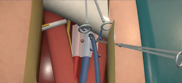
Skin flaps are raised with their fat on them, the node-bearing fat of the femoral triangle is taken off the fascia lata from below upward with the superficial and deep nodes, the great saphenous vein is kept, and the sartorius is turned over the femoral vessels.

The lessons are the flap that dies when it is raised too thin, the femoral vein under the deep dissection, and the lymph that collects under the flap.

Advanced tier FRCS urological oncology, penile cancer Interactive 3D

Operative visualisation

Schematic scene preview: Inguinal lymph node dissection
Preparing the 3D anatomy…

UrOops3D · UroRef · © Nity G

How to use this module. Play through the nine steps, or use Back and Next. Watch the flaps lift with their fat, the packet peel up off the floor round the saphenous vein, the deep nodes leave the femoral vein and the sartorius swing over the vessels. The triggers show flap necrosis, a lymphocele and a vein tear.

Click me next Section 2 of 8 Anatomy The structures this operation works between, and the one it must not cross.

The structures in the scene

Inguinal lymph node dissection: how the structures relateThe node-bearing packet of the groin, and what lies round it. Node-bearing fat of the femoral triangle: lies under them; raised in the plane at Scarpa’s fascia Skin flaps with their fat (danger). Node-bearing fat of the femoral triangle: lies between them; the lateral and medial limits Sartorius and adductor longus. Node-bearing fat of the femoral triangle: surrounds it; kept, its tributaries divided Great saphenous vein (danger). Node-bearing fat of the femoral triangle: lies on them; the deep dissection is on the vein Femoral vein and artery (danger). Node-bearing fat of the femoral triangle: lateral to the artery; the sheath is not opened there Femoral nerve (danger). Node-bearing fat of the femoral triangle: its upper end, medial to the vein at the canal Node of Cloquet, then the pelvic nodes.Node-bearing fat of thefemoral triangle!Skin flaps withtheir fatSartorius andadductor longus!Great saphenous vein!Femoral vein andartery!Femoral nerveNode of Cloquet,then the pelvicnodeslies under them;raised in the planeat Scarpa’s fascialies between them;the lateral andmedial limitssurrounds it; kept,its tributariesdividedlies on them; thedeep dissection ison the veinlateral to theartery; the sheathis not opened thereits upper end,medial to the veinat the canalTargetDangerLandmarkContext
The node-bearing packet of the groin, and what lies round it. Schematic, not to scale; relationships only.
  • Seen from in front with the head at the top, the inguinal ligament runs from the anterior superior iliac spine down and medially to the pubic tubercle.
  • Below it lies the femoral triangle, with the sartorius as its lateral border and the adductor longus as its medial border, meeting at the apex.
  • In its floor the femoral vein lies medially, the artery lateral to it and the femoral nerve lateral again and deeper.
  • The great saphenous vein comes up the medial thigh and joins the femoral vein at the fossa ovalis, and the superficial nodes lie in the fat over the triangle, clustered round that junction.
  • ›Inguinal ligament - from the anterior superior iliac spine to the pubic tubercle; the upper limit of the template and the base of the femoral triangle.
  • ›Scarpa’s fascia - the membranous layer of the subcutaneous tissue; the flaps are raised in the plane at it, with Camper’s fat kept on the skin.
  • ›Superficial inguinal nodes - in the fat between Scarpa’s fascia and the fascia lata, in five zones round the saphenofemoral junction.
  • ›Fascia lata - the deep fascia of the thigh; incised over the border muscles and lifted with the packet.
  • ›Sartorius - the lateral border of the femoral triangle; divided at its origin and turned over the vessels at the end.
  • ›Adductor longus - the medial border of the femoral triangle; the transposed sartorius is sewn to its edge.
  • ›Great saphenous vein - runs up the medial thigh and through the fossa ovalis to join the femoral vein; preserved here, its tributaries divided.
  • ›Femoral vein - in the femoral sheath, medial to the artery; the deep nodes lie beside and medial to it.
  • ›Femoral artery - lateral to the vein in the sheath, at the midpoint of the inguinal ligament.
  • ›Femoral nerve - lateral to the artery, outside the sheath, under the iliacus fascia; the reason the sheath is not opened laterally.
  • ›Node of Cloquet - the highest deep node, medial to the femoral vein at the femoral canal; marks the transition from the groin to the pelvis.
  • ›External oblique aponeurosis and spermatic cord - exposed superiorly and medially by the superior flap; the aponeurosis holds the sutures of the transposed sartorius.
Click me next Section 3 of 8 Steps The operation in order, with the landmark that guards each move.

Operative steps

  1. Mark the template Supine, with pneumatic compression devices on both legs, the hips abducted and externally rotated and the knees flexed. The abdomen, genitalia and both thighs are prepared and a urethral catheter is passed. The upper limit of the dissection is marked from the anterior superior iliac spine to the pubic tubercle; the line is carried down the thigh medially and further laterally, and the two lower ends are joined.
    where it bites: the template is the clearance: the superior limit is the inguinal ligament, the lateral and medial limits run down the thigh, and the skin over all of it will be lifted as flaps.
  2. Incision below the ligament The skin of the medial thigh is incised a short distance below the inguinal ligament and parallel to it, across the template. The incision is carried down to Scarpa’s fascia, which gives the plane for the flaps.
    where it bites: the depth of this cut sets the thickness of both flaps.
  3. Raise the flaps Superior and inferior skin flaps are developed by sharp and blunt dissection in the plane at Scarpa’s fascia, below Camper’s fascia, out to the marked limits, keeping the superficial vasculature with them. Skin hooks and padded retractors protect the flaps. Superiorly the dissection exposes the external oblique aponeurosis; medially, the spermatic cord.
    where it bites: the flap lives on the fat and the vessels in it: a flap raised thin, or handled roughly at its edge, necroses.
  4. Open the fascia lata The superficial nodes lie in the fat beneath, in five zones round the saphenofemoral junction. The fascia lata is incised longitudinally over the sartorius and over the adductor longus, which are the lateral and medial borders of the deep dissection and meet at the apex of the femoral triangle.
    where it bites: the femoral vessels lie under the fascia between the two muscles; the incisions are made over muscle, not over the vessels.
  5. Free the packet from below The dissection begins inferiorly. The lower end of the lymphatic packet is freed, lifted off its deep margin, divided and secured with clips or ties. The packet is then dissected upward over the floor of the triangle, the fascia lata coming with it, with the sartorius laterally, the adductor longus medially and the femoral vessels between them.
    where it bites: the femoral artery and vein run up the middle of the floor and come into view from below as the packet is lifted.
  6. Keep the saphenous vein The great saphenous vein is identified medially and preserved. The tributaries that run from it into the packet are dissected, clipped or tied and divided, and the surrounding nodes are freed. The packet is dissected up over the saphenofemoral junction toward the ligament.
    where it bites: the saphenous vein is divided only when the nodes are massive; its tributaries are taken flush enough to leave the main trunk intact.
  7. Deep nodes and Cloquet At the fossa ovalis the femoral sheath is incised, exposing the femoral artery and vein. The dissection continues medially to isolate the node of Cloquet at the femoral canal and the deep nodes beside the vein and between the vessels. Lateral dissection of the femoral sheath is avoided. The superficial and deep packet is secured at the ligament, divided and removed en bloc, and sent to pathology labelled by side.
    where it bites: the femoral vein is the wall of this dissection; the femoral nerve lies lateral to the artery, outside the sheath.
  8. Transpose the sartorius Haemostasis and lymphostasis are secured with clips and diathermy throughout, and the femoral artery and the femoral and saphenous veins are inspected for bleeding or injury. The sartorius is freed bluntly on its lateral side, divided at its origin at the anterior superior iliac spine, turned medially over the femoral vessels and sutured to the external oblique aponeurosis and the edge of the adductor longus.
    where it bites: the vessels supplying the sartorius enter its medial side; dissecting it medially devascularises the muscle meant to cover the vessels.
  9. Drain, check the flaps, close The wound is irrigated. A drain is placed beneath the flap and brought out through a separate inferior stab incision. The same dissection is done on the other side. Both flaps are inspected for viability before closure; each wound is closed in two layers, Scarpa’s fascia and then the skin with staples, and a pressure dressing is applied.
    where it bites: a flap that is dusky now will be necrotic in a week; it is assessed while it can still be trimmed.
Click me next Section 4 of 8 Danger zones Where this operation bites, and why it bites there.
Critical relationship

Danger zones

The spatial relationships the animation is built to make automatic. Before you read each one, ask yourself: at this point in the operation, what is about to go wrong, and what is my first move? The save for each sits in the Complications tab.

  • RiskFemoral artery and vein - under the sheath in the middle of the triangle; the deep dissection works on the wall of the vein.
  • RiskFemoral nerve - lateral to the artery, outside the sheath; the sheath is not dissected laterally.
  • RiskGreat saphenous vein - kept unless the nodes are massive; its tributaries are clipped and divided without narrowing the trunk.
  • RiskSkin-flap blood supply - carried in the fat of the flap; a thin flap, a rough edge or a tight closure loses it.
  • RiskExternal iliac and obturator vessels during pelvic dissection - the next basin when a pelvic dissection follows; the node of Cloquet marks its lower end.
Practise it Take me into the scene Opens the animation at a danger moment and asks you to predict the mechanism and your first move before it reveals.
Click me next Section 5 of 8 Complications The save for each one: the first move, the escalation trigger, the op-note line.

Complications and immediate saves

Trigger each of these live in the Animation tab, then read the save: the first move, the escalation trigger, and the line that goes in the op note.

Skin flap necrosis

  • the edge of a flap dies, usually the inferior flap at the suture line, because its blood supply was lost when the flap was raised thin or handled roughly; the wound breaks down over the vessels.
The save
  1. Look at the whole wound: how much skin is dead, whether there is pus beneath it, and how close the femoral vessels lie under the breakdown. first move
  2. Treat infection and let a dry edge demarcate; then debride the dead skin back to bleeding tissue.
  3. Dress the wound and let a small defect heal from its base, with the sartorius still covering the vessels.
  4. Exposed vessels, a large defect or bleeding from the wound: plastic surgical help for a graft or a myocutaneous flap, and vascular advice now. escalation trigger
  5. Record the extent of the necrosis, what was debrided and how the vessels are covered. document

Lymphocele or lymphorrhoea

  • lymph from the divided channels of the leg collects under the flap or leaks through the wound; the drain under the flap is the first defence, and its output is the measure.
The save
  1. Keep the drain and measure what it gives each day; the drain comes out only when its output has fallen. first move
  2. A collection after the drain is out is confirmed by ultrasound and aspirated, or drained again if it recurs.
  3. Compression of the groin and the leg, and elevation, while the lymphatics settle.
  4. A collection that is infected, keeps refilling or threatens the flap: back to the operating surgeon. escalation trigger
  5. Record the drain output, the volume aspirated and whether the fluid was infected. document

Femoral vein injury

  • the femoral vein is torn while the node of Cloquet or the deep nodes are freed from its wall; vascular injury and bleeding are listed complications of the operation.
The save
  1. Press on the tear with a finger or a swab and keep pressing. Do not clamp or diathermy blindly at the vein. first move
  2. Ask for suction, a second assistant, blood and the anaesthetist’s attention while the pressure is held.
  3. Get control above and below the tear before the pressure is released, then define the hole.
  4. Call a vascular surgeon to theatre to repair the vein; the dissection waits until it is safe. escalation trigger
  5. Record where the tear was, how it was controlled and repaired, the blood loss and the plan for thromboprophylaxis. document
Click me next Section 6 of 8 Troubleshooting When the case turns: the problem, then the next move.

Intra-operative troubleshooting

When the case turns: the problem, then the next move.

  • ActionMassive or matted nodes round the saphenous junction - the operative text ligates the saphenous vein in massive lymphadenopathy; the vein is not dissected out of tumour.
  • ActionA flap edge looks dusky at the end - its viability can be tested with intravenous fluorescein and a Wood’s lamp, as the operative text allows; dead-looking skin is trimmed before closure.
  • ActionNot enough soft tissue to cover the vessels - the operative text closes the defect with a rectus abdominis myocutaneous flap; this is planned with a plastic surgeon.
  • ActionImpalpable groins and the question is only whether there is microscopic disease - a superficial or modified dissection is the operative text’s variation; the guideline prefers dynamic sentinel node biopsy where it is available.
  • ActionDeep venous thrombosis - a listed risk after groin dissection: pneumatic compression devices are on from the start, and thromboprophylaxis and early mobilisation follow.
  • ActionBrisk bleeding from a tributary near the junction - pressure, then a clip on the tributary itself, flush enough to leave the saphenous trunk open.
Click me next Section 7 of 8 FRCS viva Say it out loud, then mark yourself against the model answer.

FRCS viva questions

The examiner stems this scene prepares you for. Try to answer out loud first, then reveal, and learn the discriminator, the detail that turns a pass into a strong pass.

QA man has a pT2 squamous cell carcinoma of the penis and impalpable groins. How do you stage his groins, and what is a modified inguinal lymphadenectomy?reveal answer
  • He is high risk: the EAU-ASCO guideline treats every tumour that is T1b or higher as high risk and offers surgical nodal staging to all of them, because only invasive staging finds micrometastases and delay costs survival.
  • I would first examine both groins and do an ultrasound with fine-needle aspiration of any abnormal node.
  • If that is negative I would offer dynamic sentinel node biopsy, done with a radioactive tracer and blue dye;
  • in experienced centres the guideline quotes a sensitivity of 92 to 96 per cent, a false-negative rate of 4 to 8 per cent and complications in 6 to 14 per cent.
  • A positive sentinel node means a completion dissection on that side.
  • If sentinel node biopsy is not available and referral is not feasible, or he prefers it, I would offer an inguinal dissection.
  • The modified template uses a shorter incision, with no dissection lateral to the femoral artery or below the fossa ovalis, and keeps the saphenous vein.
  • It therefore spares the lateral and the two inferior of Daseler’s five zones, which is reasonable because the first nodes involved lie in the superomedial and central zones and solitary spread to the inferior zones has not been seen.
  • Even so, the guideline gives the modified dissection complication rates of 35 to 49 per cent and false-negative rates of 15 to 20 per cent.
Strong pass

Say both

  • Knowing that sentinel node biopsy is the first choice for the impalpable groin
  • Being able to say exactly what the modified template leaves behind
Examiner note
Knowing that sentinel node biopsy is the first choice for the impalpable groin and being able to say exactly what the modified template leaves behind.
QThe same man returns with a mobile palpable node in the right groin. What now?reveal answer
  • I would confirm it is tumour with an image-guided biopsy before treating it, because the guideline puts the rate of metastasis in palpable nodes at only 45 to 80 per cent.
  • I would stage the pelvis and look for distant disease with FDG PET-CT or CT of the chest and abdomen.
  • For a single mobile node the guideline offers an ipsilateral fascia-sparing dissection or an open radical dissection sparing the saphenous vein if possible;
  • for bulky or bilateral mobile nodes, an open radical dissection on that side, or neoadjuvant cisplatin and taxane chemotherapy in a fit patient.
  • A fixed mass or pelvic nodes call for chemotherapy first and consolidation surgery in responders.
  • The other groin, if impalpable, is staged by sentinel node biopsy.
  • The guideline asks that the inguinal and pelvic nodal surgery is complete within three months of diagnosis.
Strong pass

Proving the node is tumour first, and matching the operation to whether the nodes are mobile, bulky or fixed.

QTalk me through a radical inguinal lymphadenectomy.reveal answer
  • With compression devices on, he lies supine with the hips abducted and externally rotated and the knees flexed, and I give antibiotics and pass a catheter.
  • I mark the upper limit from the anterior superior iliac spine to the pubic tubercle, carry it down the thigh medially and further laterally, and join the ends.
  • I make a transverse incision a short distance below the inguinal ligament and go down to Scarpa’s fascia.
  • I raise the superior and inferior flaps in that plane, keeping the fat and its vessels on the skin, up over the ligament to the aponeurosis and down to the lower limit.
  • I incise the fascia lata over the sartorius and over the adductor longus, which are my lateral and medial borders.
  • I start inferiorly: I free the end of the packet, divide it between clips and lift it off its deep margin, and work upward.
  • I find the saphenous vein medially and keep it, clipping and dividing its tributaries.
  • At the fossa ovalis I open the femoral sheath over the vessels and go medially, taking the node of Cloquet and the deep nodes between the vessels, and I do not open the sheath laterally, where the femoral nerve lies.
  • I take the packet out en bloc and label it by side.
  • After haemostasis and lymphostasis I divide the sartorius at its origin and turn it medially over the vessels, sewing it to the aponeurosis and the adductor edge.
  • I place a drain through a separate stab, check that the flaps are viable, and close Scarpa’s fascia and the skin.
Strong pass

Say all three

  • Raising the flaps in the right plane
  • Starting the dissection from below
  • Keeping off the lateral side of the sheath
Examiner note
Raising the flaps in the right plane, starting the dissection from below, and keeping off the lateral side of the sheath.
QWhat are the complications of this operation, and how do you reduce them?reveal answer
  • The operative text lists bleeding, infection, vascular injury, flap necrosis, lymphocele and lymphoedema, and I would add deep venous thrombosis.
  • The numbers are large.
  • In the series of Bevan-Thomas and colleagues from the M.D.
  • Anderson Cancer Center, 106 dissections in 53 patients, 58 per cent of patients had a complication, with skin edge necrosis in 8 per cent against 45 to 62 per cent in earlier series, and leg oedema in 23 per cent.
  • In the Netherlands Cancer Institute cohort of Stuiver and colleagues, 237 dissections in 163 patients, 58 per cent of procedures had at least one wound complication: infection in 43 per cent, seroma in 24 per cent and skin flap problems in 16 per cent.
  • Body mass index and sartorius transposition were the factors most strongly associated with the more serious ones, with an odds ratio of 2.64 for the transposition in the univariable analysis.
  • The guideline quotes 21 to 55 per cent for contemporary radical series.
  • To reduce them I would use sentinel node biopsy where it is enough, a linear transverse incision, flaps with their fat on them, saphenous preservation, careful clipping of lymphatics, a drain, compression devices and thromboprophylaxis, and antibiotics.
  • I transpose the sartorius as the operative text does, knowing that its benefit is now questioned.
Strong pass

Quoting a real series with its denominators, and knowing that sartorius transposition is associated with more, not fewer, wound complications in one large cohort.

QThe pathology shows three positive nodes in the right groin. Does he need a pelvic dissection?reveal answer
  • Yes.
  • Spread runs from the superficial to the deep inguinal nodes and then to the pelvic nodes on the same side; pelvic disease does not occur without inguinal disease, and crossover to the other pelvis is rare.
  • The guideline offers an open or minimally invasive prophylactic ipsilateral pelvic lymphadenectomy when three or more inguinal nodes are involved on one side or when there is extranodal extension, because those features carry a much higher rate of pelvic metastasis.
  • It asks that the nodal surgery is complete within three months of diagnosis.
  • The operative text’s pelvic dissection runs from the common iliac artery above to the node of Cloquet below, between the genitofemoral nerve laterally and the ureter medially, down to the obturator nerve, so the external iliac and obturator vessels are the danger structures.
  • If the pelvic nodes are involved I would discuss adjuvant treatment in the multidisciplinary meeting.
Strong pass

Say both

  • Giving the guideline’s trigger of three nodes on one side or extranodal extension
  • Knowing the pelvic template is ipsilateral
Examiner note
Giving the guideline’s trigger of three nodes on one side or extranodal extension, and knowing the pelvic template is ipsilateral.
Click me next Section 8 of 8 Evidence The guidance each of these answers is built on.

Evidence and guidance

Operative Dictations in Urologic Surgery
Armenakas NA, Fracchia JA, Golan R. Inguinal lymph node dissection. Chapter 20. Wiley, 2019
The indication (penile or scrotal wall cancer), the nine essential steps and their order, the template dictation followed here (positioning, the template from the anterior superior iliac spine to the pubic tubercle carried down the thigh, the incision below the ligament to Scarpa’s fascia, flaps below Camper’s fascia, the fascia lata incised over the sartorius and the adductor longus, the dissection from below, the saphenous vein preserved and its tributaries ligated, the femoral sheath opened at the fossa ovalis, the node of Cloquet and the deep nodes, no lateral dissection of the sheath, the packet en bloc, sartorius transposition, a drain through a separate stab, the flaps inspected, a two-layer closure and a pressure dressing), the variations (a superficial or modified dissection, the saphenous vein ligated in massive lymphadenopathy, fluorescein and a Wood’s lamp, a rectus abdominis myocutaneous flap) and the complication list. The chapter’s distances and suture materials are left off the page. The saves, the deep venous thrombosis line and the tributary line are general surgical practice; the chapter lists complications without their management. The scene is schematic: one groin, a neutral hip, the instruments at true size.
Open guideline
EAU-ASCO
EAU-ASCO Guidelines on Penile Cancer, update March 2026: sections 5.2.2, 5.2.3, 5.4 and 6.2, read 18 October 2026
Tumours T1b or higher are high risk; surgical staging is offered to all of them, by dynamic sentinel node biopsy, with ILND if it is not available or the patient prefers; sentinel node biopsy sensitivity 92 to 96%, false-negative 4 to 8%, complications 6 to 14%. The modified template: a shorter incision, no dissection lateral to the femoral artery or caudal to the fossa ovalis, the saphenous vein preserved; complications 35 to 49%, false-negative 15 to 20%. Palpable nodes are metastatic in 45 to 80%, to be confirmed by image-guided biopsy and staged with FDG PET-CT or CT. Daseler’s five zones round the saphenofemoral junction; the first nodes involved lie in the superomedial and then the central zones, with no solitary spread to the two inferior zones. cN1: fascia-sparing or open radical ILND sparing the saphenous vein if possible; cN2: open radical ILND sparing the saphenous vein if possible, or neoadjuvant chemotherapy. Radical ILND complications 21 to 55% in contemporary series. Prophylactic ipsilateral pelvic dissection for three or more involved inguinal nodes on one side or extranodal extension; nodal surgery complete within three months.
Open guideline
Journal of Urology
Bevan-Thomas R, Slaton JW, Pettaway CA. Contemporary morbidity from lymphadenectomy for penile squamous cell carcinoma: the M.D. Anderson Cancer Center experience. J Urol 2002;167:1638-1642
Abstract read 18 October 2026. 106 lymphadenectomy procedures in 53 patients: 66 prophylactic (superficial), 28 therapeutic, 12 palliative. 41 minor and 19 major complications; complications in 31 of 53 patients (58%). Skin edge necrosis 8% against 45 to 62% in three earlier series; leg oedema 23% against 50% in an earlier report from the same centre. Major complications included skin flap necrosis needing a graft and deep venous thrombosis; one death from sepsis after a palliative dissection.
Open guideline
European Urology
Stuiver MM, Djajadiningrat RS, Graafland NM, Vincent AD, Lucas C, Horenblas S. Early wound complications after inguinal lymphadenectomy in penile cancer: a historical cohort study and risk-factor analysis. Eur Urol 2013;64:486-492
Abstract read 18 October 2026. 237 ILNDs in 163 patients at the Netherlands Cancer Institute, 2003 to 2012. One complication or more in 58% of procedures; wound infection 43%, seroma 24%, skin-flap problems 16%. For grade 2 or worse complications, univariable odds ratios 1.66 for body mass index and 2.64 for sartorius muscle transposition; in the reduced multivariable model 2.12 for transposition (P = 0.06) and 1.76 for body mass index.
Open guideline

Curriculum. Supports learning relevant to the ISCP Urology Curriculum syllabus topic Penile cancer. Not a workplace-based assessment and not evidence of competence.

Two minutes

Close the lap: recall it before you leave.

Say it aloud, then reveal. Reading is not remembering.

  1. 01 The relationship Say the critical spatial relationship out loud, then reveal. reveal
    Memory frame

    Seen from in front with the head at the top, the inguinal ligament runs from the anterior superior iliac spine down and medially to the pubic tubercle.

    Why?
    Below it lies the femoral triangle, with the sartorius as its lateral border and the adductor longus as its medial border, meeting at the apex. In its floor the femoral vein lies medially, the artery lateral to it and the femoral nerve lateral again and deeper. The great saphenous vein comes up the medial thigh and joins the femoral vein at the fossa ovalis, and the superficial nodes lie in the fat over the triangle, clustered round that junction.
  2. 02 The first move Skin flap necrosis. Your first move? reveal
    First move

    Look at the whole wound: how much skin is dead, whether there is pus beneath it, and how close the femoral vessels lie under the breakdown.

  3. 03 The escalation trigger Skin flap necrosis. What makes you call for help? reveal
    Escalate when

    Exposed vessels, a large defect or bleeding from the wound: plastic surgical help for a graft or a myocutaneous flap, and vascular advice now.

  4. 04 The discriminator What turns a pass into a strong pass? reveal
    Strong pass

    Say both

    • Knowing that sentinel node biopsy is the first choice for the impalpable groin
    • Being able to say exactly what the modified template leaves behind
    Examiner note
    Knowing that sentinel node biopsy is the first choice for the impalpable groin and being able to say exactly what the modified template leaves behind.
  5. 05 The pitfall Where does this operation bite first? reveal
    Pitfall

    Femoral artery and vein

    Why?
    under the sheath in the middle of the triangle; the deep dissection works on the wall of the vein.

Revision practice with self-marked answers, not an assessment of clinical competence. Kept in this browser only; it does not sync between devices.