
UrOops3D, danger-zone atlas
Operative animation, Kidney transplantation

Recipient kidney transplantation into the iliac fossa

A kidney from a living or deceased donor placed extraperitoneally in the iliac fossa, its vein and artery joined to the external iliac vessels, reperfused, and its ureter tunnelled into the bladder over a stent.

Every step protects two things that cannot be seen: the graft vessels and the blood supply of the ureter. This page is the match, the bench, the Gibson incision, the iliac vessels, the two anastomoses, reperfusion, the Lich-Gregoir and the first night.

Advanced tier FRCS transplant and specialist urology, renal transplantation Interactive 3D

Operative visualisation

Schematic scene preview: Recipient kidney transplantation
Preparing the 3D anatomy…

UrOops3D · UroRef · © Nity G

Click me next Section 2 of 8 Anatomy The structures this operation works between, and the one it must not cross.

The structures in the scene

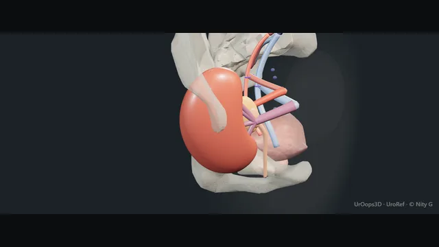
Kidney transplantation: how the structures relateRight iliac fossa, peritoneum retracted medially, the graft on the psoas. Graft renal vein and artery on its patch: join its side; a twist or kink thromboses External iliac vein and artery (danger). Graft renal vein and artery on its patch: the alternative when the external is diseased Internal iliac artery. Graft renal vein and artery on its patch: tie or clip; diathermy makes a lymphocele Lymphatics over the vessels (danger). Graft renal vein and artery on its patch: the only supply to the transplant ureter The golden triangle (danger). Graft renal vein and artery on its patch: swept medially; the graft lies behind it The peritoneum. Graft renal vein and artery on its patch: ureter tunnels into its anterolateral wall Filled bladder and JJ stent.Graft renal vein andartery on its patchExternal iliac veinand arteryInternal iliacartery!Lymphatics over thevessels!The golden triangleThe peritoneumFilled bladder andJJ stentjoin its side; atwist or kinkthrombosesthe alternativewhen the externalis diseasedtie or clip;diathermy makes alymphocelethe only supply tothe transplantureterswept medially; thegraft lies behinditureter tunnels intoits anterolateralwallTargetDangerLandmarkContext
Right iliac fossa, peritoneum retracted medially, the graft on the psoas. Schematic, not to scale; relationships only.
  • The kidney lies in the right iliac fossa on the psoas with its hilum facing medially toward the external iliac vessels, which run from the bifurcation above to the inguinal ligament below with the peritoneum retracted medially.
  • The renal vein joins the side of the external iliac vein and the artery on its patch joins the side of the external iliac artery just above it, and the ureter, kept short, curves down from the lower pole to enter the anterolateral wall of the filled bladder below.
  • The golden triangle of fat sits between the lower pole, the pelvis and the ureter.
  • ›The external iliac vein and artery - exposed from the bifurcation to the inguinal ligament; the vein more superficial on the right, which is why the right fossa is used first.
  • ›The internal iliac artery - the alternative for an end-to-end arterial anastomosis when the external iliac is diseased or the renal artery is short.
  • ›The lymphatics over the vessels - tied or clipped, not coagulated; divided lymph becomes a lymphocele.
  • ›The golden triangle - fat between the lower pole, the pelvis and the ureter, carrying the only blood supply to the transplant ureter; left intact on the bench.
  • ›The graft vessels - the renal vein, lengthened with cava on a right deceased donor kidney, and the artery on its Carrel patch with any accessory artery reconstructed on the bench.
  • ›The peritoneum - swept medially and held so the kidney lies extraperitoneally on the psoas and iliacus.
  • ›The bladder and the stent - filled to bring the anterolateral wall up; the ureter enters through a detrusor tunnel over a JJ stent for four to six weeks.
Click me next Section 3 of 8 Steps The operation in order, with the landmark that guards each move.

Operative steps

  1. Who: the recipient and the match End-stage kidney disease, on dialysis or approaching it with an eGFR under about 15, in a patient fit for a general anaesthetic and lifelong immunosuppression, assessed for cardiac disease, malignancy, infection, the iliac vessels on imaging where there is calcification, and a lower urinary tract that stores and empties. Blood group compatible or a planned incompatible programme, HLA typed, and a negative crossmatch on the day. A living donor kidney is scheduled; a deceased donor kidney arrives at any hour after brain death or circulatory death, with its cold ischaemia time on the clock, and the recipient is called in, dialysed if the potassium is high, consented and cross-matched while the kidney is on the bench. Induction immunosuppression is given before the clamps go on.
  2. The kidney on the bench In ice, in a bowl of cold perfusion solution, the kidney is prepared: the perinephric fat removed from the capsule except at the lower pole and the hilum, where the fat between the lower pole, the renal pelvis and the ureter, the golden triangle, is left untouched because it carries the whole blood supply of the transplant ureter from the renal artery. The renal vein is cleared and, on a right kidney from a deceased donor, lengthened with a cuff of vena cava; the artery is identified on its Carrel patch of aorta, any accessory artery joined to the main or kept on the patch, and small branches ligated. The kidney is flushed and checked for leaks and for any lump, and the ureter is kept long and left alone.
    where it bites: the transplant ureter has one blood supply, from the renal artery through the hilar and periureteric fat; the golden triangle is never dissected.
  3. The Gibson incision and the extraperitoneal space The right iliac fossa for a first transplant, because the external iliac vein is longer and more superficial on the right; the left for a second, or where the right vessels are diseased. A curved Gibson incision from two fingers above the anterior superior iliac spine to the midline above the pubis, through the external oblique, internal oblique and transversus, the inferior epigastric vessels divided between ties, the round ligament divided in a woman and the spermatic cord preserved and retracted medially in a man. The peritoneum is swept medially with the fingers and a self-retaining retractor holds it, so that the kidney will sit extraperitoneally on the psoas and iliacus with the peritoneum medial to it.
  4. The external iliac vessels The external iliac artery and vein are exposed from the bifurcation to the inguinal ligament, the lymphatics that cross them ligated or clipped rather than coagulated, because divided lymph makes a lymphocele that compresses the vein and the ureter. The vein is mobilised first, its tributaries ligated, until a segment long enough for a side clamp is free; the internal iliac artery is identified in case it is needed end to end. The artery is palpated for plaque and a soft segment chosen for the anastomosis. Systemic heparin is given by unit protocol before the clamps go on, and the position of the kidney is rehearsed so that the vessels lie without kinking when it is finally placed.
  5. The venous anastomosis The kidney comes to the field wrapped in ice slush, held by the assistant; the anastomosis time is the second warm ischaemia and is kept to about thirty minutes. Side-biting clamps on the external iliac vein, a venotomy the length of the renal vein, flushed with heparinised saline. The renal vein is joined end to side with 5-0 or 6-0 Prolene, two stay sutures at the apices and a running suture down each wall from inside then outside, the kidney orientated so that when it lies in the fossa the vein is not twisted or stretched. The clamps stay on. A vein that is too short on a right kidney has been lengthened on the bench with cava, not stretched here.
  6. The arterial anastomosis The external iliac artery is clamped proximally and distally, an arteriotomy made with a blade and enlarged with a punch to a circle, and the renal artery on its Carrel patch joined end to side with 6-0 Prolene, running, the patch sewn to the arteriotomy with the same care at the heel and the toe where the stenosis will form if a bite is too big. A living donor artery has no patch and is spatulated. An end-to-end anastomosis to the internal iliac artery with interrupted sutures is the alternative for a short artery or a diseased external iliac. The clamps are prepared for release in order: distal artery, proximal artery, vein.
    where it bites: a bite too large at the heel or the toe of the arterial anastomosis is a stenosis; the arteriotomy is a punched circle, not a slit.
  7. Reperfusion The venous clamps come off first, then the distal arterial clamp, then the proximal, and the kidney is watched: it should pink evenly and become firm within a minute, and urine may appear at the ureter within minutes in a living donor kidney. The anaesthetist has the patient filled, with the central venous pressure and the blood pressure kept up, mannitol or furosemide given by protocol, and the potassium and acidosis of the flushed preservation solution watched. Bleeding points at the hilum and the anastomoses are taken with fine sutures under vision, not diathermy near the vessels. A kidney that stays blue and soft has a problem with inflow or outflow that is found now: a kinked vein, a twisted artery, a clamp injury or an intimal flap.
    where it bites: a graft that does not pink and firm up on release is a vascular problem to be fixed at once; a kidney left blue is a kidney lost.
  8. The ureter: Lich-Gregoir over a stent The bladder is filled with saline through the catheter so that the dome and the anterolateral wall rise into the wound. The ureter is cut to the shortest length that reaches without tension, keeping the periureteric fat, and spatulated. A two to three centimetre incision through the detrusor to the mucosa on the anterolateral wall, the mucosa freed on each side and opened at the distal end, and the ureter sewn to the mucosa with interrupted or running 5-0 or 6-0 PDS or Monocryl over a JJ stent passed to the renal pelvis. The detrusor is closed over the ureter with two or three interrupted sutures to make a tunnel that prevents reflux, without constricting it. The stent comes out at four to six weeks.
    where it bites: a long ureter is a distal end far from its only blood supply; the ureter is cut short, and the distal centimetre is the part that necroses.
  9. Closure and the first night The kidney is placed in the fossa with the vessels lying without kink, checked once more for perfusion and for bleeding, and a drain left beside it. The muscles are closed in layers with looped PDS so that the graft is not compressed, and the skin closed. The first night is hourly urine output with fluid replaced millilitre for millilitre, potassium and haemoglobin checked, the catheter kept draining, and a duplex ultrasound within the first day or immediately if the urine stops. Anuria in a kidney that was passing urine is a catheter problem, a vascular problem or a leak, in that order of speed, and the duplex is done that hour. Immunosuppression continues, and the first creatinine tells whether the kidney is working or is delayed.
Click me next Section 4 of 8 Danger zones Where this operation bites, and why it bites there.
Critical relationship

Danger zones

The spatial relationships the animation is built to make automatic. Before you read each one, ask yourself: at this point in the operation, what is about to go wrong, and what is my first move? The save for each sits in the Complications tab.

  • RiskThe iliac vessels and their lymphatics - a diseased artery is a dissection or a stenosis, a torn vein is a haemorrhage, and lymph divided with diathermy is a lymphocele.
  • RiskThe graft renal artery and vein - a twist, a kink, a stretched vein or a bite at the heel; thrombosis in the first days is a graft lost.
  • RiskThe ureteric blood supply - from the renal artery alone through the hilar and periureteric fat; stripped on the bench or cut long, the distal ureter necroses and leaks.
  • RiskThe bladder, bowel and native ureter - a small defunctioned bladder that will not fill, peritoneum breached with bowel behind it, and the native ureter that can be mistaken for the transplant ureter.
Practise it Take me into the scene Opens the animation at a danger moment and asks you to predict the mechanism and your first move before it reveals.
Click me next Section 5 of 8 Complications The save for each one: the first move, the escalation trigger, the op-note line.

Complications and immediate saves

Trigger each of these live in the Animation tab, then read the save: the first move, the escalation trigger, and the line that goes in the op note.

Renal vein or artery thrombosis

  • the kidney that stops making urine in the first days, or never starts: a kinked or twisted vessel, a stretched vein, an intimal flap, a hypotensive night or a hypercoagulable recipient.
The save
  1. Check the catheter and flush it, then a duplex ultrasound within the hour; absent flow or reversed diastolic flow is theatre now. first move
  2. Tell the transplant consultant and the anaesthetist; the window for salvage is measured in minutes to an hour or two.
  3. Re-explore: the vessels inspected for kink, twist and thrombus, a thrombectomy and revision of the anastomosis if the kidney is salvageable.
  4. A kidney that is black, soft and does not reperfuse after thrombectomy: graft nephrectomy, and the patient back to dialysis. escalation trigger
  5. Op note: the time the urine stopped, the duplex findings, the vessel involved, what was done and the outcome for the graft. document

Haemorrhage

  • a falling haemoglobin and a swelling wound in the first hours from the anastomosis, the hilum or a bench-ligated branch, or later from a mycotic aneurysm or a biopsy.
The save
  1. Resuscitate, cross-match, reverse any heparin and tell the transplant surgeon; a tense wound over a graft is theatre. first move
  2. An ultrasound for the haematoma and the perfusion of the graft; a compressed vein thromboses.
  3. Re-explore, evacuate the haematoma and take the bleeding point with a fine suture under vision.
  4. Bleeding from an infected anastomosis or a mycotic aneurysm: vascular help, ligation of the graft vessels and graft nephrectomy if the iliac artery is at risk. escalation trigger
  5. Op note: the source, the sutures, the transfusion, and whether the graft was perfused at the end. document

Ureteric leak, necrosis and stricture

  • urine in the drain in the first two weeks from a necrotic distal ureter or the anastomosis, and months later a hydronephrosis with a rising creatinine from a stricture at the same place.
The save
  1. Keep the catheter draining, send the drain fluid for creatinine, and ultrasound the graft for a collection and hydronephrosis. first move
  2. A nephrostomy and an antegrade study to define the leak or the stricture; a stent antegrade if it passes.
  3. A small leak with a stent and a catheter is given time; a short stricture is dilated or incised and stented.
  4. A necrotic ureter, a large leak or a recurrent stricture: reoperation with reimplantation, a psoas hitch, or the native ureter to the transplant pelvis. escalation trigger
  5. Note: the level and length, the nephrostomy and stent with their dates, and the reconstruction planned. document

Lymphocele

  • a collection of lymph beside the graft weeks after the operation, from lymphatics divided over the iliac vessels: pressure on the vein and the ureter, a swollen leg, a rising creatinine, and infection if it is aspirated.
The save
  1. Ultrasound to size the collection and see the ureter and the iliac vein; a small asymptomatic lymphocele is left alone. first move
  2. Aspirate once under ultrasound for fluid creatinine to distinguish lymph from urine, and for culture.
  3. A lymphocele obstructing the ureter or the vein: a drain, or sclerotherapy through it, with the recurrence rate explained.
  4. A recurrent or large lymphocele: laparoscopic or open marsupialisation into the peritoneum, keeping the ureter and the vessels in view. escalation trigger
  5. Note: the size, its effect on the ureter and vein, the fluid creatinine and the drainage plan. document

Delayed graft function

  • the kidney that needs dialysis in the first week after a deceased donor transplant: ischaemia-reperfusion injury, a long cold time, a marginal donor, or a surgical problem hiding behind it.
The save
  1. Exclude the surgical causes first: catheter patent, duplex for flow and resistive index, no leak or collection on ultrasound. first move
  2. Dialyse for potassium, fluid and uraemia by the nephrologist; avoid hypotension and nephrotoxins.
  3. A biopsy at about a week to distinguish tubular necrosis from rejection and adjust immunosuppression.
  4. A kidney with no function at two to three weeks and a biopsy showing infarction or cortical necrosis: the graft is lost and is removed if it is symptomatic. escalation trigger
  5. Note: the cold and warm ischaemia times, the duplex findings, the biopsy and the dialysis schedule. document
Click me next Section 6 of 8 Troubleshooting When the case turns: the problem, then the next move.

Intra-operative troubleshooting

When the case turns: the problem, then the next move.

  • ActionThe renal vein is too short to reach - lengthen it on the bench with a cuff of cava on a deceased donor kidney; mobilise the iliac vein higher; do not stretch the graft vein or the kidney.
  • ActionThe external iliac artery is calcified where the anastomosis was planned - palpate for a soft segment, or go end to end onto the internal iliac artery; a clamp on plaque is a dissection.
  • ActionThe kidney is mottled and soft on reperfusion - check the vein for kink and the artery for twist, take the clamps off again in order, listen with the Doppler; a spasmed artery is given papaverine and time, a flap is revised.
  • ActionThe bladder will not fill - a small defunctioned bladder needs patience with the saline; if it truly will not rise, a longer incision, or an open ureteroneocystostomy through the bladder.
  • ActionUrine stops on the first night - flush the catheter, then duplex within the hour; a vascular cause is fixed in theatre, a leak is drained, a clot is washed out.
  • ActionThe leg on the side of the graft is cold after closure - pulses and a Doppler at once; a clamp injury, dissection or embolus in the external iliac artery needs the vascular surgeon now.
Click me next Section 7 of 8 FRCS viva Say it out loud, then mark yourself against the model answer.

FRCS viva questions

The examiner stems this scene prepares you for. Try to answer out loud first, then reveal, and learn the discriminator, the detail that turns a pass into a strong pass.

QDescribe the steps of a standard extraperitoneal kidney transplant.reveal answer
  • With the kidney prepared on the bench in ice, its fat cleared except the golden triangle at the hilum and lower pole, the vein lengthened with cava on a right deceased donor kidney and the artery on its Carrel patch, I would make a Gibson incision in the right iliac fossa, divide the inferior epigastric vessels and the round ligament, preserve the spermatic cord, and sweep the peritoneum medially.
  • I would expose the external iliac vein and artery from the bifurcation to the inguinal ligament, ligating rather than coagulating the lymphatics.
  • After systemic heparin I would clamp the vein, make a venotomy and join the renal vein end to side with running 5-0 or 6-0 Prolene, then clamp the artery, punch an arteriotomy and join the patch end to side with 6-0 Prolene.
  • I would release the venous clamps, then the arterial, and watch the kidney pink and firm.
  • I would fill the bladder and perform a Lich-Gregoir extravesical ureteroneocystostomy: the ureter cut short and spatulated, joined to the mucosa with fine absorbable suture over a JJ stent, and the detrusor closed over it as a tunnel.
  • I would place the kidney without kinking the vessels, leave a drain, close in layers and manage the first night with hourly urine output and a duplex.
Strong pass

Say all three

  • The golden triangle preserved on the bench
  • The lymphatics ligated with the reason
  • The clamps released in order with the kidney watched
Examiner note
The golden triangle preserved on the bench, the lymphatics ligated with the reason, and the clamps released in order with the kidney watched.
QA transplant recipient stops passing urine six hours after the operation. What do you do?reveal answer
  • Anuria in a kidney that was working is a surgical emergency until proven otherwise, and the causes in order of speed are the catheter, the vessels and a leak.
  • I would go to the patient, check the observations and the fluid balance, and flush the catheter to exclude a clot, which is the commonest and simplest cause.
  • If the catheter is clear I would arrange a duplex ultrasound within the hour, not the next morning: absent arterial or venous flow, or reversed diastolic flow, means thrombosis and the patient goes to theatre immediately, because the graft survives minutes to an hour or two, not longer.
  • I would also look on the ultrasound for a perinephric collection suggesting bleeding or a urine leak, and check the drain output and its creatinine.
  • I would inform the transplant consultant and the anaesthetist as soon as the duplex is requested, so that theatre is ready.
  • In theatre the vessels are inspected for kink, twist and thrombus, a thrombectomy performed and the anastomosis revised if the kidney reperfuses, and a graft that stays black is removed.
  • If the duplex is normal and there is no leak, this is delayed graft function, managed with the nephrologist by dialysis as needed and a biopsy at about a week.
Strong pass

Say both

  • The duplex requested within the hour with the finding that sends the patient to theatre
  • The surgeon and anaesthetist told before the result
Examiner note
The duplex requested within the hour with the finding that sends the patient to theatre, and the surgeon and anaesthetist told before the result.
QWhat are the urological complications of kidney transplantation and how are they prevented?reveal answer
  • The urological complications are leak, obstruction and reflux, and most trace to the blood supply of the transplant ureter, which comes only from the renal artery through the hilar and periureteric fat.
  • Leak occurs in the first two weeks, usually from necrosis of the distal ureter or the anastomosis, and presents with urine in the drain, a collection, a falling output and a rising creatinine.
  • Stricture occurs months later at the same distal segment, or from a tunnel that is too tight, and presents as hydronephrosis with a rising creatinine, since the graft is denervated and does not hurt.
  • Prevention is on the bench and at the anastomosis: the golden triangle is never dissected, the ureter is cut to the shortest length that reaches without tension so that the distal end is close to its supply, the anastomosis is spatulated and mucosa to mucosa, a detrusor tunnel of two to three centimetres is made without constricting it, and a JJ stent is placed, which roughly halves early leak and obstruction and is removed at four to six weeks with its date recorded.
  • Lymphocele is prevented by ligating the lymphatics over the iliac vessels.
  • Management of a leak or stricture is decompression with a nephrostomy, an antegrade study and a stent, then endoscopic or open reconstruction according to length.
Strong pass

Say both

  • The single blood supply of the ureter as the explanation for both leak and stricture
  • The stent given with its effect and its removal date
Examiner note
The single blood supply of the ureter as the explanation for both leak and stricture, and the stent given with its effect and its removal date.
QHow do you choose the side, the vessels and the type of arterial anastomosis?reveal answer
  • I would choose the right iliac fossa for a first transplant because the external iliac vein is longer and more superficial on the right, and the left for a second graft or where the right iliac vessels are diseased or a previous graft occupies the fossa.
  • I would assess the iliac arteries beforehand with imaging where there is diabetes or peripheral vascular disease, because a clamp on plaque causes a dissection and a diseased inflow gives a poorly perfused graft and an ischaemic leg.
  • At operation I would palpate the external iliac artery for a soft segment.
  • A deceased donor artery on a Carrel patch is joined end to side to the external iliac artery, the patch sewn to a punched circular arteriotomy.
  • A living donor artery has no patch and is spatulated end to side, or joined end to end to the internal iliac artery, which is the choice when the external iliac is heavily diseased or the artery is short, at the cost of pelvic arterial supply and erectile function.
  • Two renal arteries are reconstructed on the bench, side to side or onto a common patch, rather than joined separately.
  • The vein is joined end to side to the external iliac vein in every case, lengthened with cava on the bench if needed.
Strong pass

Say both

  • The right-side reason given as vein anatomy
  • The internal iliac end-to-end named with its cost
Examiner note
The right-side reason given as vein anatomy, and the internal iliac end-to-end named with its cost.
Click me next Section 8 of 8 Evidence The guidance each of these answers is built on.

Evidence and guidance

Two minutes

Close the lap: recall it before you leave.

Say it aloud, then reveal. Reading is not remembering.

  1. 01 The relationship Say the critical spatial relationship out loud, then reveal. reveal
    Memory frame

    The kidney lies in the right iliac fossa on the psoas with its hilum facing medially toward the external iliac vessels, which run from the bifurcation above to the inguinal ligament below with the peritoneum retracted medially.

    Why?
    The renal vein joins the side of the external iliac vein and the artery on its patch joins the side of the external iliac artery just above it, and the ureter, kept short, curves down from the lower pole to enter the anterolateral wall of the filled bladder below. The golden triangle of fat sits between the lower pole, the pelvis and the ureter.
  2. 02 The first move Renal vein or artery thrombosis. Your first move? reveal
    First move

    Check the catheter and flush it, then a duplex ultrasound within the hour; absent flow or reversed diastolic flow is theatre now.

  3. 03 The escalation trigger Renal vein or artery thrombosis. What makes you call for help? reveal
    Escalate when

    A kidney that is black, soft and does not reperfuse after thrombectomy: graft nephrectomy, and the patient back to dialysis.

  4. 04 The discriminator What turns a pass into a strong pass? reveal
    Strong pass

    Say all three

    • The golden triangle preserved on the bench
    • The lymphatics ligated with the reason
    • The clamps released in order with the kidney watched
    Examiner note
    The golden triangle preserved on the bench, the lymphatics ligated with the reason, and the clamps released in order with the kidney watched.
  5. 05 The pitfall Where does this operation bite first? reveal
    Pitfall

    The iliac vessels and their lymphatics

    Why?
    a diseased artery is a dissection or a stenosis, a torn vein is a haemorrhage, and lymph divided with diathermy is a lymphocele.

Revision practice with self-marked answers, not an assessment of clinical competence. Kept in this browser only; it does not sync between devices.