
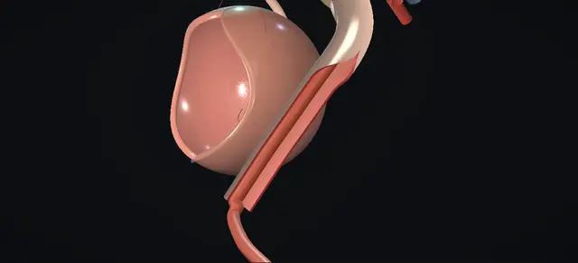
UrOops3D, danger-zone atlas

Schematic child bladder and ureter: every dimension is an authoring estimate.

Operative animation, Megaureter repair

Megaureter repair: reimplantation with excisional tapering

Indications

  • Pain, obstruction or urinary infection attributable to the megaureter.
  • Poor split function at the outset, or function that falls on repeat renography.
  • Hydroureteronephrosis that progresses on follow-up.

Alternatives

  • Observation: most primary obstructive megaureters improve or resolve without surgery.
  • In an infant: a temporary stent, a refluxing reimplant or an end cutaneous ureterostomy, with definitive repair later.
  • Endoscopic balloon dilatation of the narrow segment, in centres that offer it.
  • Reimplantation without tapering, or tapering by folding the wall in, when the ureter allows.

Risks

  • Bleeding and infection.
  • Urine leak or a urinoma.
  • Ureteric obstruction or stricture.
  • Persistent or recurrent reflux, and recurrent urinary infection.
  • Urinary retention.
  • Harder access to that ureter for a catheter in later life.

Open repair of a primary obstructive megaureter in a child, shown on the left side. The ureter is freed through the opened bladder and then outside it, with its blood supply kept on it, as far as the iliac vessels, and its narrow last segment is removed.

The dilated ureter is then tapered: a strip of its lateral wall is excised, away from the longitudinal vessels on the medial side, and the wall is closed round a catheter.

The tapered ureter is reimplanted through a submucosal tunnel across the trigone to a new opening above and lateral to the opposite orifice. The lesson is where the blood supply runs, how much wall the catheter allows to be taken, and how long a tunnel a wide ureter needs.

Advanced tier FRCS paediatric urology, megaureter, reconstruction Interactive 3D

Operative visualisation

Schematic scene preview: Megaureter repair
Preparing the 3D anatomy…

UrOops3D · UroRef · © Nity G

How to use this module. Play the nine steps, or use Back and Next. Watch the ureter leave the bladder, the narrow segment go, the lateral strip come away from round the catheter, the two suture layers close, and the tapered ureter cross the trigone under the mucosa. The triggers show a leak, a kink and a short tunnel.

Click me next Section 2 of 8 Anatomy The structures this operation works between, and the one it must not cross.

The structures in the scene

Megaureter repair: how the structures relateThe left megaureter from the pelvic brim to its new opening in the bladder. Dilated ureter: tapered, then reimplanted: supplied by them; kept out of the taper Longitudinal vessels on its medial side (danger). Dilated ureter: tapered, then reimplanted: redundant wall excised here over a catheter Lateral wall: the strip and the suture line. Dilated ureter: tapered, then reimplanted: ends in it at the bladder; removed Narrow distal segment: excised. Dilated ureter: tapered, then reimplanted: crosses them; freed to above the dilatation Iliac vessels at the pelvic brim. Dilated ureter: tapered, then reimplanted: lies in it: five times as long as it is wide Submucosal tunnel across the trigone. Dilated ureter: tapered, then reimplanted: comes back in here; a kink obstructs it Original hiatus: the turn into the wall (danger).Dilated ureter:tapered, thenreimplanted!Longitudinal vesselson its medial sideLateral wall: thestrip and the suturelineNarrow distalsegment: excisedIliac vessels at thepelvic brimSubmucosal tunnelacross the trigone!Original hiatus: theturn into the wallsupplied by them;kept out of thetaperredundant wallexcised here over acatheterends in it at thebladder; removedcrosses them; freedto above thedilatationlies in it: fivetimes as long as itis widecomes back in here;a kink obstructs itTargetDangerLandmarkContext
The left megaureter from the pelvic brim to its new opening in the bladder. Schematic, not to scale; relationships only.
  • Through the cystotomy the dome is at the top, the bladder neck at the bottom and the patient’s right on the surgeon’s left.
  • The megaureter is the left one.
  • Outside the bladder it runs down the pelvic side wall from the iliac vessels, wide and tortuous, to a narrow last segment that enters the bladder wall.
  • Its longitudinal vessels are on its medial side, toward the bladder, and the strip is taken from the lateral side.
  • After the repair the tapered ureter comes in through the original hiatus, crosses the trigone under the mucosa above the interureteric bar, and opens just beyond and above the right orifice.
  • ›Megaureter - the dilated ureter; here the left, wide and tortuous in the pelvis down to a narrow last segment.
  • ›Narrow distal segment - the short segment next to the bladder that is not dilated; it is divided and removed.
  • ›Longitudinal vessels - run on the medial aspect of the ureter; seen, kept, and never included in the strip.
  • ›Adventitia and periureteric tissue - carry the vessels of the ureter; left on it through both dissections.
  • ›Lateral wall - the redundant wall that is excised, and where the suture line of the taper then lies.
  • ›Peritoneum - behind the bladder wall and on the pelvic ureter; swept off, not opened.
  • ›Iliac vessels - the ureter crosses them at the brim; their bifurcation marks how far up the mobilisation goes.
  • ›Original orifice and hiatus - where the ureter is circumscribed and freed, and where the tapered ureter comes back into the wall.
  • ›Submucosal tunnel - roof of mucosa, floor of detrusor, from the original orifice across the trigone.
  • ›New opening - beyond and above the opposite orifice, on the far side of the trigone; the new ureteric orifice is sewn there.
  • ›Opposite orifice - normal and left alone; urine is seen to jet from it as well at the end.
  • ›Abdominal wall - skin, Scarpa’s fascia, rectus sheath and recti; the peritoneum is swept up off the bladder and stays closed.
Click me next Section 3 of 8 Steps The operation in order, with the landmark that guards each move.

Operative steps

  1. Open the bladder The child is supine with the hips abducted. A urethral catheter is passed, the bladder is filled with saline and the catheter is clamped. A curved transverse incision is made along the skin lines a fingerbreadth above the pubis and carried down to the rectus sheath, which is opened in an arc with diathermy, clear of the inguinal canals. The recti are separated in the midline and retracted. The anterior bladder wall and the bladder neck are exposed and the peritoneum is swept cephalad. A self-retaining retractor is set with padding on each blade. The anterior wall is held in Allis forceps and opened by a vertical midline cystotomy. Stay sutures are placed on each side, the bladder is emptied by suction, the urethral catheter is removed, and a figure-of-eight stitch at the lower apex stops the cut extending caudally.
    where it bites: The inferior epigastric vessels lie under the recti as they are retracted. A cystotomy that extends caudally runs into the bladder neck.
  2. Free the intramural ureter The orifice of the megaureter is identified, and a fine infant feeding tube is passed up the ureter and secured to the adjacent ureteric wall with a stitch. With gentle traction on the tube the orifice is lifted and circumscribed, with a hooked blade or diathermy. Tenotomy scissors dissect sharply on the inferior aspect of the ureter first and then free it from the detrusor all the way round. The peritoneum is swept off the back of the bladder wall under direct vision with a fine gauze pledget. The adventitia is kept on the ureter throughout.
    where it bites: The adventitia carries the blood supply of the ureter. The peritoneum lies directly behind the bladder wall where the ureter passes through it.
  3. Mobilise outside the bladder Once the ureter is completely free inside the bladder it is passed out through the bladder wall, and the dissection continues outside the bladder. The distal ureter is freed from the adjacent peritoneum, with the periureteric tissue and its blood supply left on the ureter. Mobilisation is carried proximally above the dilated segment, to the level of the bifurcation of the common iliac vessels.
    where it bites: Tissue stripped off the ureter takes its vessels with it. The iliac vessels lie behind the ureter at the pelvic brim, and in a boy the vas crosses the ureter near the bladder.
  4. Examine and prepare The width of the dilated ureter is measured, and the decision is made to taper it by excision so that its distal segment can be reimplanted without reflux. The ureter and its blood supply are examined: the longitudinal vessels run on its medial aspect and are kept. The narrow distal segment is divided and removed, and the feeding tube is replaced by a larger soft catheter, which is advanced to the renal pelvis.
    where it bites: The taper is planned on the lateral wall because the vessels are medial. A ureter that would lie in an adequate tunnel as it is does not need to be tapered.
  5. Excise the lateral strip Small Babcock forceps are applied to the lateral aspect of the dilated ureter. The redundant lateral wall is excised sharply as a single strip and sent for histology. The catheter in the lumen limits how much wall can be taken: what is left has to close round it.
    where it bites: Too wide a strip leaves too little wall to close without narrowing the lumen. A strip taken medially takes the longitudinal vessels with it.
  6. Close the taper The wall is brought together over the catheter in two layers. The first is a running locking suture through mucosa and muscle; the second is a running suture through muscle and adventitia. The catheter is then removed and a traction suture is placed in the distal ureteric wall. The ureter is examined: peristalsis is seen, and there is no injury and no bleeding.
    where it bites: A gap in the suture line leaks urine into the tissues. From here the ureter is moved by its traction suture.
  7. Tunnel and draw through Attention returns to the inside of the bladder. The mucosa above and lateral to the opposite orifice is incised. A submucosal tunnel is made by sharp and blunt dissection from the original orifice to this new opening; the ratio of its length to the width of the ureter should be five to one. The tapered ureter is drawn through the tunnel by its traction suture and placed in it without tension. Its lie is checked: it must not be obstructed or kinked. The mucosal defect at the original orifice is closed over the ureter where it enters the tunnel.
    where it bites: A tunnel too short for so wide a ureter lets reflux persist. A ureter that is tight or kinked where it enters the wall is obstructed. A wide hiatus left open behind the ureter can let a diverticulum form.
  8. Anastomose The end of the ureter is spatulated ventrally and its edges are trimmed so that the blood supply at the edge is not in doubt. Dorsally, two interrupted absorbable sutures anchor the ureter to the trigone muscle and the bladder mucosa. Similar sutures at nine, twelve and three o’clock complete the anastomosis. Jets of urine are seen from both orifices, which confirms that the ureter is neither obstructed nor kinked.
    where it bites: A pale edge is trimmed further. Sutures placed too deeply narrow the new orifice.
  9. Leave a tube and close Haemostasis is secured with diathermy. A fine feeding tube is passed through the new orifice and advanced to the renal pelvis, and anchored to the adjacent bladder mucosa with an absorbable stitch. A urethral catheter is placed. The cystotomy is closed in two layers, a running suture in mucosa and interrupted sutures in muscle, and irrigation through the catheter confirms that the closure is watertight. A drain is placed in the retropubic space away from the cystotomy. The drain and the feeding tube are brought out through a separate skin incision and secured. The retractor is removed and the wound is closed in layers: recti, rectus sheath, Scarpa’s fascia and skin.
    where it bites: A drain lying on the cystotomy closure, or a tube that is not secured, puts the repair at risk.
Click me next Section 4 of 8 Danger zones Where this operation bites, and why it bites there.
Critical relationship

Danger zones

The spatial relationships the animation is built to make automatic. Before you read each one, ask yourself: at this point in the operation, what is about to go wrong, and what is my first move? The save for each sits in the Complications tab.

  • RiskThe medial longitudinal vessels - what the reimplanted segment lives on; identified before the taper is planned and kept out of it.
  • RiskThe adventitia and periureteric tissue - left on the ureter during the dissection inside the bladder and outside it.
  • RiskThe lumen under the suture line - the taper is closed with a catheter in the lumen so that the lumen is not narrowed.
  • RiskThe turn into the tunnel - where the tapered ureter enters the bladder wall; a kink here obstructs it.
  • RiskThe length of the tunnel - the ratio asked for, length to ureteric width, is five to one; a short tunnel refluxes.
  • RiskPeritoneum behind the bladder - swept away under direct vision before the ureter is passed out.
Practise it Take me into the scene Opens the animation at a danger moment and asks you to predict the mechanism and your first move before it reveals.
Click me next Section 5 of 8 Complications The save for each one: the first move, the escalation trigger, the op-note line.

Complications and immediate saves

Trigger each of these live in the Animation tab, then read the save: the first move, the escalation trigger, and the line that goes in the op note.

Urine leak from the suture line

  • urine escapes through a gap in the suture line of the taper and collects beside the bladder as a urinoma; the operative text lists urine leak and urinoma among its complications.
The save
  1. On the table: inspect the whole suture line before the catheter comes out, and close any gap with a further suture. first move
  2. Leave the tube across the repair and the drain as planned.
  3. After surgery, a drain output that rises, pain, distension or fever prompts an ultrasound for a collection.
  4. A collection, a leak that persists or sepsis needs senior paediatric urology review and a plan for drainage. escalation trigger
  5. Op note: the two layers of the taper, the tube left across the repair and where the drain lies. document

Obstruction at the hiatus

  • the reimplanted ureter does not drain: a kink where it enters the wall, tension, or a lumen narrowed by the taper; obstruction or stricture is on the operative text’s own list.
The save
  1. On the table: if the ureter kinks as it enters the wall or lies tight, take it back out of the tunnel, free it further and pass it again. first move
  2. Before closing, look for a jet of urine from the new orifice and pass the feeding tube up it.
  3. After surgery, loin pain, vomiting, fever or increasing dilatation on ultrasound suggests obstruction.
  4. Obstruction with infection or falling function needs drainage of that kidney and senior review. escalation trigger
  5. Op note: the lie of the ureter, the jet seen, and the tube passed and left. document

Persistent reflux

  • reflux continues after the repair, here because the tunnel is short for a ureter of that width; persistent or recurrent reflux is a listed complication of this operation.
The save
  1. On the table: judge the tunnel against the tapered ureter before it is drawn through, and lengthen it across the trigone if the mucosa allows. first move
  2. After surgery, follow the child clinically and with ultrasound; a cystogram is usually reserved for a febrile urinary infection.
  3. Recurrent febrile infection with reflux on imaging goes back to the paediatric urology team for a planned decision. escalation trigger
  4. Op note: the route and length of the tunnel, and the length of ureter tapered. document
Click me next Section 6 of 8 Troubleshooting When the case turns: the problem, then the next move.

Intra-operative troubleshooting

When the case turns: the problem, then the next move.

  • ActionThe ureter is too wide for the tunnel the bladder can give - this is the reason to taper. The dictation excises the lateral wall over a catheter; the alternative it names is to taper by infolding and imbricating the wall, which removes nothing.
  • ActionThe dilatation and tortuosity run further up, as in prune belly syndrome - the textbook advises a vertical abdominal incision for exposure in that case.
  • ActionAn infant with poor function in that kidney - the textbook suggests an end cutaneous ureterostomy first, with definitive reconstruction if the function improves enough. The British consensus also describes a temporary stent or a refluxing reimplant in infancy.
  • ActionThe cut end does not look well supplied - the edges are trimmed back to tissue that is well supplied, and the reach is checked again before the first suture.
  • ActionThe tapered ureter kinks where it enters the bladder wall - it is taken back, freed further up and passed again. The dictation has the lie checked for obstruction or kinking before the anastomosis.
  • ActionWhether to leave a stent - the operative text leaves a feeding tube through the new orifice to the renal pelvis, brought out with the drain, and notes that a ureteric stent is optional.
  • ActionThe tunnel cannot be made five times the width of the ureter - Boswell’s review notes that the full ratio can be hard to achieve in a small bladder and reports series that accepted a shorter tunnel. That is a decision for the operating consultant, made before the ureter is drawn through.
Click me next Section 7 of 8 FRCS viva Say it out loud, then mark yourself against the model answer.

FRCS viva questions

The examiner stems this scene prepares you for. Try to answer out loud first, then reveal, and learn the discriminator, the detail that turns a pass into a strong pass.

QWhat is a megaureter, and which ones need an operation?reveal answer
  • A megaureter is a dilated ureter.
  • Boswell’s review gives the usual threshold as wider than 7 or 8 mm, and the British Association of Paediatric Urologists consensus defined a ureteric diameter over 7 mm as abnormal.
  • I would classify it by whether it is obstructed, refluxing, both or neither, after excluding a secondary cause such as posterior urethral valves or a neuropathic bladder.
  • For a primary obstructive megaureter the first treatment is observation, because most improve: Boswell’s review puts it at around 72 per cent improving or resolving.
  • I would offer surgery for symptoms, meaning febrile urinary infection or pain, and in a child without symptoms for loss of function.
  • The British consensus gives a differential function below 40 per cent with massive or progressive hydronephrosis, or a drop in differential function on serial renograms.
  • The operative dictation I follow lists pain, obstruction or infection attributable to the megaureter, function under 40 per cent at the outset or falling by more than 10 per cent on repeat scans, and progressive hydroureteronephrosis.
  • Delayed drainage on a renogram alone, in a well child with stable dilatation, is not an indication on Boswell’s reading of the consensus.
Strong pass

Say both

  • Starting with observation
  • Operating for symptoms or loss of function and not for delayed drainage alone
Examiner note
Starting with observation, and operating for symptoms or loss of function and not for delayed drainage alone.
QTalk me through the repair of a primary obstructive megaureter.reveal answer
  • I fill the bladder through a urethral catheter and expose it through a low transverse incision, parting the recti and sweeping the peritoneum up.
  • I open the bladder vertically in the midline, place stay sutures and an apex stitch, and set a padded ring retractor.
  • I pass a fine feeding tube up the megaureter and stitch it to the ureteric wall, lift the orifice on the tube, circumscribe it and free the intramural ureter from the detrusor, inferior aspect first, keeping the adventitia on it.
  • I then pass the ureter out through the bladder wall and mobilise it extravesically, off the peritoneum and with its periureteric tissue intact, to above the dilated segment at the iliac bifurcation.
  • I identify the longitudinal vessels on the medial side, divide and remove the narrow distal segment, and pass a larger catheter to the renal pelvis.
  • I put small Babcock forceps on the lateral wall, excise the redundant wall, and close the ureter with the catheter in place: a running locking layer in mucosa and muscle, then a running layer in muscle and adventitia.
  • I remove the catheter, place a traction suture and check peristalsis.
  • Inside the bladder I incise the mucosa above and lateral to the opposite orifice, make a submucosal tunnel from the original orifice, draw the ureter through without tension, spatulate it, anchor it dorsally with two sutures and complete the anastomosis at nine, twelve and three o’clock.
  • I look for jets from both orifices, leave a feeding tube up the new orifice, and close the bladder in two layers with a urethral catheter and a drain.
Strong pass

The order: free the ureter inside the bladder and then outside it, remove the narrow segment, taper on the lateral side with a catheter in the lumen, then tunnel, anastomose and leave a tube.

QWhy taper, and why on the lateral side over a catheter?reveal answer
  • A ureter stops refluxing when enough of it lies under the mucosa with muscle behind it, so that the filling bladder closes it.
  • How much is enough depends on its width: the operative text asks for a tunnel five times as long as the ureter is wide, a ratio that Boswell’s review attributes to Paquin.
  • A megaureter may be too wide for the tunnel a child’s bladder can give, so I would reduce its width over the length that will lie in the tunnel.
  • I take the wall from the lateral side because the longitudinal vessels run medially, and the tapered segment depends on them once it has been detached and mobilised.
  • I do it with a catheter in the lumen because the catheter fixes the calibre: the wall that is left has to close round it.
  • The alternative named in that text is to fold the wall in and imbricate it, which removes nothing.
  • I would add that tapering has a cost.
  • Boswell’s review describes a higher operative complexity and a higher complication rate after tapering, including urine leak, stricture and reflux, and argues that there is less reason to taper now than there was when the need is in question.
Strong pass

Say all three

  • Linking the taper to the width a tunnel can cover
  • The lateral side to the medial vessels
  • The catheter to the calibre of the lumen
Examiner note
Linking the taper to the width a tunnel can cover, the lateral side to the medial vessels, and the catheter to the calibre of the lumen.
QIs a tunnel five times the width of the ureter necessary?reveal answer
  • It is the traditional figure and it is the one in the dictation I follow.
  • Boswell’s review attributes it to Paquin and questions whether the full ratio is needed.
  • It notes that the ratio is hard to achieve for a megaureter in a small bladder, and it describes a series by Babu of reimplantation without tapering into a tunnel only twice as long as the ureter was wide.
  • Two of 13 children had reflux afterwards, which the review calls a similar reflux outcome to 15 children who had a classic cross-trigonal reimplant after excisional tapering, with more obstruction and more reoperation in the tapered group.
  • These are small series, and the review says that most of this literature is retrospective and from single centres.
  • In a viva I would give the ratio as the rule I work to, say that the evidence for the exact figure is weak, and say that the review treats obstruction as worse than reflux, so I would not chase the ratio at the cost of a ureter that drains poorly.
Strong pass

Say both

  • Knowing the ratio and that the evidence for the exact figure is weak
  • Weighing obstruction as the worse failure
Examiner note
Knowing the ratio and that the evidence for the exact figure is weak, and weighing obstruction as the worse failure.
QWhat can go wrong after a tapered reimplant, and what do you tell the family?reveal answer
  • The operative text lists bleeding, infection, urine leak or urinoma, persistent or recurrent reflux, ureteric obstruction or stricture, urinary retention, recurrent urinary infection, and difficulty with later catheterisation of that ureter.
  • The two that matter most for the kidney are obstruction and reflux: one from a ureter that is tight, kinked or narrowed, the other from a tunnel that is short for its width.
  • I would reduce the first by keeping the blood supply on the ureter, tapering round a catheter and checking the lie and the jet before I close, and the second by the length of the tunnel.
  • I leave a tube across the repair and a drain.
  • I would tell the family that the new orifice lies on the other side of the bladder, so passing a catheter up that ureter in later life is harder, and that the operation should be recorded in the notes that go with the child.
Strong pass

Say both

  • Naming obstruction and reflux as the two failures of the repair
  • Later difficulty with catheterising the ureter as the lasting consequence of its route
Examiner note
Naming obstruction and reflux as the two failures of the repair, and later difficulty with catheterising the ureter as the lasting consequence of its route.
Click me next Section 8 of 8 Evidence The guidance each of these answers is built on.

Evidence and guidance

Operative Dictations in Urologic Surgery
Armenakas NA, Fracchia JA, Golan R. Megaureter repair. Chapter 118. Wiley, 2019
The indications, including function under 40% at the outset or falling by more than 10% on repeat scans; the twelve essential steps and the template dictation, followed step by step here; the excisional taper on the lateral aspect with the medial longitudinal vessels preserved, closed in two layers over a catheter; the tunnel from the original orifice to a new opening above and lateral to the opposite orifice, with a length to ureteric width ratio of 5:1; the feeding tube left through the new orifice; the variations (end cutaneous ureterostomy in an infant with poor function, a vertical incision for more proximal dilatation, tapering by infolding and imbricating, an optional stent); and the complication list. The chapter’s calibres and suture sizes are kept off the page. The chapter lists complications without their management: the first, escalate and document steps, the danger lines not traceable to the dictation, and the remarks that the catheter fixes the calibre, that the vas crosses the ureter in a boy and that the reach is checked after trimming are general surgical practice and anatomy, as is reserving a cystogram for a febrile urinary infection. The closure of the mucosa at the original orifice is general practice (and chapter 125 of the same book); chapter 118 does not describe it. The scene is schematic: every dimension of the child bladder and ureter is an authoring estimate.
Open guideline
Current Urology Reports
Boswell TC. Advancements in surgical management of megaureters. Curr Urol Rep 2024;25(9):215-223
Full text read 13 October 2026. A review. Megaureter usually defined as wider than 7 or 8 mm; classification by obstruction and reflux; observation first, with around 72% improving or resolving; indications drawn from the British consensus, with delayed washout alone not an indication; reimplantation with or without tapering as the historical standard, and ureterostomy, stent, refluxing reimplant and balloon dilatation as other options; tapering carries higher complexity and a higher complication rate, including urine leak, stricture and reflux; the 5:1 ratio attributed to Paquin and questioned; Babu’s series of 13 untapered reimplants with a 2:1 tunnel (two with reflux) against 15 tapered cross-trigonal reimplants; most of the literature is retrospective and single-centre.
Open guideline
Journal of Pediatric Urology
Farrugia MK et al. British Association of Paediatric Urologists consensus statement on the management of the primary obstructive megaureter. J Pediatr Urol 2014;10(1):26-33
Abstract read 13 October 2026. A consensus of 56 consultant members. A ureteric diameter over 7 mm is abnormal; initial management is conservative; surgery for symptoms such as febrile urinary infection or pain, and in the asymptomatic child for a differential function below 40% with massive or progressive hydronephrosis or a drop in differential function on serial renograms; reimplantation recommended over 1 year of age, with a temporary JJ stent or a refluxing reimplantation proposed as alternatives in infancy.
Open guideline

Curriculum. Supports learning relevant to the ISCP Urology Curriculum syllabus topic Paediatric urology. Not a workplace-based assessment and not evidence of competence.

Two minutes

Close the lap: recall it before you leave.

Say it aloud, then reveal. Reading is not remembering.

  1. 01 The relationship Say the critical spatial relationship out loud, then reveal. reveal
    Memory frame

    Through the cystotomy the dome is at the top, the bladder neck at the bottom and the patient’s right on the surgeon’s left.

    Why?
    The megaureter is the left one. Outside the bladder it runs down the pelvic side wall from the iliac vessels, wide and tortuous, to a narrow last segment that enters the bladder wall. Its longitudinal vessels are on its medial side, toward the bladder, and the strip is taken from the lateral side. After the repair the tapered ureter comes in through the original hiatus, crosses the trigone under the mucosa above the interureteric bar, and opens just beyond and above the right orifice.
  2. 02 The first move Urine leak from the suture line. Your first move? reveal
    First move

    On the table: inspect the whole suture line before the catheter comes out, and close any gap with a further suture.

  3. 03 The escalation trigger Urine leak from the suture line. What makes you call for help? reveal
    Escalate when

    A collection, a leak that persists or sepsis needs senior paediatric urology review and a plan for drainage.

  4. 04 The discriminator What turns a pass into a strong pass? reveal
    Strong pass

    Say both

    • Starting with observation
    • Operating for symptoms or loss of function and not for delayed drainage alone
    Examiner note
    Starting with observation, and operating for symptoms or loss of function and not for delayed drainage alone.
  5. 05 The pitfall Where does this operation bite first? reveal
    Pitfall

    The medial longitudinal vessels

    Why?
    what the reimplanted segment lives on; identified before the taper is planned and kept out of it.

Revision practice with self-marked answers, not an assessment of clinical competence. Kept in this browser only; it does not sync between devices.