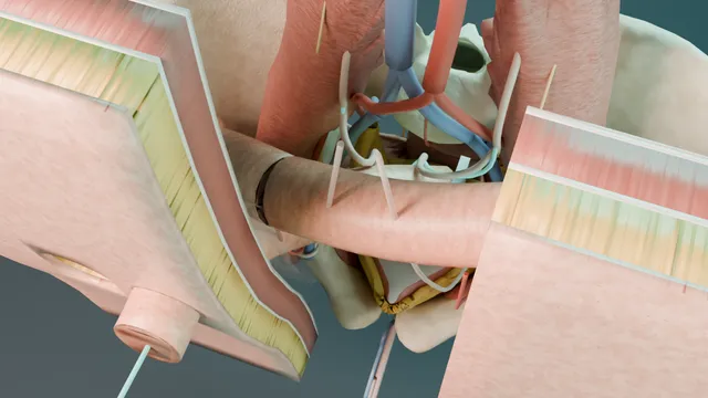
Operative animation, Open supratrigonal cystectomy + ileal conduit
Open supratrigonal cystectomy with ileal conduit for benign disease
The operation for the bladder that has failed everything: end-stage painful bladder, radiation cystitis or the contracted neuropathic bladder.
The dome and body come out through a lower midline incision, the trigone and urethra stay, and the urine is diverted through a short segment of ileum to a stoma marked before the first cut.
You will be asked to hold a Deaver, load a needle holder and cut sutures; this page tells you what you are looking at and why.
Advanced tierFRCS functional urology, reconstruction, open abdominal surgeryInteractive 3D
Lower midline wound from the assistant's side, bowel held up by the Deaver. Schematic, not to scale; relationships only.
From the assistant's side the field is a lower midline wound held open by a Balfour, packs and a Deaver keeping the bowel up.
Below the Deaver the bladder dome sits in the middle, the ureters come over the pelvic brim on either side wearing their vessel loops, and the sigmoid crosses the left side.
When the body of the bladder is gone the trigone is a small cuff closed over the catheter deep in the pelvis.
Above it, to the right, lies the conduit on its mesentery with both ureters joined to its proximal end, its far end rising through the right rectus to the stoma.
›Layers of the lower midline - skin, Camper's and Scarpa's fascia, linea alba between the anterior rectus sheaths, rectus bellies, transversalis fascia, extraperitoneal fat, parietal peritoneum.
›The trigone and interureteric ridge - the triangle between the two orifices and the bladder neck; the transection line stays above the ridge so the trigone and its innervation remain.
›Superior vesical pedicle - the patent umbilical artery running to the superolateral bladder; the only pedicle taken in a supratrigonal operation.
›Ureteric blood supply - runs in the adventitia; enters medially above the pelvic brim and laterally below it. Dissect on the far side at each level and never strip the sheath.
›Terminal ileum and its arcades - the conduit is cut on an arcade seen by transillumination, twenty centimetres proximal to the valve.
›The stoma site - through the right rectus so the muscle supports the appliance; skin disc, cruciate sheath incision, split rectus, two-finger peritoneal opening.
›Retractors - Langenbeck for the wall, Balfour or Bookwalter self-retaining for the wound, Deaver to hold the bowel up, Morris to hold the bladder down.
Before the knife: marking, consent, positionThe stoma nurse marks the site the day before with the patient sitting, standing and lying: over the right rectus, below the belt line, away from the umbilicus, scars and skin folds, where the patient can see it. On the table the patient is supine with a slight break at the umbilicus, arms out, a urethral catheter in on the sterile field, calf compression on and antibiotics given at induction. Bowel preparation is not routine. The house officer stands on the patient's left, opposite the surgeon, and will hold the retractors, cut the sutures and follow the swab count.
Incision and the layers of the abdominal wallA lower midline incision from the pubic symphysis to just above the umbilicus, curving around it to the left. Skin with a number 22 blade, then subcutaneous fat down to the linea alba with diathermy; the Langenbeck retractors go in here. The linea alba is opened in the midline with the blade and extended with scissors, the rectus muscles are swept apart with a swab, and the transversalis fascia and parietal peritoneum are picked up between two Kocher forceps and opened with scissors, checking that no bowel is caught. From skin to cavity: skin, Camper's and Scarpa's fascia, linea alba between the anterior rectus sheaths, the rectus bellies, transversalis fascia, extraperitoneal fat, parietal peritoneum. where it bites: the peritoneum is opened at the top of the wound where the bladder cannot be; low in the wound a distended bladder or a loop of bowel sits directly under it.
Retraction and packingWhen the peritoneum opens the small bowel is straight underneath it, in the way of everything, so the first job is to move it. Moist packs sweep the loops and the sigmoid up into the upper abdomen and a Deaver retractor holds them there, handed to the house officer with the instruction not to lift but to lean. A Balfour self-retaining retractor is then placed with its two lateral blades on the wound edges and the bladder blade over the pubis; on a larger patient the Bookwalter ring goes on the post instead. A Morris retractor holds the bladder dome downward while the peritoneum over it is inspected. The table is tipped head-down (Trendelenburg) so gravity keeps the bowel out of the pelvis.
Finding the ureters, and the rule about their blood supplyThe peritoneum is incised lateral to the sigmoid on the left and over the caecum on the right, and each ureter is found where it crosses the common iliac bifurcation at the pelvic brim: a pale cord under the posterior peritoneum, medial to the gonadal vessels, which stays stuck to the peritoneum when the peritoneum is lifted. Touched with a forceps and given a moment it vermiculates, a slow peristaltic wave; a vessel pulsates and does not. Its blood supply runs in the adventitia and enters from the medial side above the pelvic brim and from the lateral side below it, so the dissection stays on the far side from the supply at each level and the sheath is never stripped. The sling goes on in one sequence: a Lahey or Mixter right-angle forceps is passed beneath the ureter with its curve pointing down, hugging the ureter so nothing behind it is caught, until the tip is seen on the far side; the jaws are opened, the scrub nurse lays the tip of a vessel loop into them, the jaws close on it, and the forceps is drawn back the way it came so the loop follows around the ureter. Both ends are held in a clip. The ureter is then mobilised in its sheath down to the bladder and divided between two Ligaclips or ties of 2-0 Vicryl a centimetre from the bladder, the bladder end ligated. where it bites: above the brim the supply comes from medially, below it from laterally, and it runs in the adventitia; dissect on the wrong side or strip the sheath and the anastomosis strictures.
Dropping the bladderThe urachus and the medial umbilical ligaments are divided high between clips with the diathermy, and the bladder is dropped off the anterior abdominal wall into the retropubic space of Retzius with a swab on a stick. Laterally the superior vesical pedicle is taken between Kelly clamps and tied with 2-0 Vicryl, or sealed with a LigaSure, working from the divided ureter toward the base. The inferior pedicle and the posterior attachments below the level of the trigone are left alone: this is a supratrigonal operation and the pelvic floor is not entered.
The supratrigonal transectionThe bladder is opened on the dome with the Harmonic shears and the cystotomy is extended so the interior can be seen: both ureteric orifices, the interureteric ridge between them and the bladder neck below. The line of transection is marked about a centimetre and a half above the interureteric ridge, outside the trigone, and the body of the bladder is taken off with the Harmonic, which seals as it cuts. Rather than delivering the whole body as one piece it is split down the midline, sagittally, and comes out in two halves, each lifted with a Lane tissue forceps and sent for histology. The urethral catheter is left in place: the trigonal remnant is closed over it in two layers, a running 3-0 Vicryl on the mucosa and a second 2-0 Vicryl on the muscle, and the balloon sits inside the closed remnant and stays there, draining it, as a second drain beside the tube drain in the pelvis. where it bites: the ureteric orifices mark the trigone; cutting below the interureteric ridge takes the trigone the operation is designed to keep.
Choosing and isolating the ileal segmentThe terminal ileum is delivered and a segment of about fifteen centimetres is chosen twenty centimetres proximal to the ileocaecal junction, on a mesentery that reaches the marked stoma site without tension when the segment is lifted to the skin. The mesentery is held up against the theatre light so the arcades show through, and the segment is taken where a good arcade runs to it. Three windows are then poked through the mesentery at the proximal end of the segment and three at the distal end, between the arcades, with a fine haemostat; the Harmonic divides the mesentery between the windows and the bowel at each end, and only the segment is freed on its own mesentery. The rest of the ileum is not disturbed. A Babcock holds the conduit without crushing it. where it bites: a mesentery that is short at the moment the segment is chosen is a dusky stoma at the end of the operation.
Restoring the bowelBowel continuity is restored above the conduit with a stapled side-to-side (functional end-to-end) ileo-ileal anastomosis: the two ends are laid antimesenteric border to antimesenteric border, held with Babcocks, a GIA 75 fired along their length and the common enterotomy closed with a TA 60 or a running 3-0 PDS. The mesenteric window is closed with interrupted 3-0 Vicryl so that nothing can herniate through it, keeping the conduit and its mesentery caudal to the anastomosis. The conduit is irrigated with saline through a catheter until it runs clear.
The Wallace anastomosis over stentsThe left ureter is passed under the sigmoid mesentery to the right side through a window made with a finger, without kinking, and both ureters are brought to the open proximal end of the conduit. Each is spatulated for fifteen to twenty millimetres along its lateral wall with Potts scissors, keeping the adventitia. Their medial edges are then sewn together with a running 5-0 PDS so that the two ureters become a single plate, the Wallace technique. Before the plate goes in, a 6 French single-J stent, forty centimetres long, is passed up each ureter to the renal pelvis and fed out through the conduit, and the stents are fixed to the conduit with a fine fast-absorbing suture. The plate is then sewn into the open end of the conduit, mucosa to mucosa, in four quadrants with interrupted 4-0 PDS, so the bowel closes around the joined ureters. The Bricker alternative joins each ureter separately, end-to-side, to the conduit wall. With the plate in, the conduit is filled with 50 millilitres of saline through the distal end and the join watched; a leak is closed with an interrupted suture and the fill repeated. where it bites: a tensioned or twisted anastomosis is a planned stricture; the ureters should lie without a bend when the packs come out.
The stomaThe Balfour comes out so the wall lies flat, and at the marked site a disc of skin two to two and a half centimetres across is lifted with a Kocher and excised, the subcutaneous fat is spread rather than cut, a cruciate incision is made in the anterior rectus sheath, the rectus is split along its fibres with two fingers, and the posterior sheath and peritoneum are opened until two fingers pass. The distal end of the conduit is brought through the wall, its staple line trimmed, and the bud is matured everted as a spout with 3-0 Vicryl or Monocryl sutures taking skin edge, then seromuscular bowel, then the cut end of the bowel, so the mucosa turns out on itself. A clear appliance goes on in theatre so the bud can be watched. where it bites: a bud that is dusky before the drapes come down is revised now, not on the ward.
ClosureThe pelvis is washed and a tube drain is placed near the anastomoses, brought out through a separate stab. The swab, needle and instrument count is confirmed aloud with the scrub nurse. The linea alba is closed as a mass closure with a looped 1 PDS on a blunt needle, continuous, taking centimetre bites a centimetre apart so that the suture length is at least four times the wound length, approximating rather than strangling the tissue. Interrupted 1 PDS or nylon, the same bites tied one by one, is used instead where the sheath is poor or the abdomen is distended. Fat is not closed; a dead space is drained, not stitched. Skin is closed with staples, an Adson toothed forceps everting the two edges so the stapler sits square on them and a staple goes in every eight millimetres, or with a subcuticular 3-0 Monocryl, and the clear appliance is fitted to the finished stoma before the drapes come down.
The spatial relationships the animation is built to make automatic. Before you read each one, ask yourself: at this point in the operation, what is about to go wrong, and what is my first move? The save for each sits in the Complications tab.
RiskThe peritoneal opening low in the wound - bladder or bowel lies under it; open the peritoneum high, between Kochers, with the cavity seen.
RiskThe interureteric ridge - the lower limit of the transection; below it the trigone goes and the operation becomes a different one.
RiskThe mesentery of the conduit - short, twisted or under tension it strangles the segment; the stoma is dusky before the closure begins.
RiskThe ureteric sheath - the supply runs in the adventitia, medial above the brim and lateral below it; a stripped ureter leaks first and strictures later.
Trigger each of these live in the Animation tab, then read the save: the first move, the escalation trigger, and the line that goes in the op note.
Uretero-ileal leak
urine in the drain in the first days, a creatinine-rich output that does not fall, from a Wallace plate that was tensioned, ischaemic or not watertight.
The save
Send the drain fluid for creatinine: a level well above serum confirms urine and settles the question the volume alone cannot.first move
Keep the drain in as the controlled fistula and confirm the stents are draining; most leaks close with drainage and time.
Image with a conduitogram or CT urogram to map the leak and exclude an obstructed system.
Nephrostomy and antegrade stenting for a leak that will not close or a kidney that is obstructed.
Generalised peritonitis, worsening sepsis or a disrupted anastomosis: return to theatre for revision.escalation trigger
Op note and discharge summary: side, technique, stent plan and the imaging surveillance for the late stricture.document
Conduit ischaemia and the dusky stoma
a segment chosen on a short mesentery, twisted through the wall, or tensioned to the skin, turning dark in the first hours.
The save
Look at the bud in theatre before closure and again on the ward with a clear appliance; a dusky bud is a theatre problem, not a wound review.first move
Check the appliance is not constricting and pass a soft catheter into the conduit to test that the deeper segment is pink.
Dusky mucosa confined to the bud may be watched; a black or grey segment below the fascia is revised now.
Return to theatre before it perforates: re-mobilise or resect the segment and re-fashion the stoma, or formally divert.escalation trigger
Op note: mesenteric length, route through the wall, colour at closure and the review plan.document
Ileo-ileal anastomotic leak
pain, fever, a rising white cell count and a change in drain output from the bowel anastomosis on day three to five.
The save
Suspect it early: a patient who is not progressing after a bowel anastomosis has a leak until proven otherwise.first move
Resuscitate, antibiotics, nil by mouth and CT with oral contrast to define a contained leak from free peritonitis.
A contained collection drains radiologically and is fed and observed.
Free leak with generalised peritonitis: washout, repair or resection and diversion in theatre.escalation trigger
Op note: anastomotic technique, findings, what was done and the nutrition plan.document
Wound dehiscence and parastomal hernia
the burst abdomen in the first two weeks, and the late bulge beside the stoma in the years after.
The save
Serosanguinous discharge from the midline wound on day five to ten is dehiscence until proven otherwise; keep the patient in bed and review the wound.first move
A burst abdomen returns to theatre for mass re-closure with deep tension sutures or a mesh.
A parastomal hernia that is asymptomatic is managed with a support belt and stoma care.
Obstruction, incarceration or an appliance that will not seal: repair, with or without relocation of the stoma.escalation trigger
Op note: closure technique, suture and the 4:1 ratio; stoma site and layers traversed.document
When the case turns: the problem, then the next move.
ActionYou are asked to hold the Deaver and the bowel keeps falling in - lean the blade against the packs rather than lifting it; ask for a second pack and a head-down tilt before pulling harder.
ActionThe ureter will not reach the conduit - mobilise it higher with its adventitia intact and bring the conduit toward it; a longer segment beats a tensioned join.
ActionThe staple line bleeds - oversew with 3-0 PDS; do not diathermy a staple line.
ActionThe stoma will not reach the skin without tension - the segment is too short or the mesentery too tethered; free the mesentery or choose another segment while the abdomen is open.
ActionCount is one swab short at closure - stop, recount, look in the pelvis and behind the packs, then X-ray before the fascia is closed.
The examiner stems this scene prepares you for. Try to answer out loud first, then reveal, and learn the discriminator, the detail that turns a pass into a strong pass.
QWhat are the indications for supratrigonal cystectomy, and why keep the trigone?reveal answer
Supratrigonal cystectomy is an operation for benign end-stage bladder disease when conservative and endoscopic measures have failed: refractory bladder pain syndrome with a small-capacity fibrotic bladder, radiation cystitis with intractable haematuria or pain, the contracted tuberculous or neuropathic bladder, and occasionally the bladder devastated by intravesical therapy.
The trigone is kept because it carries the ureteric orifices and the bladder neck with its innervation, so that where an augmentation or a substitution is the reconstruction the ureters need no reimplantation and continence is preserved;
where a conduit is the diversion, as in this operation, the retained trigone and urethra avoid a deep pelvic dissection, protect the pelvic floor and the sexual nerves, and leave a small remnant that can be closed over a catheter.
The price is that the remnant can bleed or become painful in radiation cystitis and that malignancy must first be excluded, since a supratrigonal operation is never an oncological one.
The alternatives to be named are augmentation cystoplasty when capacity is the problem and the bladder is otherwise usable, and total cystectomy with diversion when the trigone itself is the source of the symptoms.
Strong pass
Say all three
Excluding malignancy first
Tying the retained trigone to the specific reconstruction
Rather than presenting supratrigonal cystectomy as a smaller version of the cancer operation
Examiner note
Excluding malignancy first and tying the retained trigone to the specific reconstruction, rather than presenting supratrigonal cystectomy as a smaller version of the cancer operation.
QDescribe the layers you go through in a lower midline laparotomy and how you close them.reveal answer
From the surface: skin, the superficial fatty layer of Camper and the membranous layer of Scarpa, then the linea alba, which below the arcuate line is the fused aponeuroses of external oblique, internal oblique and transversus forming only the anterior sheath, the rectus muscles which are swept apart rather than cut, the transversalis fascia, extraperitoneal fat and finally the parietal peritoneum.
Above the arcuate line the rectus has a posterior sheath;
below it the muscle sits directly on transversalis fascia and peritoneum, which is why a low incision must be opened with care and the bladder emptied by catheter first.
The peritoneum is opened at the upper end of the wound between two forceps with the cavity seen.
Closure is a mass closure of the linea alba with a looped 1 PDS or nylon on a blunt needle, continuous, with bites about a centimetre from the edge a centimetre apart so that the suture length is at least four times the length of the wound, which is the Jenkins ratio;
tissue is approximated, not strangled.
Fat is not routinely closed.
Skin is closed with staples or a subcuticular absorbable suture.
The stoma is brought out through a separate opening in the rectus, never through the wound.
Strong pass
Say both
The arcuate line explaining why the low peritoneum is opened with care
The four-to-one suture-to-wound ratio stated as the principle behind the mass closure
Examiner note
The arcuate line explaining why the low peritoneum is opened with care, and the four-to-one suture-to-wound ratio stated as the principle behind the mass closure.
QHow do you construct an ileal conduit, and what are the technical points that prevent complications?reveal answer
A segment of ileum about fifteen centimetres long is isolated twenty centimetres proximal to the ileocaecal valve, sparing the terminal ileum for its absorptive function, on a mesentery long enough to reach the stoma site without tension, chosen by transillumination of the arcades.
The bowel is divided with linear staplers and continuity is restored above the segment with a side-to-side stapled anastomosis, the mesenteric window closed to prevent an internal hernia, and the conduit placed caudal to the anastomosis.
The proximal end is closed.
The left ureter is brought under the sigmoid mesentery through a generous window.
Each ureter is spatulated and anastomosed end-to-side to the conduit mucosa to mucosa with fine absorbable sutures, the Bricker technique, or the two ureters are joined as a plate and sutured to the open end, the Wallace technique, over single-J stents that exit through the stoma.
The distal end is brought through the rectus at the premarked site and matured as an everted spout.
The points that prevent trouble are: no tension and no twist at either the ureteric or the bowel anastomosis, a ureter handled by its adventitia, a conduit that lies in a gentle curve, a stoma through the rectus with a two-finger aperture, and a bud that is pink before closure.
Strong pass
Say both
Naming Bricker and Wallace and the reasons for each
Treating the mesenteric window closure and the conduit position caudal to the anastomosis as deliberate steps rather than housekeeping
Examiner note
Naming Bricker and Wallace and the reasons for each, and treating the mesenteric window closure and the conduit position caudal to the anastomosis as deliberate steps rather than housekeeping.
QA house officer asks you what to watch for on the ward after an ileal conduit. What do you tell them?reveal answer
Three lanes.
The bowel lane: ileus is expected for a few days and is managed with patience, early feeding as tolerated, minimal opioids and mobilisation;
failure to progress by day five, distension, vomiting or a rising white cell count means imaging for a leak or obstruction.
The urine lane: the stents should drain into the appliance and the drain should be low and falling;
a drain that stays high is sent for creatinine, and a creatinine level far above serum is a urine leak that is managed with drainage, stent patency and imaging.
The stoma lane: the bud should be pink and moist and is looked at through a clear appliance every shift in the first two days;
a dusky or grey bud is reported at once, because ischaemia below the fascia is a theatre problem on the same day.
Beyond these, the wound is checked for the serosanguinous discharge of dehiscence, the legs for thrombosis, the electrolytes for the hyperchloraemic acidosis that a conduit can produce if urine pools, and the patient for the delirium and chest complications of major surgery in an older person.
The stents come out at a protocol date, usually around ten days, and the catheter whose balloon sits in the trigonal remnant stays as its drain until the remnant has healed.
Strong pass
Separating bowel, urine and stoma lanes and giving the house officer the specific trigger for each escalation rather than a list of things that can go wrong.
Say it aloud, then reveal. Reading is not remembering.
01The relationshipSay the critical spatial relationship out loud, then reveal.reveal
Memory frame
From the assistant's side the field is a lower midline wound held open by a Balfour, packs and a Deaver keeping the bowel up.
Why?
Below the Deaver the bladder dome sits in the middle, the ureters come over the pelvic brim on either side wearing their vessel loops, and the sigmoid crosses the left side. When the body of the bladder is gone the trigone is a small cuff closed over the catheter deep in the pelvis. Above it, to the right, lies the conduit on its mesentery with both ureters joined to its proximal end, its far end rising through the right rectus to the stoma.
02The first moveUretero-ileal leak. Your first move?reveal
First move
Send the drain fluid for creatinine: a level well above serum confirms urine and settles the question the volume alone cannot.
03The escalation triggerUretero-ileal leak. What makes you call for help?reveal
Escalate when
Generalised peritonitis, worsening sepsis or a disrupted anastomosis: return to theatre for revision.
04The discriminatorWhat turns a pass into a strong pass?reveal
Strong pass
Say all three
Excluding malignancy first
Tying the retained trigone to the specific reconstruction
Rather than presenting supratrigonal cystectomy as a smaller version of the cancer operation
Examiner note
Excluding malignancy first and tying the retained trigone to the specific reconstruction, rather than presenting supratrigonal cystectomy as a smaller version of the cancer operation.
05The pitfallWhere does this operation bite first?reveal
Pitfall
The peritoneal opening low in the wound
Why?
bladder or bowel lies under it; open the peritoneum high, between Kochers, with the cavity seen.
Revision practice with self-marked answers, not an assessment of clinical competence. Kept in
this browser only; it does not sync between devices.