
Revision sheet
Open supratrigonal cystectomy + ileal conduit
8 steps, each one the authored title and caption from the interactive animation, with the frame captured from that step. Read it before the case, or print it and work through the steps in order.
Teaching only, not for diagnosis or treatment decisions. The text on this sheet is the animation's own step copy, reproduced verbatim.
UrOops3D · UroRef · © Nity G
- 01
Lower midline: skin to peritoneum
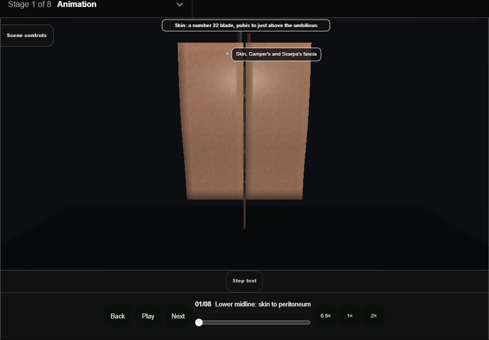
- Skin, fat, then the linea alba; the recti swept apart, transversalis and peritoneum picked up between Kochers and opened high.
- Langenbecks, then the Balfour goes in and a Deaver holds the bowel up.
- 02
Ureters at the brim: find, sling, mobilise, divide
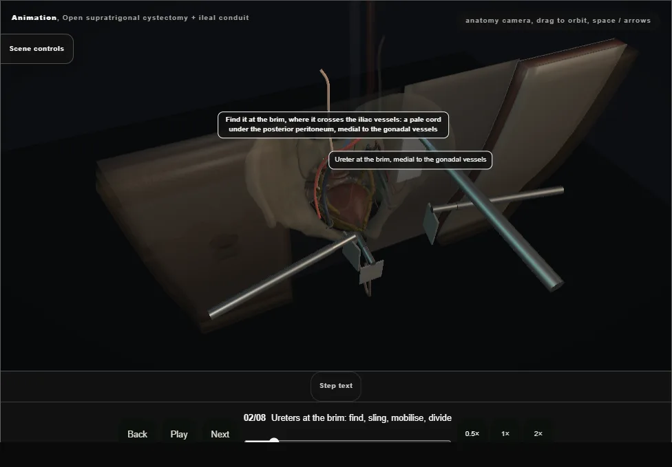
- Found where it crosses the brim, medial to the gonadal vessels, and confirmed by vermiculation.
- Its supply runs in the adventitia, entering medially above the brim and laterally below it, so the far side is worked and the sheath never stripped.
- A right-angle passes beneath it, the vessel loop is drawn round, and it is divided a centimetre from the bladder.
- 03
Drop the bladder
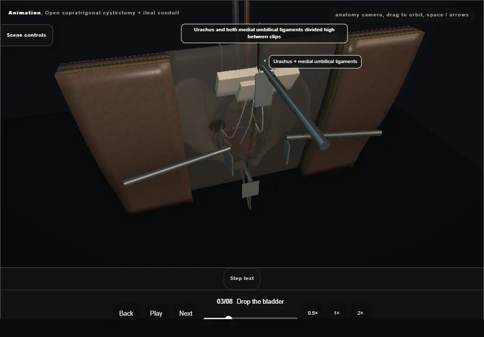
- Urachus and medial umbilical ligaments divided high, the bladder dropped into Retzius with a swab on a stick, the superior vesical pedicle taken between Kellys and tied with 2-0 Vicryl.
- The inferior pedicle and the floor are left alone.
- 04
Supratrigonal transection + remnant closure
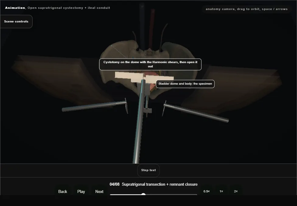
- Cystotomy with the Harmonic, both orifices and the interureteric ridge identified, the body taken a centimetre and a half above the ridge and split sagittally to come out in two halves.
- The remnant is closed in two layers over the catheter, whose balloon stays as a second drain.
- 05
Isolate the ileal segment, restore the bowel
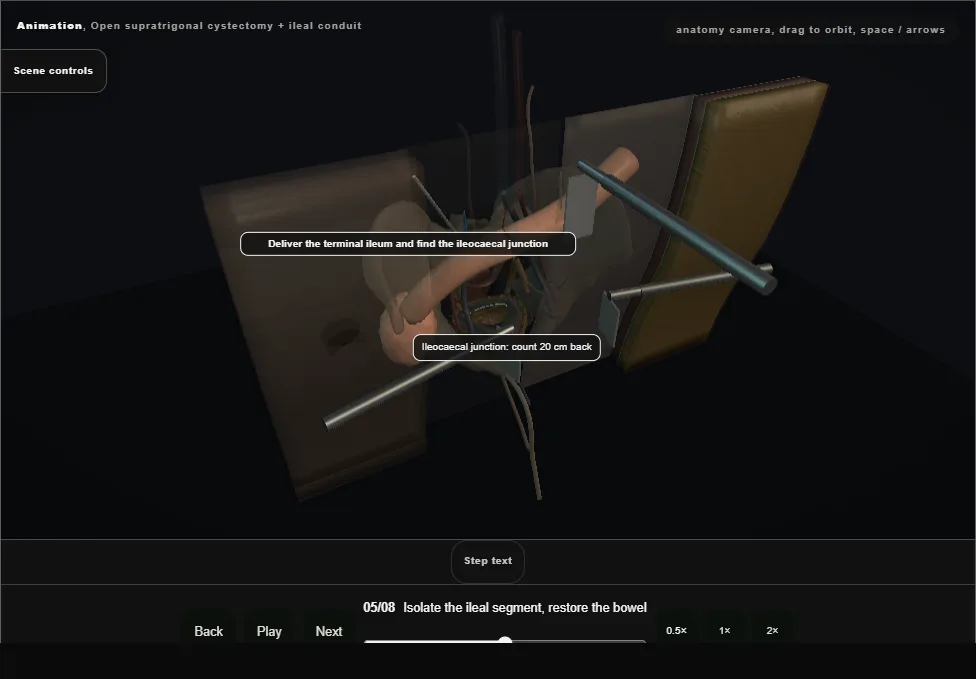
- Fifteen centimetres of ileum twenty centimetres from the ileocaecal junction, held against the light so the arcades show.
- Windows through the mesentery at each end and the Harmonic between them; the bowel itself divided with a linear cutting stapler at each end.
- Bowel rejoined above it: stapled side to side, the common opening closed across with a TA.
- 06
Wallace: the ureters joined, and sewn into the conduit
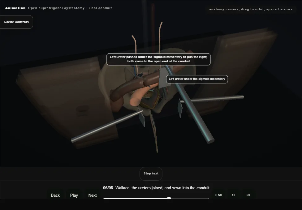
- Both ureters to the proximal end of the conduit, the left under the sigmoid mesentery.
- Each spatulated 2 to 3 cm and opened flat, the adjacent edges sewn together with 4-0 from the heel as one plate (the Wallace), 8 Fr feeding tubes up to the kidneys, and the plate sewn to the bowel end with running 3-0.
- Isoperistaltic: the distal end goes to the skin.
- 07
The stoma through the right rectus
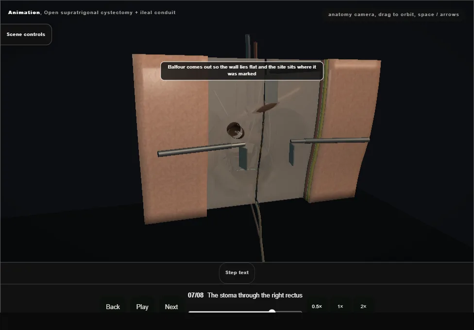
- Balfour out so the wall lies flat.
- Skin disc excised with a Kocher, cruciate incision in the anterior sheath, the rectus split, two fingers through the posterior layer.
- The conduit is brought out and matured as an everted spout with 3-0 Vicryl: skin, seromuscular bowel, cut edge.
- 08
Drain, count, mass closure
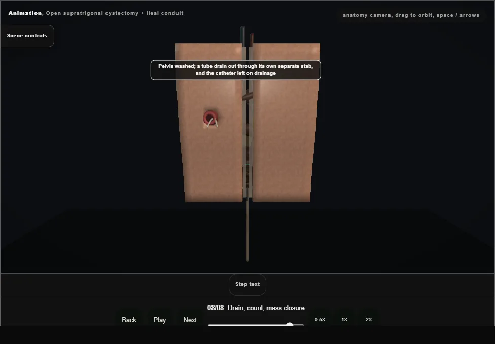
- Tube drain to the pelvis through a separate stab, count confirmed.
- Mass closure of the linea alba with looped 1 PDS, centimetre bites a centimetre apart, suture at least four times the wound length.
- Staples or subcuticular Monocryl, then the appliance goes on the finished stoma.
Derived from cystectomy/OpenCystectomyConduitScene.tsx at build time: every step title and caption above is the authored
ProcedureDef text, reproduced verbatim, and this page shares its readable-points renderer with the animation
itself. 8 of 8 steps have a captured frame.
Text fingerprint 32886ba810ff52a2. A caption edit without a re-capture fails the production
check, so the sheet and the animation cannot drift apart silently.