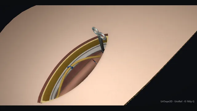
A solid intratesticular mass removed through the groin: the inguinal route protects the lymphatic drainage, the cord is ligated high at the internal ring leaving no tumour-bearing remnant, and the scrotum is never opened in cancer.
Core tierFRCS uro-oncology, testicular cancerInteractive 3D
Operative visualisation
Open on a phone to use AR
Anatomical cutaway: the abdominal wall is drawn as its layers and the scrotum is cut away to show the testis. The operative route is through the groin; the scrotal skin is not incised.
Inguinal approach, skin to internal ring, layer by layer. Schematic, not to scale; relationships only.
The anatomy is a vertical line: internal ring superiorly, external ring at the pubic tubercle, and the scrotum hanging below.
The testicular lymphatics run with the testicular vessels to the para-aortic nodes, which is why the cord is divided high at the internal ring and why the scrotum, whose skin drains to the inguinal nodes, is never opened.
›Landmarks: ASIS + pubic tubercle - the incision, about 5 cm, lies between them along the line of the inguinal canal.
›Skin, Camper's fat, Scarpa's fascia - the superficial layers, each named and divided in the line of the incision.
›External oblique aponeurosis - shiny, fibres running down and in; the superficial dissection stops on it, then it is opened along its fibres to the external ring.
›Inguinal canal + rings - the oblique corridor through the abdominal wall; the internal ring is where the cord is clamped and divided.
›Spermatic cord - vas, testicular artery and pampiniform plexus running together from ring to testis.
›Testis + tunica - a firm intratesticular mass within the tunica; delivered whole in its coverings and never breached.
›Ilioinguinal nerve - lies on the cord under the external oblique; isolated and protected before the cord is clamped.
Landmarks and incisionBefore the knife, the side is confirmed against the imaging, the markers have been sent, and a prosthesis has been discussed. The ASIS and the pubic tubercle, beside the pubic symphysis, are felt and marked. The incision, about 5 cm, lies between them along the line of the inguinal canal: over the internal ring laterally and running towards the external ring medially. One wound has to do two jobs, clamping the cord at the internal ring and delivering the testis whole, so it sits over the canal and never on the scrotum. where it bites: a scrotal incision contaminates the scrotum with tumour and changes the lymphatic drainage field.
Through the layers to the external obliqueEach layer is named as it is met. The skin is opened with the scalpel. The yellow subcutaneous fat, Camper's fascia, is divided with diathermy, and deeper the pale membranous layer, Scarpa's fascia, is divided in the same line. Then the shiny external oblique aponeurosis comes into view, its fibres running down and in. That is where the superficial dissection stops. where it bites: going through the aponeurosis before it is cleaned and seen is how the nerve beneath it is cut.
Open the external oblique; isolate the ilioinguinal nerveThe external oblique is nicked and opened along its fibres with scissors, down through the external ring, to show the spermatic cord in the canal. The ilioinguinal nerve lies on the cord under the aponeurosis. It is isolated, lifted off the cord and protected out of the way before anything is clamped or cut. where it bites: indiscriminate dissection or diathermy on the cord injures the ilioinguinal nerve.
Clamp the cord at the internal ringThe cord is freed from the floor of the canal and traced up to the internal ring. A clamp goes across it there before the testis is handled, so the venous outflow of the tumour is controlled before the testis is squeezed or pulled. where it bites: delivering the testis before the cord is clamped handles a tumour with an open venous outflow.
Evert the scrotum and deliver the testisA finger from below everts the scrotum and the testis is delivered up into the wound whole, inside its coverings, with the cord. The gubernaculum is divided as the testis comes free. Traction is on testis and cord together, never on the mass. Where the ring is tight the external oblique is opened further rather than force applied, because a large tumour forced through a tight ring is how a tunica tears. where it bites: breaching the tunica or tearing the mass spills tumour cells into the wound and scrotum.
Second clamp; divide above both clampsWith the testis held up out of the wound, a second clamp goes across the cord just above the first. The cord is divided above both clamps: the testis and cord are the specimen, and both clamps stay on the cord stump at the internal ring. Division at the ring is the oncological point of the route: a low division leaves tumour-bearing cord and lymphatics behind. where it bites: a low division leaves tumour-bearing cord behind in the remnant.
Transfix the stump; release the clamps slowlyA transfixion suture is passed through the cord and tied below the top clamp, between both clamps, and the top clamp is removed slowly while the stump is watched. A second transfixion suture is then tied below the bottom clamp, at the internal ring, and the bottom clamp is removed slowly. The stump is watched until it is dry before it is allowed to settle: a stump released bleeding is the one that is lost in the canal. Some surgeons use a non-absorbable suture on the stump so it can be found at any later retroperitoneal surgery; that is a common practice, not a rule. where it bites: releasing a clamp on an insecure tie lets the stump retract bleeding through the internal ring.
Prosthesis, if consentedA prosthesis is raised and consented before the incision, then placed when requested: the implant passes down the same inguinal route into the scrotal pouch, at the same operation or delayed until oncological treatment is complete. Delay is the usual choice where adjuvant chemotherapy or radiotherapy is likely, because infection threatens both the implant and the cancer pathway. The route never changes: a separate scrotal cut in a cancer case is forbidden. where it bites: never through a separate scrotal cut in a cancer case.
Close in layers; histopathologyThe nerve is laid back and the wound is closed in layers: the external oblique with a continuous 2-0 Vicryl, keeping the ilioinguinal nerve out of the suture line and re-forming the external ring without narrowing it; Scarpa's fascia with interrupted 3-0 Vicryl; and the skin with a subcuticular 4-0 Monocryl. Long-acting local anaesthetic goes into the wound. The specimen is sent whole and orientated by cord and testis, in a bag, because the histology of the entire orchidectomy specimen is the diagnosis; that is also why no biopsy is taken beforehand. where it bites: never biopsy a testicular mass trans-scrotally; the diagnosis is the histology of the whole orchidectomy specimen.
The spatial relationships the animation is built to make automatic. Before you read each one, ask yourself: at this point in the operation, what is about to go wrong, and what is my first move? The save for each sits in the Complications tab.
RiskDivision at the internal ring - two clamps, division above both, then two transfixion sutures: no tumour-bearing cord is left in the remnant, and each clamp comes off slowly on a secure tie.
RiskIlioinguinal nerve on the cord - injured by careless skeletonisation or broad diathermy; chronic groin pain and numbness follow.
RiskThe scrotal rule - no scrotal wounds, biopsies or drains in suspected testicular cancer; scrotal contamination alters recurrence patterns and management.
When the case turns: the problem, then the next move.
ActionTestis will not deliver through the canal - clamp the cord at the internal ring first, open the external oblique further or extend the incision, and evert the scrotum from below; never force a large tumour through a tight ring.
ActionStump bleeds as a clamp comes off - reapply the clamp, press at the internal ring, and place a further transfixion suture under vision before releasing again, slowly.
ActionIlioinguinal nerve not clearly seen - stay in the plane of the cord coverings and divide the cord deliberately at the ring rather than stripping it clean.
ActionScrotal oedema or haematoma after closure - scrotal support and pressure dressing; explore only for an expanding haematoma.
The examiner stems this scene prepares you for. Try to answer out loud first, then reveal, and learn the discriminator, the detail that turns a pass into a strong pass.
QWhy is a suspected testicular tumour removed through the groin and not the scrotum?reveal answer
Because the lymphatic drainage of the testis and of the scrotal skin go to different places.
The testis, developing intra-abdominally and descending with its vessels, drains to the para-aortic nodes at the level of the renal hilum.
The scrotal skin drains to the superficial inguinal nodes.
A scrotal approach therefore does two harmful things: it contaminates a new lymphatic field with tumour cells, and it risks leaving tumour in the scrotal skin and subcutaneous tissue, producing scrotal recurrence that is difficult to treat and alters the radiotherapy field if that is subsequently needed.
The inguinal approach also permits high ligation of the cord at the internal ring, so no tumour-bearing lymphatics remain in the remnant.
The same principle forbids trans-scrotal biopsy, needle biopsy and scrotal drains in suspected cancer.
The diagnosis is made on ultrasound, markers and the histology of the whole orchidectomy specimen.
Strong pass
Say all three
Explaining the two different lymphatic drainage fields
Para-aortic for the testis and inguinal for scrotal skin
As the mechanism rather than quoting the rule alone
Examiner note
Explaining the two different lymphatic drainage fields, para-aortic for the testis and inguinal for scrotal skin, as the mechanism rather than quoting the rule alone.
QHow do you stage a patient after radical orchidectomy?reveal answer
Staging rests on three legs: markers, imaging and histology.
Tumour markers, AFP, beta-hCG and LDH, are taken before surgery, but the decisive values are the post-operative levels repeated to confirm they fall with the expected half-life;
persistent elevation means metastatic disease even if imaging is clean.
CT of the chest, abdomen and pelvis is the staging imaging of record, the chest included because lung metastases are common and change stage and management.
Histology gives the type, seminoma or non-seminomatous, presence of lymphovascular invasion and, if applicable, rete testis involvement, which drive the decision between surveillance and adjuvant treatment.
The case then goes to the uro-oncology MDT.
Alongside staging, I would address fertility: sperm banking is offered before any further treatment, because chemotherapy and radiotherapy affect spermatogenesis.
The result is a TNM classification combined with marker status and histology, from which surveillance, chemotherapy or radiotherapy follows.
Strong pass
Say all three
Repeating markers after orchidectomy
Interpreting their half-life decay
Including the chest in the CT
Examiner note
Repeating markers after orchidectomy and interpreting their half-life decay, and including the chest in the CT.
QA young man asks about a testicular prosthesis. What do you tell him?reveal answer
I raise it before the operation, not after, because consent for orchidectomy should include the option of a prosthesis and the alternative of no prosthesis.
Insertion at the time of orchidectomy through the same inguinal incision is one option;
delay until after oncological treatment is finished is the other, and many patients and teams prefer delay when adjuvant chemotherapy or radiotherapy is likely, since infection under immunosuppression threatens both the implant and the cancer pathway.
A prosthesis never interferes with the cancer surgery itself: the oncological clearance at the internal ring is completed first and the implant simply passes down the inguinal route into the scrotal pouch, never through a separate scrotal cut.
I discuss size matching to the contralateral testis, the look and feel, and the risks, principally infection and later replacement.
Body image after orchidectomy matters to young men, and a proportion decline; the point is that it is his decision, made with real information, before the operation.
Strong pass
Say both
Timing relative to adjuvant treatment
That the prosthesis uses the same inguinal route without compromising cancer clearance
Examiner note
Timing relative to adjuvant treatment, and that the prosthesis uses the same inguinal route without compromising cancer clearance.
QAt surgery the mass looks benign, but the workup said solid intratesticular lesion. What now?reveal answer
The operation does not change on appearances.
If ultrasound and markers suggested malignancy, radical orchidectomy proceeds as consented, because the histology of the whole specimen is the diagnosis and frozen-section geography cannot replace it.
The exception is planned and rare: in a solitary testis, or where pre-operative assessment makes a benign lesion such as an epidermoid cyst genuinely likely, some centres organise intra-operative frozen section with a testis-preserving resection as the intended operation;
that decision is made before the incision, with the patient consented for orchidectomy as the fallback.
On the table, unexpected findings are documented and the specimen is still sent orientated and intact.
If histology later shows a benign diagnosis, no further treatment follows and the patient is spared oncological surveillance, which is why the pre-operative evaluation matters: tumour markers that are normal do not exclude cancer, but a mass that is solid and intratesticular on ultrasound in a young man is treated as malignant until histology says otherwise.
Strong pass
Say both
Naming the pre-planned frozen-section pathway for solitary testis or likely benign lesions
While keeping radical orchidectomy as the default and the consented fallback
Examiner note
Naming the pre-planned frozen-section pathway for solitary testis or likely benign lesions, while keeping radical orchidectomy as the default and the consented fallback.
Say it aloud, then reveal. Reading is not remembering.
01The relationshipSay the critical spatial relationship out loud, then reveal.reveal
Memory frame
The anatomy is a vertical line: internal ring superiorly, external ring at the pubic tubercle, and the scrotum hanging below.
Why?
The testicular lymphatics run with the testicular vessels to the para-aortic nodes, which is why the cord is divided high at the internal ring and why the scrotum, whose skin drains to the inguinal nodes, is never opened.
02The first moveCord retraction + haematoma. Your first move?reveal
First move
Immediate firm pressure directed at the internal ring, not the centre of the wound.
03The escalation triggerCord retraction + haematoma. What makes you call for help?reveal
Escalate when
Expanding canal or scrotal haematoma, or physiological instability → formal exploration with senior support.
04The discriminatorWhat turns a pass into a strong pass?reveal
Strong pass
Say all three
Explaining the two different lymphatic drainage fields
Para-aortic for the testis and inguinal for scrotal skin
As the mechanism rather than quoting the rule alone
Examiner note
Explaining the two different lymphatic drainage fields, para-aortic for the testis and inguinal for scrotal skin, as the mechanism rather than quoting the rule alone.
05The pitfallWhere does this operation bite first?reveal
Pitfall
Division at the internal ring
Why?
two clamps, division above both, then two transfixion sutures: no tumour-bearing cord is left in the remnant, and each clamp comes off slowly on a secure tie.
Revision practice with self-marked answers, not an assessment of clinical competence. Kept in
this browser only; it does not sync between devices.