
UrOops3D, danger-zone atlas
Operative animation, Prostatic artery embolisation

Prostatic artery embolisation for benign obstruction

The benign prostate shrunk from within by blocking its arteries with microspheres through a catheter from the groin, no scope, no cutting, no anaesthetic beyond the puncture site, in a man who wants to avoid or cannot have an operation.

The price is a long angiogram with a high radiation dose, an artery that is small and variable and shares its supply with the bladder, rectum and penis, a week of pelvic pain, and a result that is real but smaller and less durable than a resection.

This page is the selection, the map, the catheter, the check, the embolisation, and the after.

Advanced tier FRCS core urology, BPH / LUTS and bladder outlet obstruction Interactive 3D

Operative visualisation

Schematic scene preview: Prostatic artery embolisation
Preparing the 3D anatomy…

UrOops3D · UroRef · © Nity G

Click me next Section 2 of 8 Anatomy The structures this operation works between, and the one it must not cross.

The structures in the scene

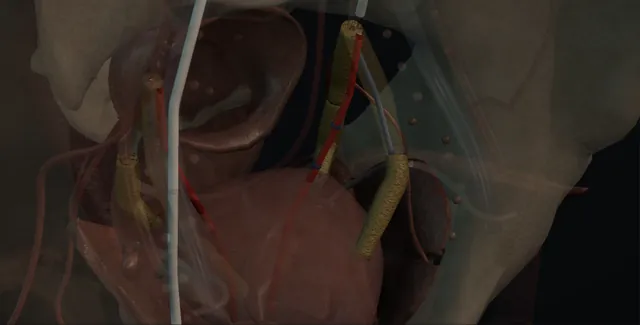
Prostatic artery embolisation: how the structures relateFluoroscopy of the pelvis from the front, groin to prostate. Common femoral artery: catheter over the bifurcation; forced, it dissects Internal iliac and its anterior division (danger). Internal iliac and its anterior division: pudendal branches supply it; inspect after Perineal and penile skin (danger). Internal iliac and its anterior division: leaves the anterior division, medially and down Prostatic artery. Prostatic artery: shares its origin; the tip sits beyond them Bladder base, rectal and penile branches (danger). Prostatic artery: particles fill the lobe until flow stops The gland blush. The gland blush: balloon contrast marks the bladder neck Catheter balloon at the bladder neck.Groin: femoral punctureTarget: the gland!Common femoral artery!Internal iliac and itsanterior division!Perineal andpenile skinProstatic artery!Bladder base,rectal and penilebranchesThe gland blushCatheter balloonat the bladderneckcatheter over the bifurcation;forced, it dissectspudendal branchessupply it; inspectafterleaves the anterior division,medially and downshares its origin;the tip sits beyondthemparticles fill the lobe untilflow stopsballoon contrastmarks the bladderneckTargetDangerLandmark
Fluoroscopy of the pelvis from the front, groin to prostate. Schematic, not to scale; relationships only.
  • On the screen the pelvis is seen from the front with the catheter rising from the groin over the bifurcation and turning down into the internal iliac, whose anterior division fans out across the sidewall with the prostatic artery leaving it and running medially and downward to the gland at the base of the bladder, marked by the catheter balloon.
  • The prostatic artery arises next to the arteries of the rectum behind, the bladder above and the penis in front and below, and the microcatheter tip has to sit beyond those branches.
  • When the particles fill the gland the blush spreads across the lobe on that side and the flow in the artery stops.
  • ›The common femoral artery - the puncture under ultrasound; the sheath, the heparin and the closure device or pressure at the end.
  • ›The internal iliac and its anterior division - the oblique run that spreads the branches; superior vesical, obturator, inferior vesical, middle rectal, internal pudendal, inferior gluteal.
  • ›The prostatic artery - a millimetre or so across, of variable origin, with a capsular and a central branch; usually one on each side, sometimes two.
  • ›The bladder base, rectal and penile branches - the non-target anastomoses seen on the selective run and the cone-beam CT; the tip is advanced beyond them or they are coiled.
  • ›The gland blush - the prostate enhancing from the microcatheter on cone-beam CT confirms the target; its disappearance at stasis is the endpoint.
  • ›The urethral catheter and balloon - a little contrast in the balloon marks the bladder neck on the screen; removed at the end unless he came in with one.
  • ›The perineal and penile skin - supplied by the pudendal branches beside the prostatic origin; inspected after the procedure for blanching or ulceration.
Click me next Section 3 of 8 Steps The operation in order, with the landmark that guards each move.

Operative steps

  1. Who chooses embolisation The man with bothersome lower urinary tract symptoms from a benign prostate, usually over about forty millilitres, assessed with a score and a flow rate, whose medication has failed or is not tolerated, and who does not want an operation or cannot safely have one: the anticoagulated, the unfit for anaesthesia, the man in retention who wants a chance of voiding, or recurrent prostatic haematuria. He is told what NICE says: it works, the gain is less than after TURP, and some need a second treatment. Cancer is excluded by PSA, examination and MRI where indicated; infection, a neuropathic bladder, severe iliac disease or aneurysm, poor renal function and contrast allergy are the reasons to say no. The decision is made with the radiologist at the meeting.
  2. CT angiography and the plan A CT angiogram of the pelvis in the arterial phase before the procedure, read by the radiologist who will do it. It shows the prostate volume, the aortoiliac disease and tortuosity that decide between the femoral and the radial route, and above all the origin of the prostatic artery on each side, which is variable: most often from the internal pudendal, otherwise from a trunk with the superior vesical, from the obturator, the inferior gluteal or directly from the anterior division, and there are often two on one side. It shows the branches that matter because they are not prostate: the rectal, vesical and penile arteries and their anastomoses with the prostatic supply. The plan, which artery on which side and which approach, is written before the man arrives.
  3. Access and the internal iliac Local anaesthetic to the right groin, or the left wrist, with sedation and analgesia as the man needs, a urethral catheter with a little contrast in the balloon to mark the bladder neck. A 5 French sheath is placed in the common femoral artery under ultrasound. A catheter is passed over the aortic bifurcation into the contralateral internal iliac, or up and over into the ipsilateral one, and an angiogram in the oblique projection opens the anterior division and shows the branches: superior vesical, obturator, inferior vesical, middle rectal, internal pudendal, inferior gluteal. The prostatic artery is found on this run and compared with the CT. The man is on heparin through the sheath, and the radiation dose is watched from the start because this is a long procedure.
    where it bites: a stiff or tortuous iliac dissects under a catheter forced round it; the route was chosen on the CT and a dissection is recognised on the run and the side abandoned.
  4. The anterior division and the prostatic artery The anterior division is entered with the catheter and a 2 French microcatheter is advanced over a microwire into the prostatic artery, which is typically a millimetre or so across and runs medially and inferiorly toward the gland, giving a capsular branch that runs around the gland and a central branch that enters it. A selective run from the microcatheter shows the gland blush, the capsular and central branches, and any branch leaving the gland: to the bladder base, a middle rectal, a branch to the penis through the internal or accessory pudendal, or an anastomosis across the midline to the other prostatic artery. Vasodilator through the microcatheter opens a spastic artery. The tip is advanced beyond any non-target branch where the artery allows it.
    where it bites: the prostatic artery shares its origin and its collaterals with the rectum, bladder and penis; the tip sits beyond those branches before any particle is injected.
  5. Microcatheter, cone-beam CT and the non-target check With the microcatheter in place a cone-beam CT is acquired during a slow injection of contrast through it: a rotational run reconstructed as a three dimensional image that shows exactly what the microcatheter tip supplies. Enhancement of the prostate on that side confirms the artery; enhancement of the bladder wall, the rectal wall, the penis or the perineal skin shows a non-target branch and is the reason to reposition the tip further in, to coil the branch first, or to leave that pedicle alone. The angle of the C-arm is set from the reconstruction so the fluoroscopy shows the tip and the gland. This step is the safety of the procedure: the syndrome and the bleeding are tolerable, a dead patch of bladder or rectum is not.
    where it bites: a bladder base or rectal branch that enhances on cone-beam CT is a non-target embolisation waiting to happen; the check is done before the first syringe of particles, not after.
  6. Embolisation to stasis Calibrated microspheres, 100 to 500 micrometres according to the unit protocol with the smaller sizes reaching the centre of the gland, are suspended in contrast and injected slowly through the microcatheter under fluoroscopy, watching the blush of the gland and, more importantly, watching for reflux back along the catheter into the parent artery, which sends particles somewhere else. The injection continues until the flow in the artery stops, near stasis, and a check run shows no more gland enhancement and no new non-target branch opened by the change in pressure. Some units then advance the microcatheter into the gland and embolise again, so that the capsular and central branches are both filled. The particles are counted and recorded.
    where it bites: reflux of particles from a stagnant artery goes to whatever the parent artery supplies next; the injection is slowed as stasis approaches and stopped before reflux is seen.
  7. The other side The catheter is withdrawn and the contralateral internal iliac catheterised, from the same femoral puncture by the crossover or by re-forming the catheter, and the sequence is repeated: anterior division run, microcatheter into the prostatic artery, cone-beam CT, embolisation to stasis. Bilateral embolisation is the aim because the result is better; the second side is sometimes not possible for anatomy, spasm or dissection, and a unilateral procedure is accepted and named as such rather than forced. A final run of each internal iliac confirms the non-target branches are open. The sheath is removed, the puncture closed with a device or pressure, and the total fluoroscopy time and dose are written in the note because the skin dose can be high enough to matter.
  8. After: the post-embolisation syndrome He goes home the same day or the next with the catheter out, unless he came in with one, which stays a week or two before a trial. For up to a week most men have the post-embolisation syndrome: pelvic and perineal pain, dysuria, frequency and urgency, a low-grade fever, nausea, and some blood in the urine, semen or stool. Paracetamol, a non-steroidal, an alpha blocker and antibiotics by protocol; he is told which symptoms are the syndrome and which are not. Retention in the first week is a catheter and a trial in one to two weeks. A patch of penile or perineal skin that blanches or ulcerates, rectal bleeding beyond a streak, or a persistent fever are reviewed the same day, because those are non-target.
  9. Outcomes and re-treatment By three months the gland has shrunk by about a third on imaging and the symptom score has fallen by around ten points in most men, with a modest gain in flow rate; the effect is real, and it is less than after TURP, which is what NICE says he should be told. Ejaculation and erection are usually preserved, and that is the reason many men choose it. Review at three and twelve months with symptom score, flow and residual. A proportion of men, about one in five over the following years on current teaching, need a second treatment: repeat embolisation after new CT angiography, since the supply has changed, or a TURP or enucleation, which is not made harder by the embolisation.
Click me next Section 4 of 8 Danger zones Where this operation bites, and why it bites there.
Critical relationship

Danger zones

The spatial relationships the animation is built to make automatic. Before you read each one, ask yourself: at this point in the operation, what is about to go wrong, and what is my first move? The save for each sits in the Complications tab.

  • RiskThe bladder, rectal and penile collaterals - the non-target branches and anastomoses; particles here mean bladder wall ischaemia, a rectal ulcer or penile skin necrosis.
  • RiskThe internal iliac and the access site - dissection of a tortuous or atheromatous iliac under a forced catheter; haematoma or pseudoaneurysm at the femoral puncture.
  • RiskThe prostatic capsular anastomoses - the capsular branch joins the other side and the vesical supply; reflux or over-embolisation sends particles across.
  • RiskContrast and radiation - a long procedure with a high skin dose and a large contrast load in a man who is often old with poor kidneys.
Practise it Take me into the scene Opens the animation at a danger moment and asks you to predict the mechanism and your first move before it reveals.
Click me next Section 5 of 8 Complications The save for each one: the first move, the escalation trigger, the op-note line.

Complications and immediate saves

Trigger each of these live in the Animation tab, then read the save: the first move, the escalation trigger, and the line that goes in the op note.

Non-target embolisation

  • particles in a branch that was not prostate: a patch of bladder wall, the rectal mucosa, the penile or perineal skin, seen as pain, bleeding, an ulcer or a blanched area in the first days.
The save
  1. Prevent it: selective run and cone-beam CT from the microcatheter before every injection, the tip beyond the non-target branch or the branch coiled, and the injection stopped before reflux. first move
  2. A blanched penile or perineal patch, rectal bleeding or severe dysuria in the first days: examine, and cystoscopy or flexible sigmoidoscopy to see the extent.
  3. Most ischaemic patches heal with analgesia, catheter drainage for bladder ulcers, laxatives for rectal ulcers, and time; skin is dressed and kept clean.
  4. A full-thickness bladder or rectal wall necrosis, a fistula, or skin necrosis that extends: admit, image, and involve the radiologist and the surgical specialty for that organ. escalation trigger
  5. Procedure note: the cone-beam findings on each side, the branches coiled, the particle size and volume, and any reflux seen; the ward note names the non-target sign and who reviewed it. document

Access-site bleeding, pseudoaneurysm or arterial injury

  • a groin haematoma or a pulsatile lump after the sheath, or a dissection of the iliac under the catheter in an atheromatous pelvis.
The save
  1. Pressure on the puncture site for a timed period or a closure device, then bed rest by protocol and the groin and distal pulses checked with the observations. first move
  2. A lump that expands or a bruit: duplex ultrasound for pseudoaneurysm, which is treated with compression or thrombin injection by the radiologist.
  3. A dissection seen on the run: the side is abandoned, the true lumen confirmed, and the leg observed; most are managed conservatively.
  4. A cold or painful leg, a falling haemoglobin or a retroperitoneal haematoma on CT: vascular surgery and transfusion now. escalation trigger
  5. Note: the access site and closure method, the heparin given, any dissection and its management, and the pulse chart. document

Post-embolisation syndrome

  • the expected week: pelvic and perineal pain, dysuria, frequency, low fever, nausea and some blood in urine, semen or stool from an ischaemic gland.
The save
  1. Tell him before it happens: regular paracetamol and a non-steroidal, an alpha blocker, antibiotics by protocol, and the list of what is normal for a week. first move
  2. Fluids and a bladder scan if he is not passing urine well; a catheter if the residual is large.
  3. Fever above 38 or pain that is not settling by a week: urine and blood cultures, examination of the perineum and rectum, and antibiotics changed by the result.
  4. Rigors, sepsis, a fluctuant gland on examination or a collection on imaging: admit for intravenous antibiotics and drainage of a prostatic abscess. escalation trigger
  5. Note: the analgesia and alpha blocker, the safety-net symptoms given in writing, and the review date. document

Urinary retention and infection

  • oedema of an ischaemic gland in a man who was obstructed to begin with: retention in the first week, or infection in the necrotic tissue.
The save
  1. A bladder scan before he goes home and a catheter if he cannot void; the man who came in with a catheter keeps it for a week or two. first move
  2. An alpha blocker from the day of the procedure and a trial without catheter at one to two weeks.
  3. Infection is cultured and treated; a catheter in situ is changed.
  4. A second failed trial, or retention with a rising creatinine: a longer catheter period and the meeting again, since some men will need a resection or enucleation. escalation trigger
  5. Note: the residual at discharge, the catheter and alpha blocker, the trial date, and the plan if it fails. document

Incomplete response and re-treatment

  • the man whose score and flow have not moved at three months, or who has drifted back after two years: a unilateral procedure, a missed second artery, or recanalisation.
The save
  1. Measure, do not assume: symptom score, flow rate, residual and volume at three and twelve months against the baseline. first move
  2. Check the procedure note: was it bilateral, was a second prostatic artery on either side seen and treated, was stasis reached.
  3. A new CT angiogram if repeat embolisation is considered, because the supply will have changed and collaterals will have opened.
  4. No response, or recurrence with a large gland or retention: TURP, enucleation or simple prostatectomy, which the embolisation does not make harder. escalation trigger
  5. Note: the baseline and follow-up scores and flows side by side, the sides treated, and the man's stated preference for the next step. document
Click me next Section 6 of 8 Troubleshooting When the case turns: the problem, then the next move.

Intra-operative troubleshooting

When the case turns: the problem, then the next move.

  • ActionThe prostatic artery cannot be found on the anterior division run - go back to the CT angiogram; look at the internal pudendal and the superior vesical trunk in a steeper oblique, and give a vasodilator for spasm before calling the side impossible.
  • ActionCone-beam CT shows the rectum enhancing from the microcatheter - advance the tip beyond the rectal branch if the artery allows, coil the branch if it does not, and if neither is possible leave that pedicle and treat the other side.
  • ActionReflux appears around the microcatheter during injection - stop at once; the artery is at stasis already; a check run, and no further particles down that pedicle.
  • ActionThe iliac is too tortuous for the crossover - an ipsilateral up-and-over approach, a radial approach, or a stiffer guide catheter; a dissection on a forced catheter costs more than a second puncture.
  • ActionHe cannot pass urine on the ward after the procedure - bladder scan, catheter if the residual is large, alpha blocker, and a trial without catheter at one to two weeks; he is not sent home in retention.
  • ActionA blanched patch on the glans the next morning - non-target embolisation of a penile branch until proved otherwise; photograph it, examine the perineum and rectum, tell the radiologist the same day, and dress and review it daily.
Click me next Section 7 of 8 FRCS viva Say it out loud, then mark yourself against the model answer.

FRCS viva questions

The examiner stems this scene prepares you for. Try to answer out loud first, then reveal, and learn the discriminator, the detail that turns a pass into a strong pass.

QWho would you consider for prostatic artery embolisation, and what would you tell him?reveal answer
  • I would consider it for a man with bothersome lower urinary tract symptoms from benign prostatic enlargement, usually a gland over about forty millilitres, whose symptoms have been objectively assessed with a symptom score, flow rate and residual, and in whom medical treatment has failed or is not tolerated.
  • The men who gain most are those who want to avoid an operation or cannot safely have one: the anticoagulated, the unfit for anaesthesia, the man in retention who wants a chance of voiding, and the man with recurrent prostatic haematuria.
  • I would exclude prostate cancer, active infection, a neuropathic bladder, severe iliac disease or aneurysm, poor renal function and contrast allergy, and I would have the CT angiogram read by the interventional radiologist before agreeing.
  • I would tell him what NICE interventional procedures guidance says: that the procedure works, that the improvement in symptom score and flow is smaller than after TURP, and that a proportion of men need a second treatment;
  • that he will have a week of pelvic pain, dysuria and bleeding;
  • that the radiation dose is high;
  • and that the serious but rare risk is embolisation of the bladder, rectum or penis.
  • I would also tell him that ejaculation and erection are usually preserved, which is often why he is asking.
Strong pass

Say all three

  • The comparison with TURP stated as NICE states it
  • Less benefit and a re-treatment rate
  • Alongside the sexual function preservation that drives the choice
Examiner note
The comparison with TURP stated as NICE states it, less benefit and a re-treatment rate, alongside the sexual function preservation that drives the choice.
QDescribe the steps of a prostatic artery embolisation and where the urologist fits in.reveal answer
  • The urologist selects the man, excludes cancer and the contraindications, consents him, and manages him afterwards; the interventional radiologist performs the procedure, and both attend the planning meeting.
  • After a pre-procedure CT angiogram to map the prostatic artery origin on each side and the non-target branches, the man has a urethral catheter with contrast in the balloon to mark the bladder neck and a common femoral or radial puncture under local anaesthetic and sedation.
  • A catheter is passed into the internal iliac and an oblique run opens the anterior division.
  • A microcatheter is advanced into the prostatic artery, a selective run shows the gland and any branch to bladder, rectum or penis, and a cone-beam CT during injection confirms that the tip supplies prostate and nothing else;
  • the tip is advanced beyond, or the branch coiled, if it does not.
  • Calibrated microspheres are injected slowly to near stasis, watching for reflux, with a check run at the end.
  • The contralateral side is treated the same way, bilateral being the aim.
  • The sheath is removed and the puncture closed, and the fluoroscopy time and dose are recorded.
  • He goes home the same or the next day on analgesia, a non-steroidal and an alpha blocker, with a written list of the syndrome symptoms and of the warning signs of non-target embolisation.
Strong pass

Say both

  • Cone-beam CT from the microcatheter as the non-target check before the first injection
  • Stasis without reflux as the endpoint
Examiner note
Cone-beam CT from the microcatheter as the non-target check before the first injection, and stasis without reflux as the endpoint.
QWhat are the complications of prostatic artery embolisation?reveal answer
  • I would divide them into the expected, the access-related, the non-target and the failures.
  • Expected: the post-embolisation syndrome in most men for about a week, with pelvic pain, dysuria, frequency, low-grade fever and blood in the urine, semen or stool, managed with analgesia, a non-steroidal, an alpha blocker and reassurance;
  • acute retention in a few per cent, and urinary infection or rarely a prostatic abscess in the necrotic gland.
  • Access-related: groin haematoma, pseudoaneurysm and, in the atheromatous pelvis, iliac dissection; contrast nephropathy and allergy; and a high radiation dose with a small risk of skin injury from a long procedure.
  • Non-target: particles in the bladder base, the rectal wall or the penile or perineal skin through the collaterals that the prostatic artery shares, causing ischaemic ulcers, bleeding, pain and rarely a fistula or skin necrosis, which is why the cone-beam CT check is done before every injection.
  • Failures: technical failure on one or both sides from anatomy or spasm, a unilateral result, and clinical failure or recurrence in a proportion of men, about one in five on current teaching over the following years, treated by repeat embolisation after new imaging or by an outlet operation.
  • Sexual function is usually preserved, and retrograde ejaculation is uncommon compared with resection.
Strong pass

Say both

  • Non-target embolisation explained by the shared collaterals and linked to the cone-beam CT check
  • Rather than listed as a bare risk
Examiner note
Non-target embolisation explained by the shared collaterals and linked to the cone-beam CT check, rather than listed as a bare risk.
QA man is unhappy with the result a year after embolisation. What are his options?reveal answer
  • I would first measure rather than accept his account: repeat the symptom score, flow rate, residual and prostate volume against the baseline, and exclude other causes such as infection, a stricture, bladder dysfunction or a stone.
  • I would read the procedure report for whether the embolisation was bilateral, whether a second prostatic artery on either side was found and treated, whether stasis was reached and whether there was any non-target embolisation, and I would repeat the PSA and examination, since cancer must still be excluded in a gland that was never sampled.
  • If the gland was treated on one side only, or a large vascular gland has recanalised, repeat embolisation is reasonable after a new CT angiogram, because the supply and the collaterals will have changed and the first map is out of date.
  • If the gland remains large and obstructing with a poor flow and residual, or he is in retention, I would offer a definitive outlet operation, a TURP for the moderate gland or enucleation or simple prostatectomy for the large one, and I would tell him that the embolisation does not make that operation harder or its result worse.
  • If he is fit for neither and his residual is safe, medication or a catheter strategy remain.
  • The decision goes back to the meeting and is his.
Strong pass

Say both

  • Measuring the failure and re-reading the procedure note before choosing
  • The point that embolisation does not compromise a later resection
Examiner note
Measuring the failure and re-reading the procedure note before choosing, and the point that embolisation does not compromise a later resection.
Click me next Section 8 of 8 Evidence The guidance each of these answers is built on.

Evidence and guidance

Two minutes

Close the lap: recall it before you leave.

Say it aloud, then reveal. Reading is not remembering.

  1. 01 The relationship Say the critical spatial relationship out loud, then reveal. reveal
    Memory frame

    On the screen the pelvis is seen from the front with the catheter rising from the groin over the bifurcation and turning down into the internal iliac, whose anterior division fans out across the sidewall with the prostatic artery leaving it and running medially and downward to the gland at the base of the bladder, marked by the catheter balloon.

    Why?
    The prostatic artery arises next to the arteries of the rectum behind, the bladder above and the penis in front and below, and the microcatheter tip has to sit beyond those branches. When the particles fill the gland the blush spreads across the lobe on that side and the flow in the artery stops.
  2. 02 The first move Non-target embolisation. Your first move? reveal
    First move

    Prevent it: selective run and cone-beam CT from the microcatheter before every injection, the tip beyond the non-target branch or the branch coiled, and the injection stopped before reflux.

  3. 03 The escalation trigger Non-target embolisation. What makes you call for help? reveal
    Escalate when

    A full-thickness bladder or rectal wall necrosis, a fistula, or skin necrosis that extends

    More detail
    A full-thickness bladder or rectal wall necrosis, a fistula, or skin necrosis that extends: admit, image, and involve the radiologist and the surgical specialty for that organ.
  4. 04 The discriminator What turns a pass into a strong pass? reveal
    Strong pass

    Say all three

    • The comparison with TURP stated as NICE states it
    • Less benefit and a re-treatment rate
    • Alongside the sexual function preservation that drives the choice
    Examiner note
    The comparison with TURP stated as NICE states it, less benefit and a re-treatment rate, alongside the sexual function preservation that drives the choice.
  5. 05 The pitfall Where does this operation bite first? reveal
    Pitfall

    The bladder, rectal and penile collaterals

    Why?
    the non-target branches and anastomoses; particles here mean bladder wall ischaemia, a rectal ulcer or penile skin necrosis.

Revision practice with self-marked answers, not an assessment of clinical competence. Kept in this browser only; it does not sync between devices.