UrOops3D, danger-zone atlas

Schematic infant groin: every dimension is an authoring estimate.

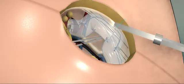
Operative animation, Paediatric inguinal hernia repair

Open inguinal hernia repair in an infant: high ligation of the sac

Indications

  • An intermittent bulge in the groin, with or without a reducible mass.
  • An incarcerated or symptomatic hernia.

Alternatives

  • Laparoscopic repair with percutaneous ligation of the processus.
  • High ligation without opening the external oblique aponeurosis, by age, length of canal and preference.
  • In older patients, a tension-free repair with mesh.

Risks

  • Bleeding, and swelling of the groin or scrotum afterwards.
  • Injury to the ilioinguinal nerve.
  • Injury to the testis or the vas deferens.
  • Bowel injury.
  • Recurrent hernia.

Herniotomy for the inguinal hernia of infancy: a patent processus vaginalis wide enough to let bowel or omentum into the groin.

Through a skin crease incision over the superficial ring the canal is opened along the fibres of the external oblique, the ilioinguinal nerve is found and kept, and the sac is dissected off the vas and the testicular vessels.

The sac is tied at the deep ring; the floor of the canal is repaired only for a large hernia or abnormal tissue. The lesson is where the sac lies on the cord, what may be held and what may not, and what marks the level of the tie.

Core tier FRCS paediatric urology, inguinoscrotal surgery Interactive 3D

Operative visualisation

Schematic scene preview: Paediatric inguinal hernia repair
Preparing the 3D anatomy…

UrOops3D · UroRef · © Nity G

How to use this module. Play the eight steps, or use Back and Next. Watch each layer open in turn, the nerve drawn aside, the sac come clear of the vas and the vessels, the tie at the deep ring and the testis drawn back down. The three triggers show a divided nerve, a crushed vas and a tie placed too low.

Click me next Section 2 of 8 Anatomy The structures this operation works between, and the one it must not cross.

The structures in the scene

Paediatric inguinal hernia repair: how the structures relateThe right inguinal canal of an infant, opened from in front. Hernia sac, on the front of the cord: swept off it under vision, never held Vas deferens, against its back wall (danger). Hernia sac, on the front of the cord: separated from it with the vas Testicular vessels, beside the vas (danger). Hernia sac, on the front of the cord: outside the cremaster; kept out of the field Ilioinguinal nerve, on the cord (danger). Hernia sac, on the front of the cord: tied here, flush, on the sac alone Deep ring: the level of the tie. Hernia sac, on the front of the cord: seen at the neck: the tie is high enough Preperitoneal fat round the neck. Hernia sac, on the front of the cord: opened along its fibres to reach it External oblique aponeurosis, in front.Hernia sac, on thefront of the cord!Vas deferens,against its backwall!Testicular vessels,beside the vas!Ilioinguinal nerve,on the cordDeep ring: the levelof the tiePreperitoneal fatround the neckExternal obliqueaponeurosis, infrontswept off it undervision, never heldseparated from itwith the vasoutside thecremaster; kept outof the fieldtied here, flush,on the sac aloneseen at the neck:the tie is highenoughopened along itsfibres to reach itTargetDangerLandmarkContext
The right inguinal canal of an infant, opened from in front. Schematic, not to scale; relationships only.
  • Seen from in front with the head at the top, the right groin runs from the anterior superior iliac spine, above and lateral, down to the pubic tubercle beside the midline.
  • The canal lies just above that line: the deep ring lateral and deep, the superficial ring beside the tubercle.
  • The cord runs between them under the external oblique aponeurosis with the ilioinguinal nerve on its surface, then turns down over the pubis to the testis.
  • Inside the cremaster the sac lies in front and medial, the vas and the vessels behind it.
  • At the deep ring the sac becomes peritoneum within a collar of fat, the vessels run up, the vas turns medially, and the inferior epigastric vessels pass just medial to the ring.
  • ›Skin crease and its two landmarks - the incision lies in the crease between the pubic tubercle and the anterior superior iliac spine, over the superficial ring.
  • ›Scarpa’s fascia - a distinct membrane in the fat of an infant’s groin; divided with diathermy before the aponeurosis is reached.
  • ›External oblique aponeurosis - the front wall of the canal; its fibres run down and medially to the superficial ring, and it is opened in their line.
  • ›Superficial and deep rings - the two ends of the canal, close together in an infant; the cord leaves through the superficial ring and the sac is tied at the deep ring.
  • ›Ilioinguinal nerve - on the cord directly under the aponeurosis, leaving through the superficial ring; found, kept out of the field and replaced.
  • ›Cremaster - the muscular covering of the cord; the sac and the cord lie inside it.
  • ›Hernia sac - the patent processus vaginalis: a thin-walled tube on the front and inner side of the cord, continuous with the peritoneum at the deep ring.
  • ›Vas deferens - firm and white, against the back wall of the sac; above the deep ring it turns medially toward the pelvis.
  • ›Testicular vessels - the artery and veins of the testis, beside the vas behind the sac; above the deep ring they run cephalad.
  • ›Preperitoneal fat and inferior epigastric vessels - the fat appears round the neck of the sac at the deep ring; the vessels lie just medial to the ring.
  • ›Testis - in the scrotum throughout; it can ride up when the cord is lifted and is checked before the closure.
Click me next Section 3 of 8 Steps The operation in order, with the landmark that guards each move.

Operative steps

  1. Skin crease incision The child is supine and the lower abdomen and external genitalia are prepared and draped. The incision is made in the skin crease between the pubic tubercle and the anterior superior iliac spine, over the superficial ring, in the line of the skin tension lines. Scarpa’s fascia is divided with diathermy and the external oblique aponeurosis is seen in the floor of the wound.
    where it bites: The cord and the sac lie directly under the aponeurosis: the wound is deepened one named layer at a time.
  2. Open the canal The aponeurosis is cleaned until its fibres and the superficial ring are seen, with the cord leaving the canal through the ring. The ring is opened toward the deep ring in the line of the fibres. The ilioinguinal nerve lies on the cord directly beneath the aponeurosis: it is identified, freed and retracted out of the operative field.
    where it bites: The ilioinguinal nerve is cut with the aponeurosis if it is not looked for first.
  3. Lift the cord, find the sac The cord is lifted into the wound, which confirms the hernia sac. The sac lies on the front and inner side of the cord, inside the cremaster. The testicular vessels and the vas deferens are identified before any dissection begins.
    where it bites: Nothing is separated until the vas and the vessels have been seen.
  4. Free the sac from the cord The sac is dissected off the cord under direct vision, the vas and the vessels being separated from its back wall until the sac is clear all the way round. The sac is then inspected: no bowel and no omentum lie within it.
    where it bites: The vas and the vessels are swept off the sac and are not picked up: forceps on the vas can crush it.
  5. Follow the sac to the deep ring The sac is followed up to the deep ring, where it becomes peritoneum. Preperitoneal fat appears round its neck at this level, and the inferior epigastric vessels lie just medial to the ring.
    where it bites: A sac that is not followed to the ring is tied low.
  6. High ligation A high ligation of the sac is performed at the deep ring with an absorbable suture. The tie takes the sac alone, with the vas and the vessels outside it.
    where it bites: A tie that includes the vas or the vessels, or one that sits below the ring.
  7. Replace the cord and the nerve The cord is replaced in the canal in its normal position and orientation. The canal is irrigated with saline and inspected, and bleeding points are controlled with diathermy. The ilioinguinal nerve is inspected, seen to be intact and replaced beneath the aponeurosis. The testis is checked in the scrotum before the wound is closed.
    where it bites: A cord laid back twisted, or a testis left high in the scrotum by the handling of the cord.
  8. Close in layers The external oblique aponeurosis is closed with a running absorbable suture, then Scarpa’s fascia, then the skin with a subcuticular suture. The wound is supported with adhesive strips and a dressing.
    where it bites: A stitch in the aponeurosis that catches the nerve, or a closure that narrows the superficial ring on the cord.
Click me next Section 4 of 8 Danger zones Where this operation bites, and why it bites there.
Critical relationship

Danger zones

The spatial relationships the animation is built to make automatic. Before you read each one, ask yourself: at this point in the operation, what is about to go wrong, and what is my first move? The save for each sits in the Complications tab.

  • RiskUnder the aponeurosis - the ilioinguinal nerve lies on the cord exactly where the scissors open the ring.
  • RiskThe back wall of the sac - the vas and the testicular vessels lie against it; they are separated under vision and never held.
  • RiskThe neck of the sac - the tie belongs at the deep ring, where the fat appears; lower than that, sac is left behind.
  • RiskInside the sac - bowel or omentum may lie within it; the sac is seen to be empty before it is tied.
  • RiskThe closure - the running stitch in the aponeurosis passes close to the nerve and the cord.
Practise it Take me into the scene Opens the animation at a danger moment and asks you to predict the mechanism and your first move before it reveals.
Click me next Section 5 of 8 Complications The save for each one: the first move, the escalation trigger, the op-note line.

Complications and immediate saves

Trigger each of these live in the Animation tab, then read the save: the first move, the escalation trigger, and the line that goes in the op note.

Ilioinguinal nerve injury

  • the nerve is divided as the aponeurosis is opened, or caught in its closure.
The save
  1. Look for the nerve as the ring is opened, free it and keep it out of the field. first move
  2. Inspect it before the closure and place the stitches clear of it.
  3. A nerve found divided is shown to a senior colleague before the wound is closed. escalation trigger
  4. Operation note: nerve identified, protected and seen intact at closure, or the injury and its site. document

Vas deferens injury

  • the vas or the testicular vessels are crushed, stripped or divided while the sac is being freed.
The save
  1. Stop. Find the vas and the vessels above and below the place and see what has been done. first move
  2. No diathermy on the cord; from here on only the sac is held.
  3. A crushed or divided vas, or a testis that looks dusky at the end, goes to a senior paediatric surgeon or urologist before the child leaves theatre. escalation trigger
  4. Operation note: what was injured, where, and who was told. The family is told the same day. document

Recurrent hernia

  • the sac was tied below the deep ring, or the tie did not hold; a bulge returns in the groin.
The save
  1. On the table: if no fat has been seen at the neck, free the sac higher and tie it again at the ring. first move
  2. A large hernia or a connective tissue disorder is a reason to consider repair of the floor of the canal.
  3. A bulge that returns after repair is referred back for assessment and a planned second operation. escalation trigger
  4. Operation note: the level of the tie and what was seen at the neck. document
Click me next Section 6 of 8 Troubleshooting When the case turns: the problem, then the next move.

Intra-operative troubleshooting

When the case turns: the problem, then the next move.

  • ActionThe sac tears while it is being freed - its edges are picked up with mosquitoes, the vas and the vessels are identified again, and the dissection is carried above the tear so that the tie sits on intact sac at the ring.
  • ActionBowel or omentum lies in the sac - it is returned to the abdomen before the neck is tied; the sac is seen to be empty first.
  • ActionThe child is a girl - the sac lies on the round ligament and is dissected from the structures beside it; the ligation is the same.
  • ActionA hydrocele on the same side - the testis can be delivered and the tunica vaginalis opened at the same operation.
  • ActionA large hernia, or a child with a connective tissue disorder - repair of the floor of the canal may be added to the ligation.
  • ActionA small infant with a short canal - the sac can be tied without opening the external oblique aponeurosis, according to age, the length of the canal and the surgeon’s preference.
  • ActionThe other side - contralateral exploration is done when indicated, by open exploration or with a laparoscope passed through the opened sac.
  • ActionThe testis sits high at the end - gentle traction on the scrotum brings it down, and it is seen at the floor of the scrotum before the skin is closed.
Click me next Section 7 of 8 FRCS viva Say it out loud, then mark yourself against the model answer.

FRCS viva questions

The examiner stems this scene prepares you for. Try to answer out loud first, then reveal, and learn the discriminator, the detail that turns a pass into a strong pass.

QDescribe an open inguinal hernia repair in an infant boy.reveal answer
  • I would position the child supine, prepare the lower abdomen and genitalia, and make a short incision in the skin crease between the pubic tubercle and the anterior superior iliac spine, over the superficial ring.
  • I divide Scarpa’s fascia with diathermy and clean the external oblique aponeurosis until I can see its fibres and the ring.
  • I open the ring toward the deep ring along the fibres, and I find the ilioinguinal nerve on the cord, free it and keep it out of the field.
  • I lift the cord into the wound, which shows me the sac on its front and inner side, and I identify the vas and the testicular vessels before I separate anything.
  • I dissect the sac off them under vision, handling only the sac, until it is free on every side, and I check that it is empty.
  • I follow it to the deep ring, where preperitoneal fat appears at its neck, and I tie it there with an absorbable suture, with nothing but sac in the tie.
  • I replace the cord in its own orientation, check for bleeding, see that the nerve is intact and put it back under the aponeurosis, and check the testis in the scrotum.
  • I close the aponeurosis with a running absorbable suture, then Scarpa’s fascia and the skin.
Strong pass

Say all three

  • Naming the nerve before the cord is touched
  • Saying that only the sac is held
  • Giving the preperitoneal fat at the deep ring as the level of the tie
Examiner note
Naming the nerve before the cord is touched, saying that only the sac is held, and giving the preperitoneal fat at the deep ring as the level of the tie.
QHow does a hernia differ from a communicating hydrocele, and does that change the operation?reveal answer
  • Both are a patent processus vaginalis.
  • In a communicating hydrocele only peritoneal fluid passes down it; the European paediatric urology guideline defines a hernia as a processus wide enough to let abdominal viscera or omentum through.
  • That difference changes the timing more than the technique.
  • The same guideline advises observing most infant hydroceles before considering surgery, and operating early when a hernia is suspected.
  • The operation is the same in principle: through an inguinal incision I would ligate the patent processus, and the guideline describes leaving the distal stump open.
  • Its recommendation is worded as closing the processus vaginalis at the inguinal ring, which is the high ligation of the hernia operation.
Strong pass

Say both

  • Defining the hernia by what the processus admits
  • Saying that the suspicion of a hernia moves the operation forward
Examiner note
Defining the hernia by what the processus admits, and saying that the suspicion of a hernia moves the operation forward.
QWhile you are freeing the sac, your assistant picks up the vas in toothed forceps. What do you do?reveal answer
  • I would stop and ask for the forceps to be released, then look.
  • I identify the vas above and below the place it was held and see whether it is intact, flattened or divided, and I look at the testicular vessels beside it.
  • I would not use diathermy near it.
  • If the vas has only been touched, I note it and carry on, sweeping the cord structures off the sac and holding nothing but the sac.
  • If it is crushed or divided I would call a senior paediatric surgeon or urologist to theatre before I go further, because what is done next is their decision and not one to improvise.
  • I finish the ligation safely, record exactly what happened and where, and tell the family the same day.
  • Injury to the testis or the vas is among the complications I would have discussed at consent.
  • The European guideline states that in expert hands testicular damage during hydrocele or hernia repair is very low; I would quote that as their statement and give no figure of my own.
Strong pass

Say all three

  • Stopping to define the injury
  • Calling for senior help for a crushed or divided vas
  • Telling the family on the day
Examiner note
Stopping to define the injury, calling for senior help for a crushed or divided vas, and telling the family on the day.
QA bulge returns in the same groin some months after the repair. Why does it happen and how is it prevented?reveal answer
  • A recurrent hernia is one of the recognised complications of this operation.
  • The cause I would look for first is technical: a sac tied below the deep ring leaves a funnel of peritoneum above the tie, and the hernia comes back through it.
  • A tie that slips, or a sac that was torn and not closed at its neck, does the same.
  • I prevent it by following the sac until I see preperitoneal fat round its neck, which tells me I am at the ring, and tying there.
  • In a large hernia, or in a child with a connective tissue disorder, I would consider repairing the floor of the canal as well.
  • For the child in front of me I would examine both groins and the testis, explain to the family that the first operation has not held, and plan the second repair with a senior colleague, because it is a dissection through scar.
  • The European Pediatric Surgeons’ Association guideline found no approach definitely superior and graded the available evidence as moderate to low in quality, so I would present my prevention as surgical principle.
Strong pass

Say both

  • Giving the low tie as the technical cause
  • The preperitoneal fat at the neck as the check that prevents it
Examiner note
Giving the low tie as the technical cause, and the preperitoneal fat at the neck as the check that prevents it.
QWhen would you look at the other side?reveal answer
  • I would not explore the other groin as a routine.
  • The operative text I follow says that contralateral exploration may be performed when indicated, by open exploration or laparoscopically, and it describes passing a laparoscope through the opened sac to look at the opposite internal ring.
  • The European paediatric urology guideline says that laparoscopic correction of a contralateral patent processus may be considered, but it cites a meta-analysis that found insufficient evidence to recommend it for inguinal hernia.
  • The European Pediatric Surgeons’ Association guideline examined contralateral exploration and described the evidence on it as less conclusive than for its main findings.
  • So I would examine both sides in clinic, raise the question with the family, and reserve a look at the other side for a child in whom there is a clinical reason.
Strong pass

Say both

  • Saying "when indicated" and not "always" or "never"
  • Knowing that a laparoscope can be passed through the opened sac
Examiner note
Saying "when indicated" and not "always" or "never", and knowing that a laparoscope can be passed through the opened sac.
Click me next Section 8 of 8 Evidence The guidance each of these answers is built on.

Evidence and guidance

Operative Dictations in Urologic Surgery
Armenakas NA, Fracchia JA, Golan R. Open inguinal hernia repair. Chapter 119, paediatric surgery section. Wiley, 2019
Read in full on 13 October 2026. Indications; the six essential steps (incision over the external ring; the external oblique and the ring identified; the ring opened toward the internal ring with the ilioinguinal nerve identified and avoided; the sac dissected from the cord vessels and vas, or the round ligament; high ligation; the external oblique and Scarpa’s fascia reapproximated); the variations (hydrocelectomy, repair of the canal floor, ligation without opening the external oblique, mesh in older patients, contralateral exploration); the list of complications; the order of the dictation. Its incision length and suture sizes are kept off screen. Not in the chapter, and drawn or written here from general surgical practice: opening the cremaster, the side of the cord the sac lies on, dividing and twisting the sac, transfixion, the preperitoneal fat as the landmark, the open distal sac, the check on the testis, and every danger line and management step.
Open guideline
EAU Guidelines
Paediatric Urology, chapter 7 Hydrocele: sections 7.1, 7.3 and 7.4
Web edition read on 13 October 2026. A hernia is a processus vaginalis wide enough to pass abdominal viscera or omentum; in children the operation is ligation of the patent processus through an inguinal incision with the distal stump left open; "close the processus vaginalis at the inguinal ring" and early surgery where a concomitant hernia is suspected are both strong recommendations; laparoscopic percutaneous ligation is named as an alternative to open herniorrhaphy; testicular damage during hydrocele or hernia repair is stated to be very low in expert hands, with no figure; laparoscopic correction of a contralateral patent processus may be considered, with a cited meta-analysis that found insufficient evidence to recommend it for inguinal hernia.
Open guideline
European Journal of Pediatric Surgery
Morini F, Dreuning KMA, Janssen Lok MJH, et al. Surgical management of pediatric inguinal hernia: a systematic review and guideline from the European Pediatric Surgeons’ Association Evidence and Guideline Committee. Eur J Pediatr Surg 32(3):219-232
Abstract read on PubMed on 13 October 2026; the full text was not opened. It addressed six questions, among them open against laparoscopic repair, contralateral exploration and the timing of surgery. No approach was found definitely superior, the available evidence was graded moderate to low in quality, the evidence on the topics outside its stated findings was described as less conclusive, and a tailored approach was recommended. Used for two viva answers; no figure is taken from it.
Open guideline

Curriculum. Supports learning relevant to the ISCP Urology Curriculum syllabus topic Paediatric urology. Not a workplace-based assessment and not evidence of competence.

Two minutes

Close the lap: recall it before you leave.

Say it aloud, then reveal. Reading is not remembering.

  1. 01 The relationship Say the critical spatial relationship out loud, then reveal. reveal
    Memory frame

    Seen from in front with the head at the top, the right groin runs from the anterior superior iliac spine, above and lateral, down to the pubic tubercle beside the midline.

    Why?
    The canal lies just above that line: the deep ring lateral and deep, the superficial ring beside the tubercle. The cord runs between them under the external oblique aponeurosis with the ilioinguinal nerve on its surface, then turns down over the pubis to the testis. Inside the cremaster the sac lies in front and medial, the vas and the vessels behind it. At the deep ring the sac becomes peritoneum within a collar of fat, the vessels run up, the vas turns medially, and the inferior epigastric vessels pass just medial to the ring.
  2. 02 The first move Ilioinguinal nerve injury. Your first move? reveal
    First move

    Look for the nerve as the ring is opened, free it and keep it out of the field.

  3. 03 The escalation trigger Ilioinguinal nerve injury. What makes you call for help? reveal
    Escalate when

    A nerve found divided is shown to a senior colleague before the wound is closed.

  4. 04 The discriminator What turns a pass into a strong pass? reveal
    Strong pass

    Say all three

    • Naming the nerve before the cord is touched
    • Saying that only the sac is held
    • Giving the preperitoneal fat at the deep ring as the level of the tie
    Examiner note
    Naming the nerve before the cord is touched, saying that only the sac is held, and giving the preperitoneal fat at the deep ring as the level of the tie.
  5. 05 The pitfall Where does this operation bite first? reveal
    Pitfall

    Under the aponeurosis

    Why?
    the ilioinguinal nerve lies on the cord exactly where the scissors open the ring.

Revision practice with self-marked answers, not an assessment of clinical competence. Kept in this browser only; it does not sync between devices.