UrOops3D, danger-zone atlas

Schematic model.

Operative animation, Paediatric nephrectomy and heminephrectomy

Paediatric nephrectomy and upper-pole heminephrectomy for benign disease

Removal of a kidney, or of one moiety of a duplex kidney, that no longer works in a child.

The total nephrectomy is done laparoscopically through three small ports: the colon reflected, the ureter traced to the hilum, the small hilar vessels clipped and divided, and the kidney taken out in a bag through the umbilicus.

The heminephrectomy removes a non-functioning upper moiety and its dilated ureter. The lesson is the lower moiety that stays: its vessels and its collecting system lie beside the line of division.

Advanced tier FRCS paediatric urology, the non-functioning kidney and the duplex system Interactive 3D

Operative visualisation

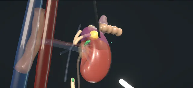
Schematic scene preview: Paediatric nephrectomy and heminephrectomy
Preparing the 3D anatomy…

UrOops3D · UroRef · © Nity G

How to use this module. Play through the ten steps, or use Back and Next. Steps one to seven are the nephrectomy: the ureter leads to the hilum and the clips go on. Steps eight to ten are the heminephrectomy, the lower pole marked as kept. The triggers show bowel injury, bleeding, an infected stump and an opened calyx.

Click me next Section 2 of 8 Anatomy The structures this operation works between, and the one it must not cross.

The structures in the scene

Paediatric nephrectomy and heminephrectomy: how the structures relateThe non-functioning upper moiety of a duplex kidney, and what lies around it. Upper moiety: removed with its ureter: beside its own vessels; only those to it are taken Segmental vessels to the lower moiety (danger). Upper moiety: removed with its ureter: divided above them, along the demarcation Lower-moiety calyces (danger). Upper moiety: removed with its ureter: drains by it; divided low, used to lift the pole Upper-moiety ureter, behind the hilum. Upper moiety: removed with its ureter: above and medial; freed off the upper pole Adrenal, spleen and pancreatic tail (danger). Upper moiety: removed with its ureter: in front until the colon is reflected Colon, reflected medially. Upper moiety: removed with its ureter: its ureter may end there; left alone Bladder neck round an ectopic ureter (danger).Upper moiety: removedwith its ureter!Segmental vessels tothe lower moiety!Lower-moiety calycesUpper-moiety ureter,behind the hilum!Adrenal, spleen andpancreatic tailColon, reflectedmedially!Bladder neck roundan ectopic ureterbeside its ownvessels; only thoseto it are takendivided above them,along thedemarcationdrains by it;divided low, usedto lift the poleabove and medial;freed off the upperpolein front until thecolon is reflectedits ureter may endthere; left aloneTargetDangerLandmarkContext
The non-functioning upper moiety of a duplex kidney, and what lies around it. Schematic, not to scale; relationships only.
  • Seen from the umbilicus with the child on the side, the colon lies over the front of the kidney until it is reflected medially.
  • The ureter runs down the psoas below the lower pole with the gonadal vein beside it.
  • At the hilum the renal vein is in front and the artery behind it, and an accessory artery often runs to the lower pole.
  • In a duplex kidney the upper-moiety ureter runs medial to the lower-moiety ureter and behind the hilar vessels.
  • ›Non-functioning kidney - pale, small and scarred after reflux or obstruction, or cystic in a multicystic dysplastic kidney; its share of function measured by DMSA.
  • ›Descending colon and the white line of Toldt - incised along the white line and reflected medially to expose Gerota's fascia.
  • ›Spleen and the tail of the pancreas - above and medial on the left; freed with the splenic flexure and kept in view.
  • ›Ureter and the gonadal vein - the ureter on the psoas, the gonadal vein beside it; followed up to the hilum.
  • ›Renal vein and renal artery - the vein in front and the artery behind; small in a child, each clipped and divided, the artery first.
  • ›Accessory lower-pole artery - common; found before it is torn, clipped and divided in a nephrectomy, preserved in a heminephrectomy if it supplies the lower pole.
  • ›Adrenal - on the upper pole; freed off it and left in place.
  • ›Upper-moiety ureter - wide and thin-walled, medial to the lower-moiety ureter; divided low and passed behind the hilum to lift the upper pole.
  • ›Upper-pole segmental vessels - clipped and divided close to the pole; the pole darkens to a line of demarcation.
  • ›Lower-moiety vessels and collecting system - the tissue that is kept: its segmental vessels and its calyces lie beside the plane of division.
Click me next Section 3 of 8 Steps The operation in order, with the landmark that guards each move.

Operative steps

  1. Who: the kidney that no longer works A child with a kidney that contributes little or nothing: scarred by reflux, destroyed by obstruction, or a multicystic dysplastic kidney. A DMSA scan gives its share of the total function. Ultrasound confirms the other kidney, and a cystogram shows whether the ureter refluxes, which decides how low it is taken. The decision to remove is made with the family, against the option of reconstruction.
    where it bites: the other kidney is the child's whole renal reserve; it is confirmed healthy before this one is taken.
  2. Position and ports General anaesthesia, a urethral catheter, and the child placed on the side with the table broken a little and every pressure point padded. The camera port goes in at the umbilicus by open entry under vision, and two working ports follow under vision: fewer and smaller than the adult set. The abdomen is inspected for injury before anything else is done.
    where it bites: a child's abdominal wall is thin and the bowel lies directly under it; entry is the moment of bowel and vessel injury.
  3. Reflecting the colon The parietal peritoneum is incised along the white line of Toldt and the descending colon is reflected medially off Gerota's fascia. On the left the splenorenal attachments are divided so that the spleen and the tail of the pancreas fall away; on the right the hepatic flexure and the duodenum are moved medially.
    where it bites: the spleen, the tail of the pancreas, the liver and the bowel are all within reach of the energy device.
  4. The ureter to the hilum The ureter is found on the psoas below the lower pole, with the gonadal vein beside it. It is lifted and followed upward to the renal pelvis, and the dissection is carried along it to the hilum.
    where it bites: on the left the gonadal vein runs beside the ureter up to the renal vein; it is a landmark, not a plane to tear.
  5. The hilar vessels The renal vein lies in front and the artery behind it. A child's vessels are small, and an accessory vessel, often to the lower pole, is common. It is looked for and found before it is torn, then clipped and divided. The renal artery is clipped and divided first, with two clips left on the patient side, and then the renal vein in the same way.
    where it bites: an accessory or lower-pole vessel torn in the dissection retracts and bleeds into a small field.
  6. Freeing the kidney and the ureter Gerota's fascia is opened and the kidney freed within it with the energy device. The upper pole is freed off the adrenal, which is left in place. Where reflux was the cause the ureter is followed down toward the bladder, clipped and divided as low as it can be reached, so that little refluxing stump is left.
    where it bites: the adrenal is attached to the upper pole; a refluxing ureteric stump left long can become infected later.
  7. Extraction and inspection The kidney is placed in a retrieval bag, the bag is closed and it is drawn out through the umbilical port site, opened a little as needed. The pneumoperitoneum is reduced and the renal bed, the clips and the stumps are inspected for bleeding and for injury to the surrounding organs. The ports are withdrawn under vision and the fascia is closed where it needs to be.
    where it bites: a vessel that is quiet under full pressure can bleed when the pressure falls; the low-pressure look is the check.
  8. Heminephrectomy: the upper-moiety ureter A second operation: a duplex kidney whose upper moiety no longer works and drains through its own dilated ureter, often to an ectopic ureter or a ureterocele. Through the same exposure the dilated upper-moiety ureter is found medial to the normal lower-moiety ureter, clipped and divided low, and its upper end is passed behind the hilar vessels and drawn up to lift the upper pole.
    where it bites: the two ureters run together; the one divided must be the dilated upper-moiety ureter, and the pelvic structures round a distal ectopic ureter are left alone.
  9. The upper-pole vessels With the upper pole lifted, the vessels running to it are found, clipped and divided close to the pole: the upper segmental artery, then its vein. The vessels to the lower pole are not dissected. As the upper vessels are taken the upper pole darkens, and the line between the dark pole and the pink lower pole marks the plane of division.
    where it bites: the segmental vessels to the lower moiety lie beside those to the upper; handled or stretched, they go into spasm and the lower pole can be lost.
  10. Dividing the parenchyma and closing The parenchyma is divided along the line of demarcation with the energy device, keeping on the upper-pole side and away from the lower-pole calyces. The upper pole leaves with its sac and its ureter in a retrieval bag through the umbilical port site. The cut face is checked for bleeding and for any opened lower-pole calyx and closed with stitches across it. Afterwards the child is followed with ultrasound, and with a DMSA scan when the ultrasound or the course raises a doubt.
    where it bites: an opened lower-pole calyx missed at this point becomes a urinoma.
Click me next Section 4 of 8 Danger zones Where this operation bites, and why it bites there.
Critical relationship

Danger zones

The spatial relationships the animation is built to make automatic. Before you read each one, ask yourself: at this point in the operation, what is about to go wrong, and what is my first move? The save for each sits in the Complications tab.

  • RiskSegmental vessels to the retained moiety - the lower-pole vessels lie beside the upper-pole vessels; only those running to the upper pole are taken, close to it.
  • RiskCollecting system of the remnant kidney - the lower-pole calyces lie under the line of division; the parenchyma is divided on the upper-pole side of the demarcation.
  • RiskAdrenal, spleen, liver, pancreas and bowel - within reach at entry, at the colon reflection and as the upper pole is freed.
  • RiskPelvic structures around a distal ectopic ureter - the upper-moiety ureter is divided above the pelvis unless it refluxes; its ectopic end lies among the bladder neck, the urethra and the genital tract.
  • RiskAccessory and small hilar vessels - a child's vessels are small and an accessory vessel is common; each is found before it is divided.
Practise it Take me into the scene Opens the animation at a danger moment and asks you to predict the mechanism and your first move before it reveals.
Click me next Section 5 of 8 Complications The save for each one: the first move, the escalation trigger, the op-note line.

Complications and immediate saves

Trigger each of these live in the Animation tab, then read the save: the first move, the escalation trigger, and the line that goes in the op note.

Bowel injury on entry

  • the first look round after entry finds a hole in the bowel under the entry site; in a child the bowel lies directly under a thin wall.
The save
  1. Leave the instrument or port where it is: it marks the injury. Insufflate no further through it. first move
  2. Inspect the whole length of bowel near the entry for a second hole, the back wall as well as the front.
  3. Call a paediatric surgical colleague to theatre: the injury is repaired now, laparoscopically or open, by whoever is most able. escalation trigger
  4. Whether the nephrectomy goes ahead at the same sitting is decided together, after the repair.
  5. Op note: the entry method, the site and size of the injury, how it was repaired and by whom; the family is told. document

Bleeding from an accessory or lower-pole vessel

  • an accessory artery, or a lower-pole vessel in a heminephrectomy, torn before it was seen; the small field fills with blood.
The save
  1. Pressure on the bleeding point with a grasper and a swab, and the insufflation pressure raised a little. No clip is placed blind. first move
  2. Tell the anaesthetist; in a small child a modest volume of blood is a large share of the circulating volume.
  3. Suction, then the vessel end found and clipped under vision. In a heminephrectomy a lower-pole vessel is repaired or preserved if it can be, not clipped by reflex.
  4. A field that cannot be cleared, or a falling pressure, is a conversion to open with the senior surgeon present. escalation trigger
  5. Op note: the vessel, the estimated loss, what was done and whether the lower pole looked perfused at the end. document

Infected ureteric stump

  • months after a nephrectomy for reflux, or a heminephrectomy for a refluxing upper-moiety ureter, fever and infected urine come from the stump left behind.
The save
  1. Treat the infection first: urine culture and antibiotics to that culture. first move
  2. Image the stump with ultrasound, and a cystogram to show whether it refluxes.
  3. Recurrent infection from a refluxing stump is an indication for its removal; the paediatric urologist plans the operation, with the bladder neck and the other ureter in mind. escalation trigger
  4. Antibiotic prophylaxis may be used until the stump is dealt with.
  5. Clinic letter: the cultures, the imaging and the plan for the stump. document

Lower-pole collecting system opened

  • the division drifts below the line of demarcation and opens a calyx of the lower moiety; urine wells up on the cut face.
The save
  1. Stop dividing and find the opening on the cut face. first move
  2. Close the calyx with a fine absorbable stitch and check the closure; a drain may be left to the cut face.
  3. A large opening, or doubt about the lower pole's drainage, is a decision for the senior surgeon now, including whether a stent is needed. escalation trigger
  4. After the operation, a fluid collection or prolonged drainage is imaged; most small leaks settle.
  5. Op note: the calyx opened, how it was closed and whether a drain or stent was placed. document
Click me next Section 6 of 8 Troubleshooting When the case turns: the problem, then the next move.

Intra-operative troubleshooting

When the case turns: the problem, then the next move.

  • ActionPrevious abdominal surgery, or a variation of the renal vessels - the operative text recommends the retroperitoneal approach for these patients, with the space developed fully before instruments go in.
  • ActionThe kidney is scarred and stuck after infection - the operative text allows a subcapsular dissection when the scarring or inflammation is severe.
  • ActionThe two ureters cannot be told apart - the upper-moiety ureter is the dilated one; a stent or catheter placed in the lower-moiety ureter at the start makes it easy to feel and to keep.
  • ActionThe upper-moiety pelvis is grossly dilated and the lower pole lies thinned over it - decompress the dilated system first, and identify each vessel before it is clipped: these are the operations in which the lower pole has been lost.
  • ActionThe upper-moiety ureter refluxes - it is taken down as low as can be reached, and the family is told a stump may still need removal later.
  • ActionThe upper moiety still has worthwhile function - a reconstruction such as ureteroureterostomy preserves it; the EAU guideline advises considering reconstruction where the moiety has function worth preserving.
Click me next Section 7 of 8 FRCS viva Say it out loud, then mark yourself against the model answer.

FRCS viva questions

The examiner stems this scene prepares you for. Try to answer out loud first, then reveal, and learn the discriminator, the detail that turns a pass into a strong pass.

QA three-year-old has recurrent urinary infections and a DMSA scan shows the left kidney scarred and small. When would you remove it?reveal answer
  • I would first confirm what the left kidney contributes and that the right kidney is healthy, because the right kidney is now the whole of her renal reserve.
  • The DMSA gives the split function, a cystogram tells me whether there is reflux, and ultrasound shows the other side.
  • The EAU guideline does not give a single threshold for removal.
  • For pelvi-ureteric junction obstruction it recommends surgery for a split function under 40 per cent or a fall of more than 10 percentage points.
  • It calls the management of a poorly functioning kidney, arbitrarily defined as under 20 per cent, controversial.
  • The chance of improvement appears limited, and primary pyeloplasty or nephrectomy should be discussed with the family.
  • So I would offer removal for a kidney that contributes very little and is causing infection, pain or hypertension, after that discussion.
  • If reflux is the cause I would take the ureter down as low as I could reach it.
Strong pass

Say both

  • Knowing that the guideline gives working figures for reconstruction and calls the poorly functioning kidney controversial
  • Rather than quoting a single cut-off for removal
Examiner note
Knowing that the guideline gives working figures for reconstruction and calls the poorly functioning kidney controversial, rather than quoting a single cut-off for removal.
QTalk me through a laparoscopic nephrectomy in a child.reveal answer
  • General anaesthesia, a catheter, and the child on the side with the table broken a little and padded.
  • I put the camera port in at the umbilicus by open entry and place two working ports under vision, and I look round for injury.
  • I incise the white line of Toldt and reflect the colon medially, freeing the splenic flexure on the left so the spleen and the tail of the pancreas fall away.
  • I find the ureter on the psoas, lift it and follow it up to the hilum.
  • There I look for an accessory vessel before anything else, because one is common, and clip and divide it.
  • Then the artery, with two clips on the patient side, and then the vein.
  • I free the kidney within Gerota's fascia and off the adrenal, which I leave.
  • I divide the ureter between clips, low if there was reflux.
  • The kidney goes into a bag and out through the umbilicus.
  • I lower the pressure and inspect the bed and the stumps before I close.
Strong pass

Say all four

  • Following the ureter to the hilum
  • Looking for the accessory vessel first
  • Clips on small vessels
  • The artery before the vein
Examiner note
Following the ureter to the hilum and looking for the accessory vessel first; clips on small vessels, the artery before the vein.
QWould you go in transperitoneally or through the retroperitoneum?reveal answer
  • Either is acceptable and I would use the one my unit does best.
  • The operative text describes the transperitoneal route and recommends the retroperitoneal one for a child with previous abdominal surgery or a vascular variation.
  • The retroperitoneoscopic approach keeps out of the peritoneal cavity and goes straight to the hilum from behind, but the space is small in a young child.
  • In the multicentre series of Jayram and colleagues, 142 retroperitoneoscopic heminephrectomies, 7.7 per cent were converted to open, most of them before 2000.
  • The transperitoneal approach gives a larger working space and a familiar view.
  • In the small randomised comparison of Golebiewski and colleagues, transperitoneal laparoscopic upper-pole heminephroureterectomy needed less analgesia and a shorter stay than open retroperitoneal surgery, and the remnant pole kept its function in all of them.
Strong pass

Say both

  • Choosing on the child and the unit
  • Knowing the reason the operative text gives for the retroperitoneal route
Examiner note
Choosing on the child and the unit, and knowing the reason the operative text gives for the retroperitoneal route.
QYou are doing an upper-pole heminephrectomy. What is the danger to the lower pole, and how often is it lost?reveal answer
  • The lower moiety is what the operation is meant to save.
  • Its segmental vessels run beside those to the upper pole, and its calyces lie just under the line of division.
  • Handling or stretching them can cause vasospasm or injury, and the lower pole can be lost.
  • In the multicentre series of Jayram and colleagues, 142 children, 7 lost significant function in the remaining moiety at a median of 4.5 years;
  • 6 of the 7 had had an upper-pole operation and 3 needed a completion nephrectomy.
  • Postoperative urinoma occurred in 4.9 per cent.
  • In the Manchester series of Wadham and colleagues, 78 retroperitoneoscopic operations, the remnant moiety was lost in 4, all after upper-pole surgery, and each had been recorded as difficult because the upper-moiety ureter and pelvis were grossly dilated, sometimes with the lower moiety thinned over them.
  • Loss was also commoner under a year of age, 2 of 8 children.
  • So I take only vessels I have traced to the upper pole, close to it, and I divide on the dark side of the line.
Strong pass

Say both

  • Quoting a remnant loss of about 5 per cent with its source
  • Knowing that the grossly dilated upper moiety and the infant are the risk
Examiner note
Quoting a remnant loss of about 5 per cent with its source, and knowing that the grossly dilated upper moiety and the infant are the risk.
QDo you remove the whole ureter in a heminephrectomy or a nephrectomy for reflux?reveal answer
  • It depends on whether the ureter refluxes.
  • A non-refluxing upper-moiety ureter can be divided low through the same exposure and the stump left.
  • A refluxing ureter left long can fill with infected urine, so I take it as low as I can reach and I warn the family that the stump may still need removing.
  • The figures are small.
  • In Jayram's series one child of 142 needed a ureterectomy for urinary infection.
  • In the robotic series of Malik and colleagues, 2 of 16 needed a secondary ureterectomy for recurrent infection with a refluxing stump.
  • In the Leeds robotic series of Di Fabrizio and colleagues, 21 of 32 children were left with a stump and 3 girls later needed it excised for recurrent infection.
  • If the stump does become infected I would treat it, image it with a cystogram and remove it if the infections recur.
Strong pass

Say both

  • Making the decision on reflux
  • Giving the family an honest figure for a second operation
Examiner note
Making the decision on reflux, and giving the family an honest figure for a second operation.
Click me next Section 8 of 8 Evidence The guidance each of these answers is built on.

Evidence and guidance

Operative Dictations in Urologic Surgery
Armenakas NA, Fracchia JA, Golan R. Laparoscopic Simple Nephrectomy. Chapter 108. Wiley, 2019
Adult chapter; the book has no paediatric nephrectomy or heminephrectomy chapter. Taken from it: the indications (a nonfunctioning kidney, infection), the six essential steps and their order (the colon reflected, adjacent organs mobilised, early hilar control with the tributary veins, the kidney mobilised within Gerota's fascia and off the adrenal, the ureter divided, inspection), the left-sided route along the gonadal vein and ureter to the hilum, the artery before the vein, the specimen bag, inspection at reduced pressure, the variations (Veress, open or optical entry; the retroperitoneal approach for prior surgery or vascular variation; subcapsular dissection; periumbilical extraction; fascial closure) and the complication list (bleeding, infection, intra-abdominal organ injury, pneumothorax, ileus). Departures: the chapter staples the renal vessels in the adult; the child's small vessels are clipped here, which is UK paediatric practice, and the chapter leaves the entry method open, so open entry at the umbilicus is shown as the UK paediatric standard. The heminephrectomy steps, the low division of a refluxing ureter and the first, escalate and document steps are general paediatric urological practice and the papers below.
Open guideline
European Association of Urology
EAU Guidelines on Paediatric Urology, 2026. Chapter 16, dilatation of the upper urinary tract (PUJ obstruction), section 16.3.2 and recommendations 16.4, read 19 October 2026
Surgery for impaired split renal function under 40 per cent or a fall of more than 10 percentage points on subsequent studies; the management of poorly functioning kidneys, arbitrarily defined as under 20 per cent differential function, is controversial and the chance of improvement appears limited; the options of primary pyeloplasty or nephrectomy should be discussed with the family. Read through a fetch tool that returns quotations from the page. The reflux chapter read the same day gives no threshold for nephrectomy; the multicystic dysplastic kidney is named among the indications from the hub data and general practice, not from these chapters.
Open guideline
European Association of Urology
EAU Guidelines on Paediatric Urology, 2026. Chapter 19, ureterocele and ectopic ureter, sections 19.1.4, 19.2.4 and 19.3, read 19 October 2026
Upper-approach options include partial nephroureterectomy; open, laparoscopic and robot-assisted approaches give comparable results; reconstruction should be considered where the upper moiety has function worth preserving; imaging to assess moiety function is a strong recommendation. The chapter gives no figure for loss of the lower pole.
Open guideline
Journal of Pediatric Urology
Jayram G, Roberts J, Hernandez A, et al. Outcomes and fate of the remnant moiety following laparoscopic heminephrectomy for duplex kidney: a multicenter review. J Pediatr Urol 2011;7:272-275
Abstract read 19 October 2026. 142 retroperitoneoscopic heminephrectomies across 4 institutions, median age 11.4 months; conversion in 11 (7.7%), 8 of them before 2000; postoperative urinoma in 7 (4.9%); 1 ureterectomy for urinary infection. At a median follow-up of 4.5 years, 7 children (4.9%) had significant loss of function in the remaining moiety, 6 of 7 after upper-pole heminephrectomy, 3 needing completion nephrectomy.
Open guideline
Journal of Pediatric Urology
Wadham B, DeSilva A, Connolly T, et al. The place of retroperitoneoscopic hemi-nephroureterectomy for duplex kidney in children; risk of damage to the remnant moiety and strategies to reduce the risk. J Pediatr Urol 2021;17:708.e1-708.e8
Abstract read 19 October 2026. 78 retroperitoneoscopic operations in 75 children, Manchester, 2005 to 2018; no conversions. Remnant moiety loss (postoperative function under 10%) in 4 of 78 (5.1%), all after upper-moiety surgery, all recorded as difficult because of a grossly dilated upper-moiety ureter and pelvis, two with a thinned lower moiety over the dilated pelvis; concern about damage to lower-moiety vessels in 3. Loss related to age under 1 year (2 of 8 under 1 year, 1 of 25 aged 1 to 2, 1 of 45 aged 2 or over). 7 needed further procedures, 5 for a ureterocele or ureteric stump.
Open guideline
Journal of Laparoendoscopic and Advanced Surgical Techniques
Golebiewski A, Losin M, Murawski M, Komasara L, Czauderna P. Laparoscopic versus open upper pole heminephroureterectomy for the treatment of duplex kidneys in children. J Laparoendosc Adv Surg Tech A 2013;23:942-945
Abstract read 19 October 2026. 27 children randomised to transperitoneal laparoscopic (15) or open retroperitoneal (12) upper-pole heminephroureterectomy, three or four ports. Operating time not significantly different; analgesic requirement and hospital stay shorter after laparoscopy; no conversions and no major complications; remnant pole function preserved in all.
Open guideline
Journal of Endourology
Malik RD, Pariser JJ, Gundeti MS. Outcomes in pediatric robot-assisted laparoscopic heminephrectomy compared with contemporary open and laparoscopic series. J Endourol 2015;29:1346-1352
Abstract read 19 October 2026. 16 children; 2 needed secondary ureterectomy for recurrent urinary infection with a refluxing ureteric stump; no remnant moiety lost; self-limiting urinoma in 2 (13%).
Open guideline
International Journal of Medical Robotics and Computer Assisted Surgery
Di Fabrizio D, Cundy TP, Najmaldin AS. Paediatric robotic transperitoneal heminephroureterectomy in complete duplicated systems: early and long-term outcomes. Int J Med Robot 2025;21:e70082
Abstract read 19 October 2026. 32 heminephroureterectomies (28 upper, 4 lower), Leeds; no conversions and no remnant moiety function loss. 11 had a total ureterectomy and 21 were left with a ureteric stump; 3 girls with a residual stump later had surgery for recurrent infection.
Open guideline
Journal of Pediatric Urology
Kawal T, Srinivasan AK, Talwar R, et al. Ipsilateral ureteroureterostomy: does function of the obstructed moiety matter? J Pediatr Urol 2019;15:50.e1-50.e6
Abstract read 19 October 2026. States that ablative upper-pole surgery confers a risk of functional loss to the remnant moiety through vasospasm or vascular injury; used for that mechanism and for reconstruction as the alternative.
Open guideline

Curriculum. Supports learning relevant to the ISCP Urology Curriculum syllabus topic Paediatric urology. Not a workplace-based assessment and not evidence of competence.

Two minutes

Close the lap: recall it before you leave.

Say it aloud, then reveal. Reading is not remembering.

  1. 01 The relationship Say the critical spatial relationship out loud, then reveal. reveal
    Memory frame

    Seen from the umbilicus with the child on the side, the colon lies over the front of the kidney until it is reflected medially.

    Why?
    The ureter runs down the psoas below the lower pole with the gonadal vein beside it. At the hilum the renal vein is in front and the artery behind it, and an accessory artery often runs to the lower pole. In a duplex kidney the upper-moiety ureter runs medial to the lower-moiety ureter and behind the hilar vessels.
  2. 02 The first move Bowel injury on entry. Your first move? reveal
    First move

    Leave the instrument or port where it is: it marks the injury.

    More detail
    Insufflate no further through it.
  3. 03 The escalation trigger Bowel injury on entry. What makes you call for help? reveal
    Escalate when

    Call a paediatric surgical colleague to theatre: the injury is repaired now, laparoscopically or open, by whoever is most able.

  4. 04 The discriminator What turns a pass into a strong pass? reveal
    Strong pass

    Say both

    • Knowing that the guideline gives working figures for reconstruction and calls the poorly functioning kidney controversial
    • Rather than quoting a single cut-off for removal
    Examiner note
    Knowing that the guideline gives working figures for reconstruction and calls the poorly functioning kidney controversial, rather than quoting a single cut-off for removal.
  5. 05 The pitfall Where does this operation bite first? reveal
    Pitfall

    Segmental vessels to the retained moiety

    Why?
    the lower-pole vessels lie beside the upper-pole vessels; only those running to the upper pole are taken, close to it.

Revision practice with self-marked answers, not an assessment of clinical competence. Kept in this browser only; it does not sync between devices.