
Revision sheet
Paediatric nephrectomy and heminephrectomy
10 steps, each one the authored title and caption from the interactive animation, with the frame captured from that step. Read it before the case, or print it and work through the steps in order.
Teaching only, not for diagnosis or treatment decisions. The text on this sheet is the animation's own step copy, reproduced verbatim.
UrOops3D · UroRef · © Nity G
- 01
Who: the kidney that no longer works
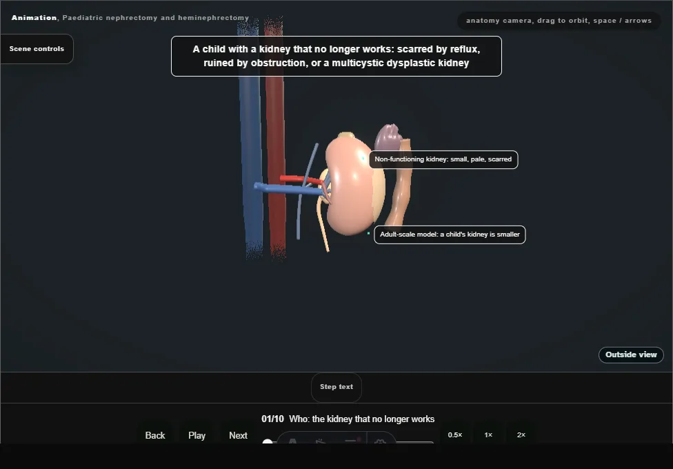
- A non-functioning kidney in a child: scarred by reflux, ruined by obstruction, or a multicystic dysplastic kidney.
- A DMSA scan gives its share of the total function, and the other kidney is confirmed healthy before this one is taken.
- 02
Position and ports
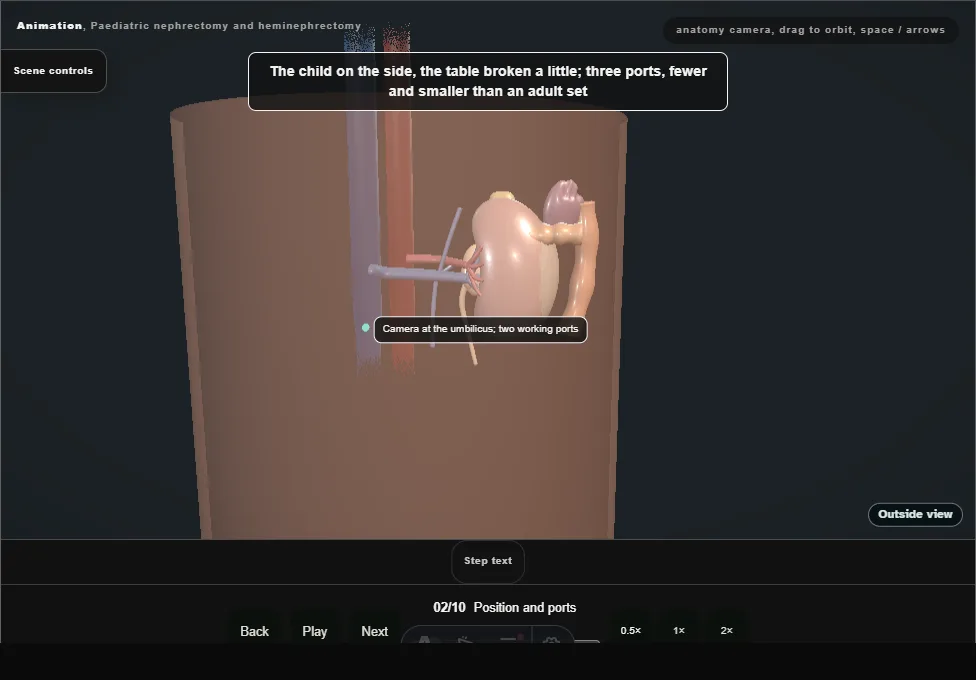
- The child on the side with the table broken a little.
- Three ports, fewer and smaller than an adult set: the camera at the umbilicus by open entry under vision, and two working ports.
- The bowel lies just under a thin wall.
- 03
Reflecting the colon
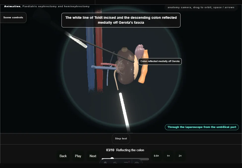
- The white line of Toldt incised and the descending colon reflected medially off Gerota's fascia, the splenic flexure freed, with the spleen and the tail of the pancreas kept in view.
- 04
The ureter to the hilum
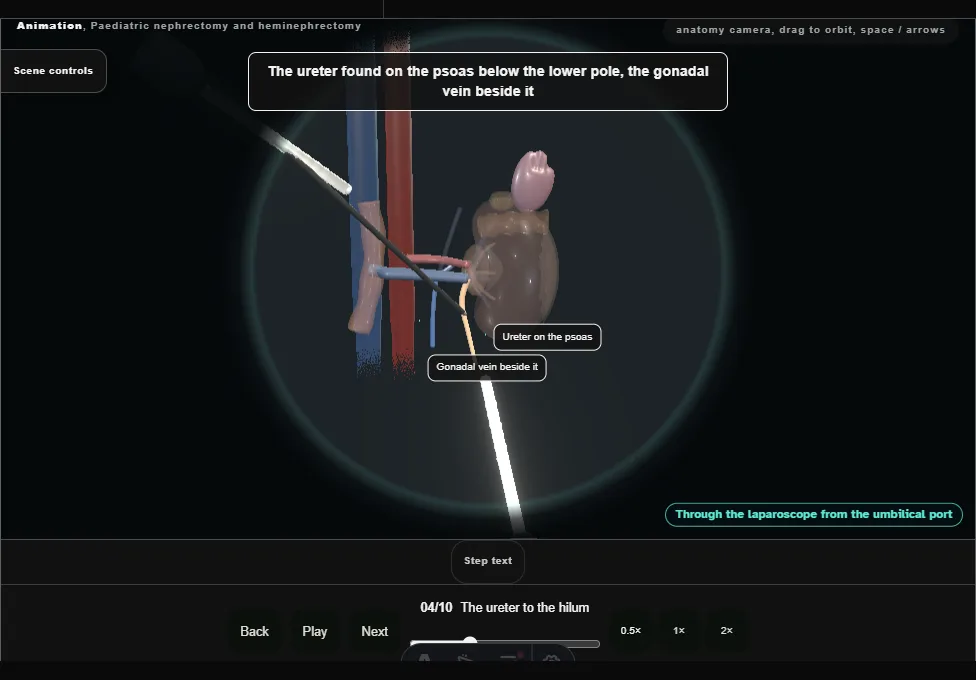
- The ureter found on the psoas below the lower pole with the gonadal vein beside it, lifted, and followed up to the renal pelvis: it leads the dissection to the hilum.
- 05
The hilar vessels
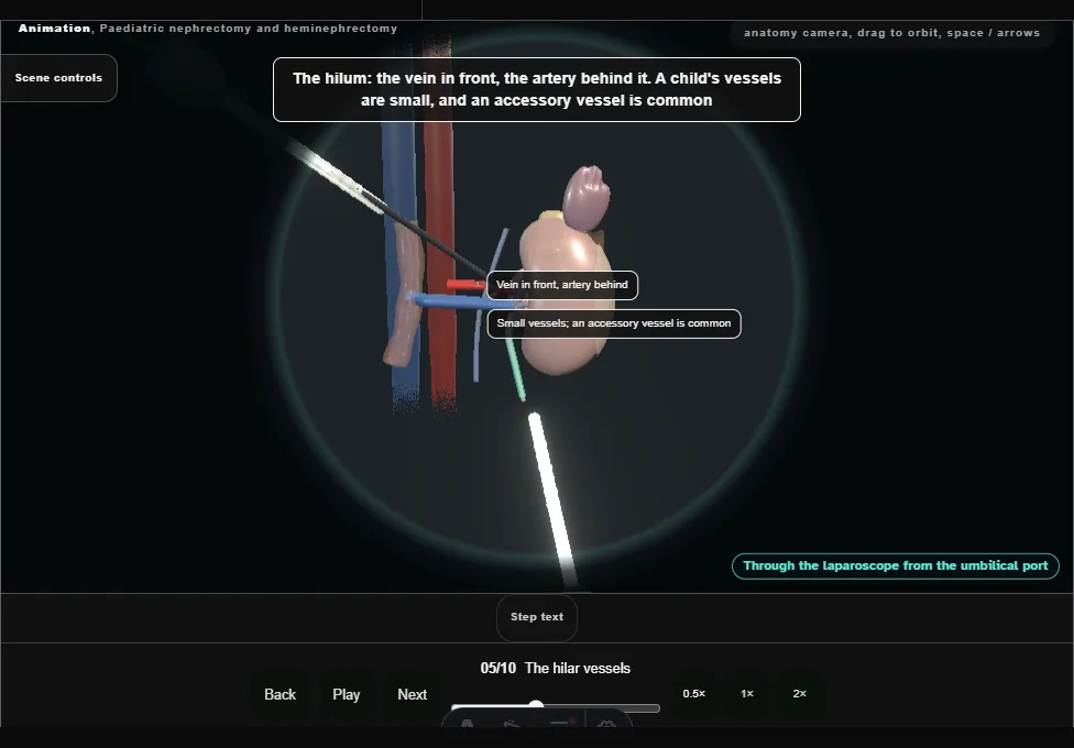
- The vein in front, the artery behind it.
- A child's vessels are small and an accessory vessel is common: the accessory lower-pole artery is found, clipped and divided; then the renal artery, then the vein, each between clips with two left on the patient side.
- 06
Freeing the kidney and the ureter
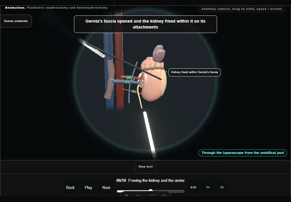
- Gerota's fascia opened and the kidney freed within it, the upper pole off the adrenal, which stays.
- When reflux was the cause the ureter is followed down toward the bladder and clipped and divided as low as it can be reached.
- 07
Extraction and inspection
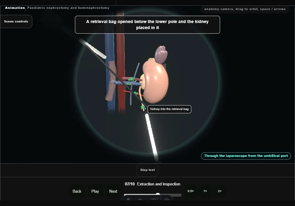
- The kidney placed in a retrieval bag, the bag closed and drawn out through the umbilical port site.
- The pressure lowered and the renal bed, the clips and the stumps inspected before the ports come out.
- 08
Heminephrectomy: the upper-moiety ureter
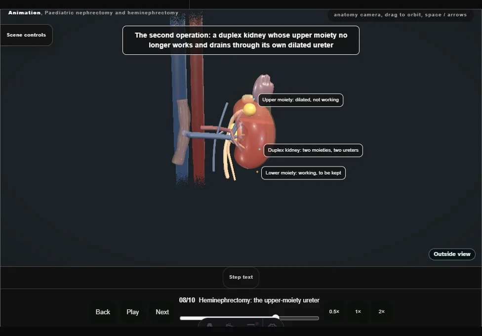
- A second child, a duplex kidney: the upper moiety no longer works and drains through its own dilated ureter.
- That ureter is found medial to the lower-moiety ureter, clipped and divided low, and its upper end passed behind the hilar vessels to lift the upper pole.
- 09
The upper-pole vessels
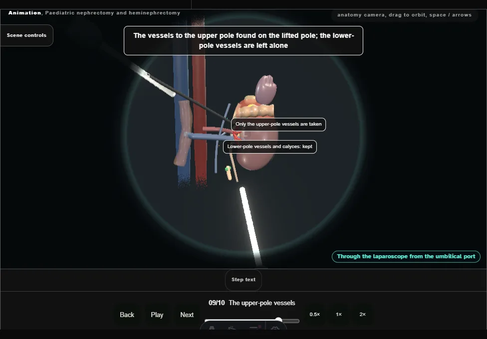
- On the lifted pole the vessels to the upper pole are clipped and divided close to it, the artery and then its vein.
- The lower-pole vessels are left alone.
- The upper pole darkens to a line of demarcation.
- 10
Dividing the parenchyma and closing
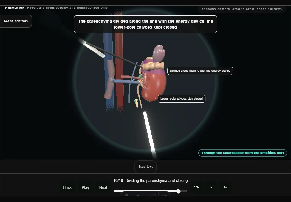
- The parenchyma divided along the line of demarcation with the energy device, the lower-pole calyces kept closed.
- The upper pole leaves in its own bag and the cut face is closed with stitches across it.
Derived from kidney/PaediatricNephrectomyScene.tsx at build time: every step title and caption above is the authored
ProcedureDef text, reproduced verbatim, and this page shares its readable-points renderer with the animation
itself. 10 of 10 steps have a captured frame.
Text fingerprint 1fd5f67f27e01f04. A caption edit without a re-capture fails the production
check, so the sheet and the animation cannot drift apart silently.