
UrOops3D, danger-zone atlas
Operative animation, Pelvic exenteration

Salvage pelvic surgery and pelvic exenteration

The pelvis emptied of bladder, rectum and the organs between them, en bloc, for a cancer that has recurred or advanced after radiotherapy and has nowhere else to be treated. Two or three specialties, ten hours, two stomas and a flap in the hole.

This page is the selection and the MDT that owns it, the sidewall on the MRI, the dissection that never enters the tumour, the internal iliac vessels, the perineal phase, the conduit and colostomy, and the empty pelvis that must be filled.

Advanced tier FRCS uro-oncology, salvage pelvic surgery Interactive 3D

Operative visualisation

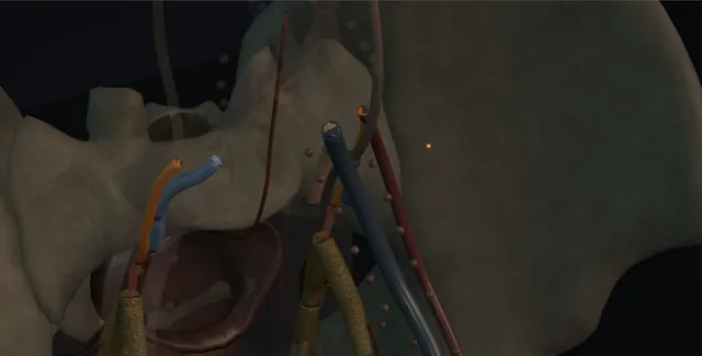
Schematic scene preview: Pelvic exenteration
Preparing the 3D anatomy…

UrOops3D · UroRef · © Nity G

Click me next Section 2 of 8 Anatomy The structures this operation works between, and the one it must not cross.

The structures in the scene

Pelvic exenteration: how the structures relateFrom the head of the table, the pelvis and the block within it. En bloc bladder, uterus or prostate, rectum: lateral limit; the lumbosacral trunk is the leg Pelvic sidewall and its nerves (danger). En bloc bladder, uterus or prostate, rectum: run along the sidewall; kept on a sling External iliac vessels. En bloc bladder, uterus or prostate, rectum: taken en bloc; the vein tears toward the sacrum Internal iliac artery and vein (danger). En bloc bladder, uterus or prostate, rectum: divided high at the brim for frozen section The ureters. En bloc bladder, uterus or prostate, rectum: levators divided from below to close the circle Pelvic floor and perineum. En bloc bladder, uterus or prostate, rectum: fills the empty pelvis the block leaves behind Rectus abdominis flap.En bloc bladder, uterusor prostate, rectum!Pelvic sidewall andits nerves!External iliacvessels!Internal iliacartery and vein!The uretersPelvic floor andperineumRectus abdominisflaplateral limit; thelumbosacral trunkis the legrun along thesidewall; kept on aslingtaken en bloc; thevein tears towardthe sacrumdivided high at thebrim for frozensectionlevators dividedfrom below to closethe circlefills the emptypelvis the blockleaves behindTargetDangerLandmarkContext
From the head of the table, the pelvis and the block within it. Schematic, not to scale; relationships only.
  • From the head of the table the pelvis is seen with the external iliac vessels running along each sidewall and the internal iliac vessels diving behind them toward the specimen, the ureters crossing at the brim, and the bladder, uterus or prostate and rectum mobilised as one block toward the pelvic floor.
  • Below the levators the perineal team is closing the circle from the skin.
  • When the specimen is out the pelvis is a bowl of bone and sidewall muscle with the sacrum behind, and the rectus flap drops into it from above.
  • ›The pelvic sidewall - internal iliac vessels, obturator internus and its fascia, the obturator nerve, and behind them the sacral plexus and the sciatic nerve; the lateral limit of the operation.
  • ›The external iliac vessels - cleared and protected on a sling; encasement is a vascular reconstruction or a bar to surgery.
  • ›The internal iliac artery and vein - taken en bloc with lateral disease; the vein and its tributaries are thin and tear toward the sacrum.
  • ›The ureters - divided high at the brim, the margin sent for frozen section, and joined to the conduit over stents.
  • ›The pelvic floor and the perineum - the levators divided from below to meet the abdominal dissection; the perineal wound closed with the flap.
  • ›The ileal conduit and the stomas - urostomy on the right, colostomy on the left, sites marked the day before with the patient standing and sitting.
  • ›The rectus abdominis flap - raised on the inferior epigastric pedicle from the side without a stoma, tunnelled through the pelvis to fill it and close the perineum.
Click me next Section 3 of 8 Steps The operation in order, with the landmark that guards each move.

Operative steps

  1. Who, and the MDT Locally advanced or recurrent pelvic cancer that can be removed with a clear margin and in a patient who can survive the operation and live with two stomas: rectal cancer recurring after chemoradiotherapy, cervical cancer recurring centrally after radiotherapy, bladder or prostate cancer fixed to the rectum, and the irradiated pelvis with an intractable fistula. The decision belongs to a specialist exenteration MDT with colorectal, urological, gynaecological, plastic and vascular surgeons, oncologists, radiologists, anaesthetists and a stoma nurse in the room. Curative intent means an R0 resection is achievable on the imaging; palliative intent means the aim is symptom control and is said so. Staging is CT of chest, abdomen and pelvis, pelvic MRI and PET-CT, and metastatic disease outside the pelvis is the usual bar.
  2. Planning the sidewall The pelvic MRI is read compartment by compartment: anterior, central, posterior and lateral, with the sacrum. Central disease with bladder and rectum together is a total exenteration; disease confined to bladder, uterus and vagina is an anterior exenteration with the rectum kept. The lateral compartment decides resectability: tumour at the internal iliac vessels can be taken with them, tumour on the obturator internus and the pelvic sidewall fascia can be taken with the muscle, tumour encasing the external iliac vessels needs a vascular surgeon and a graft, and tumour at the sciatic notch or the lumbosacral trunk is the edge of what can be done. The sacrum below S2 can be taken with a spinal surgeon. The ureters are mapped, and the kidneys checked for obstruction and function.
  3. Position, incision and the stoma sites The stoma nurse marks both sites the day before with the patient sitting, standing and lying: the urostomy on the right and the colostomy on the left, each on the summit of the rectus and away from creases, scars and the belt. Lloyd-Davies position with the legs in stirrups and the arms tucked, padding at every pressure point because the operation is long, a warming blanket, arterial and central lines, an epidural or a spinal with morphine, and blood cross-matched in quantity. Antibiotics with anaerobic cover and thromboprophylaxis. A long midline incision from the xiphisternum to the pubis, the abdomen explored for peritoneal or liver disease that would stop the operation, and a fixed ring retractor set.
  4. The en bloc dissection The principle is that the tumour is never seen, entered or handled: the dissection runs outside every plane the tumour occupies, and the specimen leaves in one piece. The sigmoid is divided with a stapler at the level the colorectal surgeon chooses, the ureters are found at the brim and divided high with a clip so they can be sent for a frozen section of the margin, and the plane is developed along the pelvic sidewall on both sides, lateral to the internal iliac vessels if they are to be taken. The peritoneum is incised around the pelvis and the bladder, uterus and rectum are mobilised as one block from front and sides toward the pelvic floor. Anything the tumour touches comes with it.
    where it bites: the dissection never enters the tumour; a plane that opens into it is a positive margin and an operation that has lost its purpose.
  5. The pelvic sidewall and the internal iliac vessels The common iliac artery is followed to its bifurcation and the external iliac artery and vein are cleared and protected on a sling. The internal iliac artery is ligated and divided beyond the origin of the superior gluteal branch where it can be spared, or at its origin where the tumour demands, and the internal iliac vein is taken between ligatures with its tributaries, the bloodiest part of the day: the veins are thin and tear toward the sacrum. The obturator nerve is preserved if it is free of tumour and sacrificed if it is not, with the patient warned about adduction. The lumbosacral trunk and the sciatic nerve are the posterior limit. The sidewall muscle comes with the specimen where the tumour is on it.
    where it bites: the internal iliac vein and its tributaries tear toward the sacrum and the pre-sacral plexus; control is proximal ligation and packing, not a clamp put on blind.
  6. The perineal phase A second team works from below while the abdominal team finishes the pelvic floor: an elliptical incision around the anus, in a woman taking the vaginal introitus and in a man the perineal skin to the bulb of the urethra, deepened through the ischiorectal fat to the levators, which are divided from below and meet the abdominal dissection at the pelvic floor. The urethra is divided at the membranous level from below or above. The specimen is delivered through the perineum or the abdomen, oriented, marked and sent fresh. The perineal wound is the size of a hand and will not close on its own in irradiated skin, which is why the flap has been planned before the incision was made.
  7. Reconstruction: conduit and colostomy The urological reconstruction is an ileal conduit in nearly every case: fifteen centimetres of ileum taken twenty centimetres from the ileocaecal valve, the ureters spatulated and joined end-to-side over single-J stents, Bricker fashion, or as a Wallace plate, with the left ureter brought under the sigmoid mesentery, and the stoma everted at the right-sided mark. Irradiated ileum near the pelvis is avoided and a segment from higher up, or transverse colon, is used instead. The colorectal surgeon brings out an end colostomy on the left. Where the rectum has been preserved in an anterior exenteration a defunctioning stoma is still usual. The stents and the drains are labelled, because two stomas, two stents and three drains become confused on the ward.
  8. Filling the pelvis An empty irradiated pelvis fills with small bowel that adheres, obstructs, fistulates and perforates, and its perineal wound breaks down; the dead space is therefore filled at the same operation. The vertical rectus abdominis myocutaneous flap on the inferior epigastric pedicle is the usual choice, raised from the side that will not carry a stoma, tunnelled through the pelvis and inset into the perineum, giving bulk below and skin at the wound. Where the rectus has been used or is needed, gracilis flaps from the thighs or an omental pedicle are the alternatives, and a mesh or a biological sling across the pelvic inlet holds the bowel out. The abdomen is closed with the stomas matured last.
    where it bites: small bowel that drops into an irradiated, empty pelvis obstructs and fistulates; the pelvis is filled at the operation, not at the reoperation.
  9. After Critical care for the first days: fluid, transfusion, the epidural, hourly urine from the conduit, and the stents flushed if they block. Stomas are checked for colour twice a day, because a dusky conduit is an ischaemic one. Nutrition is started early. The perineal flap is inspected daily for colour and the drains stay until dry. The stents come out at ten to fourteen days, the drains after them, and a loopogram is done if there is doubt about a uretero-ileal join. Histology and margins go back to the MDT. The patient meets the stoma nurse daily, the physiotherapist for the leg if the obturator was taken, and a psychologist, because two stomas and a perineal scar are a different life.
Click me next Section 4 of 8 Danger zones Where this operation bites, and why it bites there.
Critical relationship

Danger zones

The spatial relationships the animation is built to make automatic. Before you read each one, ask yourself: at this point in the operation, what is about to go wrong, and what is my first move? The save for each sits in the Complications tab.

  • RiskThe iliac vessels and the sidewall - the internal iliac vein tears toward the sacrum, the external iliac vessels are the limb, and both are scarred by radiotherapy.
  • RiskThe obturator nerve and the lumbosacral trunk - the obturator is sacrificed with lateral disease at the cost of adduction; the lumbosacral trunk and sciatic nerve are the leg and are not.
  • RiskThe ureters, bowel and pelvic floor - divided high, joined to non-irradiated bowel, and kept out of an empty pelvis.
  • RiskIrradiated tissue and the empty pelvis - every join in irradiated tissue leaks more, and dead space in an irradiated pelvis fills with bowel that obstructs and fistulates.
Practise it Take me into the scene Opens the animation at a danger moment and asks you to predict the mechanism and your first move before it reveals.
Click me next Section 5 of 8 Complications The save for each one: the first move, the escalation trigger, the op-note line.

Complications and immediate saves

Trigger each of these live in the Animation tab, then read the save: the first move, the escalation trigger, and the line that goes in the op note.

Massive haemorrhage from the sidewall or the sacrum

  • the internal iliac vein, its tributaries and the pre-sacral plexus in a scarred, irradiated pelvis: litres in minutes, and a clamp put on blind makes it worse.
The save
  1. Pack the pelvis and press, tell the anaesthetist, activate the major haemorrhage protocol and call the vascular surgeon. first move
  2. Proximal control: the common and internal iliac artery slung and ligated, the pressure in the pelvis falls.
  3. Pre-sacral bleeding: pack, a thumbtack or a bone wax into the sacrum, or a muscle fragment welded with diathermy; sutures tear it further.
  4. Bleeding not controlled with packing and ligation: leave the packs in, close, correct the coagulopathy on critical care and return at 48 hours. escalation trigger
  5. Op note: the vessel, the control, the blood loss and the products, and whether packs were left in and when they will come out. document

Urinary leak and fistula

  • urine in the drain or the perineal wound from a uretero-ileal join or the conduit in irradiated tissue, and later a fistula between the urinary tract, the bowel and the perineum.
The save
  1. Check the stents and the conduit are draining and flush them; drain fluid creatinine confirms urine. first move
  2. CT urogram with delayed phase or a loopogram to find the leak; a collection is drained percutaneously.
  3. A drained leak with stents across the joins is given time; the stents stay longer.
  4. Sepsis, a growing collection, a stent not across the leak, or a fistula to the bowel or perineum: nephrostomies to divert, and a return to theatre with the flap surgeon. escalation trigger
  5. Note: the join involved, the drains and stents, the imaging and the date it is repeated. document

Empty pelvis syndrome and pelvic sepsis

  • small bowel descended into the dead space: obstruction, a collection or abscess in the pelvis, a perineal discharge, and a fistula from bowel to perineum or to the urinary tract.
The save
  1. CT with oral and intravenous contrast for the collection and the bowel; cultures and antibiotics with anaerobic cover. first move
  2. Percutaneous or transperineal drainage of the collection, and nasogastric decompression of an obstructed bowel.
  3. Nutrition, parenteral if the gut has stopped, and the stomas checked for output and colour.
  4. Bowel that will not settle, a perforation or an enteric fistula: laparotomy, the bowel lifted out of the pelvis and the space filled with omentum or a flap. escalation trigger
  5. Note: what filled the pelvis at the operation, the collection and its drainage, and the nutritional plan. document

Flap and wound failure

  • the perineal or abdominal wound in irradiated skin, or the rectus flap that is dusky, congested or has lost its pedicle: dehiscence, a chronic perineal sinus, and an open pelvis.
The save
  1. Inspect the flap for colour, capillary return and temperature at every round in the first three days; a congested flap is reported now. first move
  2. Release tight sutures, keep the patient off the flap, and keep the drains until the perineum is dry.
  3. A partial loss is debrided and dressed with negative pressure; a sinus is imaged for a fistula behind it.
  4. A flap losing its pedicle, or a dehiscence exposing the pelvis: back to theatre with plastics for revision or a second flap. escalation trigger
  5. Note: the flap, its pedicle and the side it came from, the wound at each review, and the dressing plan. document

Nerve injury, pain and the leg

  • the obturator nerve taken with lateral disease, or the lumbosacral trunk or sciatic nerve stretched or divided at the sidewall: a leg that will not adduct, a foot that drops, and chronic neuropathic pelvic pain.
The save
  1. Examine the legs on the first day, adduction, hip flexion, knee and ankle, and record it, so that a deficit is known before the patient stands. first move
  2. Physiotherapy from the first days; adduction loss from the obturator is compensated and walking is preserved.
  3. Neuropathic pain is treated early with the pain team, with the epidural then gabapentinoids and amitriptyline.
  4. A foot drop or a sciatic deficit not expected from the operation: exclude a haematoma or a positioning injury with imaging, and involve neurology. escalation trigger
  5. Note: the nerves seen, preserved or sacrificed and why, the deficit on day one, and what the patient was told before the operation. document
Click me next Section 6 of 8 Troubleshooting When the case turns: the problem, then the next move.

Intra-operative troubleshooting

When the case turns: the problem, then the next move.

  • ActionPeritoneal nodules found on opening - biopsy and frozen section; confirmed peritoneal disease stops a curative exenteration and the MDT is told rather than the operation continued.
  • ActionThe tumour is on the external iliac vein - stop and call the vascular surgeon; the vein is resected and replaced, or the operation is palliative and does not go there.
  • ActionBleeding from the internal iliac vein that will not stop - pack and press, ligate the internal iliac artery proximally, then take the vein between ligatures under vision; packs left in and a return at 48 hours if it still bleeds.
  • ActionThe ureteric margin is positive on frozen section - trim the ureter higher and send again; a long ureter is joined to a conduit brought higher or with a psoas hitch of the conduit, not left with tumour.
  • ActionThe ileum near the pelvis is thick and irradiated - go higher for the conduit segment, or use transverse colon; irradiated ileum leaks at the join and strictures at the stoma.
  • ActionThe rectus flap will not reach the perineum - lengthen the pedicle by taking the muscle to its origin and releasing the skin paddle; if it still will not reach, bilateral gracilis with omentum in the pelvis.
Click me next Section 7 of 8 FRCS viva Say it out loud, then mark yourself against the model answer.

FRCS viva questions

The examiner stems this scene prepares you for. Try to answer out loud first, then reveal, and learn the discriminator, the detail that turns a pass into a strong pass.

QWho should be considered for pelvic exenteration, and how is that decision made?reveal answer
  • I would consider exenteration for locally advanced or recurrent pelvic malignancy that is confined to the pelvis and can be removed with a clear margin in a patient fit enough for a long operation and able to live with the consequences: recurrent rectal cancer after chemoradiotherapy, central recurrence of cervical cancer after radiotherapy, bladder or prostate cancer fixed to the rectum, and the irradiated pelvis with an intractable fistula.
  • The decision is not mine alone; it belongs to a specialist exenteration MDT that includes colorectal, urological, gynaecological, plastic and vascular surgeons, oncologists, radiologists, anaesthetists and a stoma nurse.
  • Staging is CT of chest, abdomen and pelvis, pelvic MRI read compartment by compartment for the sidewall, and PET-CT, and disease outside the pelvis usually excludes curative surgery.
  • The MDT states whether intent is curative, which means an R0 resection is achievable on imaging, or palliative, where the aim is control of bleeding, fistula, obstruction or pain.
  • The patient meets the stoma nurse and the psychologist before consenting, and is told the mortality, morbidity and the two stomas plainly.
Strong pass

The compartmental reading of the MRI as the resectability decision and the curative or palliative intent stated by the MDT before the operation.

QWhat decides resectability at the pelvic sidewall?reveal answer
  • The lateral compartment is where exenteration becomes possible or not, and I would read it on the MRI structure by structure from medial to lateral.
  • Tumour on the internal iliac vessels can be resected with them: the internal iliac artery is ligated, ideally beyond the superior gluteal origin, and the vein taken between ligatures, which is the bloodiest part of the operation.
  • Tumour on the obturator internus and the sidewall fascia can be taken with the muscle.
  • Tumour involving the obturator nerve is resected with it at the cost of adduction, which the patient is warned about.
  • Tumour encasing the external iliac artery or vein needs a vascular surgeon and a planned resection with a graft, and in most patients that is the limit of what is offered.
  • Tumour at the sciatic notch, the lumbosacral trunk or the sciatic nerve is beyond a resection compatible with a functioning leg.
  • The sacrum can be taken below S2 with a spinal surgeon; above that the deficit is not acceptable.
  • The other lateral determinant is the ureter, which is divided high and its margin sent for frozen section.
  • I would present each of these as a named structure to the MDT rather than as a general impression of fixity.
Strong pass

Say both

  • Naming the structures in order from internal iliac vessels to the lumbosacral trunk
  • Giving the limit at each
Examiner note
Naming the structures in order from internal iliac vessels to the lumbosacral trunk and giving the limit at each.
QDescribe the urological contribution to a total pelvic exenteration.reveal answer
  • The urologist has three parts.
  • Before the operation: the assessment of the kidneys and the ureters on the staging imaging, with decompression of an obstructed kidney if function is at risk, and the marking of the urostomy site with the stoma nurse.
  • During the resection: the identification of the ureters at the brim and their division high with the margin sent for frozen section, the bladder and prostate or urethra mobilised as part of the en bloc specimen without entering the tumour, and the division of the urethra at the membranous level.
  • After the specimen is out: the urinary diversion, which is almost always an ileal conduit, fifteen centimetres of non-irradiated ileum taken well away from the pelvis or, where the ileum is damaged, transverse colon, with the ureters spatulated and joined end-to-side over single-J stents and the left brought under the sigmoid mesentery.
  • A continent diversion or a neobladder is rarely appropriate after radiotherapy and with a colostomy beside it.
  • After the operation I would watch the conduit colour, the stents and the drains, flush the stents if they block, remove them at ten to fourteen days with a loopogram if there is doubt, and follow the kidneys with ultrasound for stricture at the uretero-ileal joins for life.
Strong pass

The high ureteric division with frozen section and the conduit made from non-irradiated bowel with the reason given.

QWhat is the empty pelvis syndrome and how is it prevented?reveal answer
  • After exenteration the pelvis is a bowl of bone and sidewall muscle with no organs, and in an irradiated patient small bowel descends into it, adheres to the raw surfaces and the sacrum, obstructs, forms collections and abscesses, fistulates to the perineum or the urinary reconstruction, and can perforate;
  • the perineal wound above it breaks down and becomes a chronic sinus.
  • That is the empty pelvis syndrome, and it is the commonest cause of reoperation and prolonged illness after exenteration.
  • Prevention is part of the operation: the dead space is filled at the time with vascularised tissue.
  • The vertical rectus abdominis myocutaneous flap on the inferior epigastric pedicle, raised from the side without a stoma and tunnelled into the pelvis, gives bulk below and closes the perineal skin with non-irradiated tissue.
  • Where the rectus is not available, bilateral gracilis flaps or an omental pedicle fill the pelvis, and a mesh or biological sling across the inlet keeps the bowel above it.
  • When it occurs I would image with CT, drain collections percutaneously or transperineally, decompress and feed the patient, and reoperate only for a fistula, a perforation or an obstruction that does not settle, lifting the bowel out and filling the pelvis with whatever tissue remains.
Strong pass

Say both

  • The flap named with its pedicle and the reason it is done at the first operation
  • The perineal breakdown linked to the same cause
Examiner note
The flap named with its pedicle and the reason it is done at the first operation, and the perineal breakdown linked to the same cause.
Click me next Section 8 of 8 Evidence The guidance each of these answers is built on.

Evidence and guidance

Two minutes

Close the lap: recall it before you leave.

Say it aloud, then reveal. Reading is not remembering.

  1. 01 The relationship Say the critical spatial relationship out loud, then reveal. reveal
    Memory frame

    From the head of the table the pelvis is seen with the external iliac vessels running along each sidewall and the internal iliac vessels diving behind them toward the specimen, the ureters crossing at the brim, and the bladder, uterus or prostate and rectum mobilised as one block toward the pelvic floor.

    Why?
    Below the levators the perineal team is closing the circle from the skin. When the specimen is out the pelvis is a bowl of bone and sidewall muscle with the sacrum behind, and the rectus flap drops into it from above.
  2. 02 The first move Massive haemorrhage from the sidewall or the sacrum. Your first move? reveal
    First move

    Pack the pelvis and press, tell the anaesthetist, activate the major haemorrhage protocol and call the vascular surgeon.

  3. 03 The escalation trigger Massive haemorrhage from the sidewall or the sacrum. What makes you call for help? reveal
    Escalate when

    Bleeding not controlled with packing and ligation: leave the packs in, close, correct the coagulopathy on critical care and return at 48 hours.

  4. 04 The discriminator What turns a pass into a strong pass? reveal
    Strong pass

    The compartmental reading of the MRI as the resectability decision and the curative or palliative intent stated by the MDT before the operation.

  5. 05 The pitfall Where does this operation bite first? reveal
    Pitfall

    The iliac vessels and the sidewall

    Why?
    the internal iliac vein tears toward the sacrum, the external iliac vessels are the limb, and both are scarred by radiotherapy.

Revision practice with self-marked answers, not an assessment of clinical competence. Kept in this browser only; it does not sync between devices.