UrOops3D, danger-zone atlas

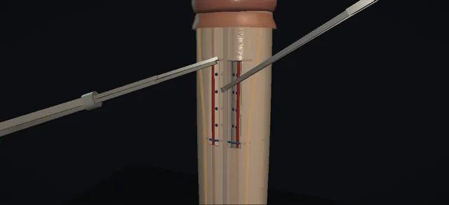
Schematic model; the dot pattern and the position of the sets relative to the dorsal arteries are authored from the book and the Gholami and Lue abstract.

Operative animation, Penile plication

Penile curvature: plication, Lue sixteen-dot technique

Four sutures, sixteen dots, through tunica only. An artificial erection shows where the bend is;

the convex face is marked, Buck's fascia is opened in two paramedian windows beside the deep dorsal vein, and each suture goes in, out, in, out through the tunica so the knots gather it into a fold. A second erection proves the correction before anything is closed.

The lesson is the bites: tunica, never the cavernosal tissue, never the bundle.

Core tier FRCS andrology, reconstruction, penile curvature Interactive 3D

Operative visualisation

Schematic scene preview: Penile plication: Lue sixteen-dot
Preparing the 3D anatomy…

UrOops3D · UroRef · © Nity G

How to use this module. Play through the eight steps, or use Back and Next. Watch the bend on the first erection, the dots, the windows beside the vein, each suture through tunica, the folds as the knots tighten and the straight shaft at the end. The triggers show a residual bend, a caught nerve and a deep bite.

Click me next Section 2 of 8 Anatomy The structures this operation works between, and the one it must not cross.

The structures in the scene

Penile plication: how the structures relateThe dorsum of the erect shaft from the degloving incision, fascia and bundle beneath. Subcoronal incision and degloving sleeve: the avascular plane leads onto it Shaft fascia: two paramedian windows. Subcoronal incision and degloving sleeve: white through each window; bites here only Tunica: sixteen dots, four sutures. Subcoronal incision and degloving sleeve: the windows sit either side of it Deep dorsal vein, midline, retracted. Subcoronal incision and degloving sleeve: each window stops at the artery Dorsal arteries, paramedian (danger). Subcoronal incision and degloving sleeve: lateral and untouched Dorsal nerves, lateral, under fascia (danger). Subcoronal incision and degloving sleeve: the plication is on the opposite face Spongiosum and urethra, concave side.Subcoronal incision anddegloving sleeveShaft fascia: twoparamedian windowsTunica: sixteendots, four suturesDeep dorsal vein,midline, retracted!Dorsal arteries,paramedian!Dorsal nerves,lateral, underfasciaSpongiosum andurethra, concavesidethe avascular planeleads onto itwhite through eachwindow; bites hereonlythe windows siteither side of iteach window stopsat the arterylateral anduntouchedthe plication is onthe opposite faceTargetDangerLandmarkContext
The dorsum of the erect shaft from the degloving incision, fascia and bundle beneath. Schematic, not to scale; relationships only.
  • The bend is on the erect shaft and the correction goes on its convex face: for a ventral bend that is the dorsum, where the deep dorsal vein runs in the midline with an artery and then a nerve lateral to it on each side.
  • The dots and the bites lie in the strip between the vein and the arteries; the windows in Buck's fascia are the same width, and nothing lateral is disturbed.
  • ›Corpora cavernosa and tunica albuginea - the plication is in the tunica; the cavernosal tissue beneath is never entered.
  • ›Corpus spongiosum and urethra - ventral, on the concave side of a ventral bend; inspected before closure.
  • ›Deep dorsal vein - midline under Buck's fascia; the two sets of dots sit either side of it; retracted, never divided.
  • ›Dorsal arteries - paramedian, lateral to the dots; the windows stop at them.
  • ›Dorsal nerves - lateral to the arteries, fanning toward the sides distally; untouched under intact fascia.
  • ›Buck's fascia - opened only as two paramedian windows over the marks, closed over the folds at the end.
  • ›Dartos and shaft skin - the degloving sleeve, raised in the avascular plane from a subcoronal incision and returned.
  • ›Glans - held on a traction stitch; the meatus is orthotopic in this adult case.
Click me next Section 3 of 8 Steps The operation in order, with the landmark that guards each move.

Operative steps

  1. Measure the bend Supine, prepped and draped. A tourniquet goes round the base and saline is injected through a butterfly needle into one corpus until the shaft is rigid. The bend declares itself: ventral here, with the urethra on the concave side. Its angle is measured against a straight edge and written down, because the same measurement is repeated at the end.
    where it bites: the operation is planned on the erect shaft, not the flaccid one; a bend measured poorly is corrected poorly.
  2. Deglove A traction stitch through the glans. A circumferential incision a little below the corona is taken through dartos and the superficial layer of Buck's fascia, and the shaft skin is degloved proximally to the root in the avascular plane. Haemostasis as the plane opens. The sleeve keeps its own blood supply and goes back at the end.
    where it bites: a plane that wanders deep bleeds from the bundle; one that wanders superficial devascularises the skin.
  3. Mark the convex face A second artificial erection finds the point of maximal curvature. The pen marks sixteen dots on the convex, dorsal face: two lateral sets either side of the deep dorsal vein, each set two rows of four, straddling the point of maximal bend. The dots sit on tunica between the vein and the dorsal arteries; the nerves run lateral to them.
    where it bites: dots placed over the bundle commit the sutures to it; the sets are paramedian, beside the vein.
  4. Open Buck's fascia Toothed forceps lift Buck's fascia beside the vein over the marks and scissors open a paramedian window on each side; the white tunica shows through. The vein is retracted and not divided. Nothing lateral is touched: the dorsal nerves stay where they are, under intact fascia.
    where it bites: the dorsal nerves lie lateral to the arteries; elevating the whole bundle to reach the tunica trades a simple plication for a sensory complaint.
  5. Place the bites Each of the four sutures has two entry and two exit points: in, out, in, out along its row, through the tunica albuginea only. The needle takes a full-thickness bite of tunica and comes out before it reaches the cavernosal tissue beneath. The sutures are left untied until all four are placed, so the tension can be judged together.
    where it bites: a bite into the cavernosal tissue bleeds, blooms a haematoma under Buck's fascia and risks the veno-occlusive mechanism.
  6. Tie and bury The sutures are tied one by one. As each knot tightens the tunica between its dots gathers into a raised fold and the convex face shortens, so the bend reduces. The knot is turned under the fold. The tension is enough to gather, no more.
    where it bites: over-tension cuts through tunica; a knot left proud is what the patient feels for years.
  7. Reassess Tourniquet and butterfly again and the shaft rises straight against the same guide that measured the bend. The urethra and the dorsal bundle are inspected for injury. A residual bend is answered with a further parallel pair at the new point of maximal bend, not by pulling the tied knots tighter.
    where it bites: a pair tied tighter tears; a second pair corrects.
  8. Close and counsel Bipolar to any bleeding point. Buck's fascia closes over the folds, the sleeve returns to the corona and dartos and skin are reapproximated with interrupted absorbable sutures. Traction stitch out, a light non-adherent dressing. The counselling was done before: some shortening, palpable knots and a possible change in sensation are expected consequences of the design, not complications.
Click me next Section 4 of 8 Danger zones Where this operation bites, and why it bites there.
Critical relationship

Danger zones

The spatial relationships the animation is built to make automatic. Before you read each one, ask yourself: at this point in the operation, what is about to go wrong, and what is my first move? The save for each sits in the Complications tab.

  • RiskThe dorsal nerves - lateral to the arteries under Buck's fascia. A bite placed lateral to the window, or a window carried lateral, catches a nerve; the technique is designed so the bundle is never elevated.
  • RiskDepth of the bite - tunica only. The needle through into cavernosal tissue bleeds, raises a haematoma and threatens the veno-occlusive mechanism that erection depends on.
  • RiskThe urethra - on the concave side of a ventral bend and far from the dorsal sutures here; in a dorsal bend the plication would be ventral and the urethra becomes the structure at risk. A catheter is one way to find it.
  • RiskTension - enough to gather the fold and no more; a pair over-tightened tears tunica and a residual bend is answered with another pair.
Practise it Take me into the scene Opens the animation at a danger moment and asks you to predict the mechanism and your first move before it reveals.
Click me next Section 5 of 8 Complications The save for each one: the first move, the escalation trigger, the op-note line.

Complications and immediate saves

Trigger each of these live in the Animation tab, then read the save: the first move, the escalation trigger, and the line that goes in the op note.

Residual curvature

  • the check erection after tying is still bent, because the sets were too few or sat away from the point of maximal bend.
  • The book lists persistent curvature among the complications; plication corrects by shortening the long side, so the correction has a limit.
The save
  1. Release the tourniquet and look at where the bend now sits; do not pull the tied knots tighter. first move
  2. Mark and place a further parallel pair at the new point of maximal bend, through tunica only, and tie.
  3. Repeat the artificial erection against the same guide.
  4. A complex, hinged or hourglass deformity is not a plication problem: grafting territory and a senior reconstructive surgeon. escalation trigger
  5. Op note: the angle before and after, the number of sutures and where each pair sits. document

Dorsal nerve injury

  • a bite placed lateral to the window, or a window carried lateral, catches a dorsal nerve.
  • The sixteen-dot design avoids elevating the bundle for exactly this reason; sensory change after bundle mobilisation is usually temporary (EAU).
The save
  1. Cut and remove that suture as soon as it is recognised. first move
  2. Re-site the bite medially, beside the vein, within the window.
  3. Do not answer the problem with more dissection of the bundle; a nerve clearly divided or crushed is a senior conversation about repair and about what the patient is told. escalation trigger
  4. Op note: the event, the re-siting and the state of the nerve as seen. document

Bite into cavernosal tissue

  • the needle goes through the tunica into the cavernosal tissue beneath: bleeding at the bite and a haematoma blooming under Buck's fascia.
  • Plication without a tunical incision is meant to protect the veno-occlusive mechanism; a deep bite is the way to lose that protection.
The save
  1. Remove the suture and re-pass it through tunica only. first move
  2. Bipolar to the bleeding point; press.
  3. Release the tourniquet and watch: a haematoma that expands after release needs a senior opinion now, not at the end of the list. escalation trigger
  4. Op note: the deep bite, its site, the re-pass and the haemostasis. document
Click me next Section 6 of 8 Troubleshooting When the case turns: the problem, then the next move.

Intra-operative troubleshooting

When the case turns: the problem, then the next move.

  • ActionThe artificial erection will not hold - check the tourniquet is snug at the base and the butterfly is in the corpus, not the subcutaneous plane; saline runs out through the glans into the spongiosum and back past a loose tourniquet.
  • ActionBleeding on degloving - the plane has strayed; come back onto the avascular dartos plane and use bipolar, not blind diathermy near the bundle.
  • ActionThe dots sit over the vein - the sets are paramedian; re-mark beside the vein. Dots over the midline put the sutures through the bundle.
  • ActionThe window keeps opening laterally - stop at the artery; the nerve is lateral to it. Widen medially toward the vein if more tunica is needed.
  • ActionStill bent after four sutures - another parallel pair at the new point of maximal bend. The book says more than two pairs may be needed for a larger bend; the figure is in the evidence list.
Click me next Section 7 of 8 FRCS viva Say it out loud, then mark yourself against the model answer.

FRCS viva questions

The examiner stems this scene prepares you for. Try to answer out loud first, then reveal, and learn the discriminator, the detail that turns a pass into a strong pass.

QA man in his twenties has a congenital ventral curvature that stops him having intercourse. Take me through your options and your operation.reveal answer
  • Congenital curvature is a tunical disproportion with a normal erection, and surgery waits until after puberty; it is offered for a bend that interferes with intercourse.
  • The options are shortening procedures on the convex side and, for the complex or hourglass deformity, grafting, which is Peyronie territory.
  • The shortening procedures are the Nesbit, an elliptical excision of tunica closed transversely;
  • the Yachia, a longitudinal tunical incision closed transversely on the Heineke-Mikulicz principle;
  • and plication without a tunical incision, of which the Lue sixteen-dot technique is the one I would use.
  • Under general anaesthesia and with a tourniquet at the base, an artificial saline erection shows the point of maximal curvature and the angle is measured.
  • Through a subcoronal circumferential incision I deglove the shaft to the root.
  • On the convex, dorsal face I mark two lateral sets of dots either side of the deep dorsal vein, open Buck's fascia in a paramedian window over each set without elevating the bundle, and place two parallel non-absorbable sutures on each side, each with two entry and two exit points through the tunica only.
  • I tie, bury the knots, and repeat the artificial erection to confirm the shaft is straight.
  • I inspect the urethra and the bundle, close Buck's fascia and the skin, and apply a light dressing.
Strong pass

Say both

  • Naming the alternative procedures with their principles
  • Saying unprompted that the artificial erection is repeated after tying
Examiner note
Naming the alternative procedures with their principles, and saying unprompted that the artificial erection is repeated after tying.
QWhat do you tell this man before the operation?reveal answer
  • That a plication straightens by shortening the long side, so some loss of erect length is part of the design and the commonest thing patients mind afterwards;
  • that the knots are non-absorbable and may be palpable under the skin;
  • that sensation on the glans may change, usually temporarily, because the dorsal nerves are close to the field;
  • that curvature can persist or recur and a further procedure is sometimes needed;
  • and that bleeding, infection and injury to the urethra or the bundle are the operative risks.
  • I measure and document stretched length and the angle before surgery and again afterwards, and I give him the erection photographs back with the plan drawn on them.
Strong pass

Say all three

  • Putting shortening first
  • Framing it as a consequence of the method
  • With length measured before and after
Examiner note
Putting shortening first and framing it as a consequence of the method, with length measured before and after.
QWhy sixteen dots, and why through tunica only?reveal answer
  • Each of the four sutures has two entry and two exit points: in, out, in, out along its row, so four dots a suture and sixteen in all.
  • Two sutures on each side of the vein make two pairs of parallel plications, each pair placed just proximal and distal to the point of maximal curvature, so the correction is distributed over a length of tunica rather than concentrated at one fold.
  • Taking the tunica only matters for two reasons: the cavernosal tissue beneath bleeds and raises a haematoma under Buck's fascia if the needle enters it, and plication without a tunical incision is chosen precisely because it leaves the veno-occlusive mechanism alone.
  • Placing the sets beside the vein, medial to the arteries, means the bundle is never elevated, which is why nerve injury is uncommon with this design.
Strong pass

Say both

  • Explaining the arithmetic of the dots
  • Linking the tunica-only bite to the veno-occlusive mechanism and the haematoma
Examiner note
Explaining the arithmetic of the dots and linking the tunica-only bite to the veno-occlusive mechanism and the haematoma.
QThe check erection is still bent after you have tied all four sutures. What now?reveal answer
  • I do not pull the tied knots tighter: that tears tunica and gains nothing.
  • I release the tourniquet, look at where the bend now sits, and mark a further parallel pair at the new point of maximal curvature, again through tunica only and beside the vein, then repeat the erection.
  • The book notes that more than two pairs may be needed for a larger bend.
  • If the deformity turns out to be hinged or hourglass, or the residual bend is one more plications would only shorten him for, I stop, close, and discuss grafting with a senior reconstructive colleague rather than persist.
  • I document the angle before and after and the number and position of the sutures.
Strong pass

Say all three

  • Refusing to over-tighten
  • Adding a pair instead
  • Knowing when the answer is a different operation
Examiner note
Refusing to over-tighten, adding a pair instead, and knowing when the answer is a different operation.
Click me next Section 8 of 8 Evidence The guidance each of these answers is built on.

Evidence and guidance

Book, chapter 29
Armenakas NA, Fracchia JA, Golan R. Operative Dictations in Urologic Surgery, first edition. Wiley, 2019. Chapter 29, Plication for Penile Curvature (Lue "16-Dot" Technique), printed pages 117 to 118
Indication (curvature of 60 degrees or less), the five essential steps, the variations (Nesbit, Yachia, penoscrotal or direct incision, more than two pairs for curvature over 30 degrees, a catheter to protect the urethra) and the template dictation: a 2-0 polypropylene glans traction suture, a circumferential incision 1 cm below the coronal sulcus, a 21-gauge butterfly for the saline erection, Buck's fascia elevated over the marking, two lateral sets of 2-0 non-absorbable polyester sutures each with two entry and two exit points taking about a 1 cm full-thickness segment of tunica, dartos and skin closed with 4-0 interrupted chromic sutures. These figures are book figures and appear nowhere else on this page.
Open guideline
EAU Guidelines
Sexual and Reproductive Health: penile curvature, sections on congenital penile curvature (8.1.3, 8.1.4) and on surgical treatment of Peyronie's disease, tunical shortening procedures (8.2.3.b, 8.2.3.b.1)
Congenital curvature: defer surgery until after puberty; Nesbit, Yachia or plication with or without bundle dissection as the operations. The 16-dot technique described as two pairs of parallel Essed-Schroeder plications; shortening as the commonest adverse outcome, palpable knots, measure length before and after. Read 8 October 2026.
Open guideline
J Urol 2002
Gholami SS, Lue TF. Correction of penile curvature using the 16-dot plication technique: a review of 132 patients. J Urol 2002;167(5):2066-2069
Abstract only, read 8 October 2026: multiple parallel plications under an induced erection, avoiding the neurovascular bundle; patients counselled about shortening and palpable knots. The full text, including the exact position of the dorsal sets relative to the dorsal arteries, has not been read.
Open guideline

Curriculum. Supports learning relevant to the ISCP Urology Curriculum syllabus topics Penile curvature and Andrology. Not a workplace-based assessment and not evidence of competence.

Two minutes

Close the lap: recall it before you leave.

Say it aloud, then reveal. Reading is not remembering.

  1. 01 The relationship Say the critical spatial relationship out loud, then reveal. reveal
    Memory frame

    The bend is on the erect shaft and the correction goes on its convex face

    Why?
    The bend is on the erect shaft and the correction goes on its convex face: for a ventral bend that is the dorsum, where the deep dorsal vein runs in the midline with an artery and then a nerve lateral to it on each side. The dots and the bites lie in the strip between the vein and the arteries; the windows in Buck's fascia are the same width, and nothing lateral is disturbed.
  2. 02 The first move Residual curvature. Your first move? reveal
    First move

    Release the tourniquet and look at where the bend now sits; do not pull the tied knots tighter.

  3. 03 The escalation trigger Residual curvature. What makes you call for help? reveal
    Escalate when

    A complex, hinged or hourglass deformity is not a plication problem: grafting territory and a senior reconstructive surgeon.

  4. 04 The discriminator What turns a pass into a strong pass? reveal
    Strong pass

    Say both

    • Naming the alternative procedures with their principles
    • Saying unprompted that the artificial erection is repeated after tying
    Examiner note
    Naming the alternative procedures with their principles, and saying unprompted that the artificial erection is repeated after tying.
  5. 05 The pitfall Where does this operation bite first? reveal
    Pitfall

    The dorsal nerves

    Why?
    lateral to the arteries under Buck's fascia. A bite placed lateral to the window, or a window carried lateral, catches a nerve; the technique is designed so the bundle is never elevated.

Revision practice with self-marked answers, not an assessment of clinical competence. Kept in this browser only; it does not sync between devices.