
Revision sheet
Penile curvature: Lue sixteen-dot plication
8 steps, each one the authored title and caption from the interactive animation, with the frame captured from that step. Read it before the case, or print it and work through the steps in order.
Teaching only, not for diagnosis or treatment decisions. The text on this sheet is the animation's own step copy, reproduced verbatim.
UrOops3D · UroRef · © Nity G
- 01
Measure the bend
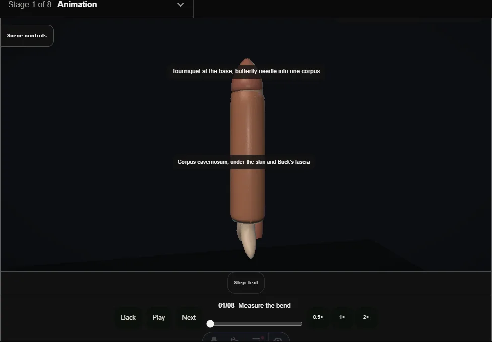
- A tourniquet at the base and saline through a butterfly needle into one corpus give an artificial erection.
- The shaft bends ventrally; the urethra sits on the concave side, so the plication goes on the opposite, convex face.
- The angle is measured against a straight edge and recorded.
- 02
Deglove
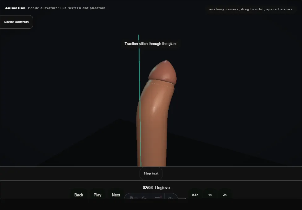
- A traction stitch through the glans, then a circumferential incision below the corona through dartos.
- The skin sleeve rolls back to the root in the avascular plane and keeps its own blood supply.
- 03
Mark the convex face
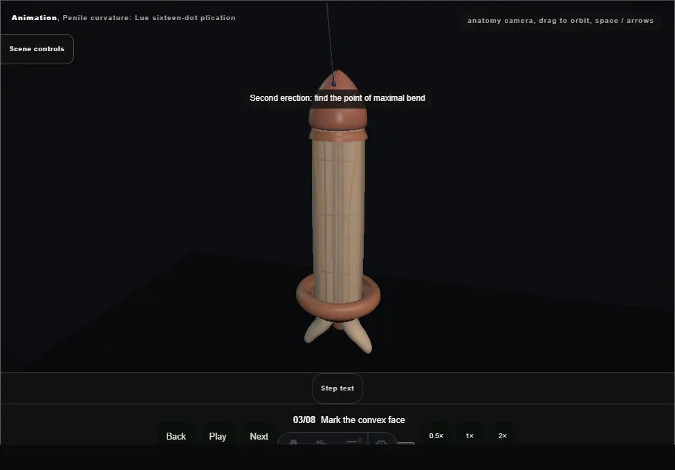
- A second erection finds the point of maximal bend.
- The pen places sixteen dots on the dorsal face, two rows of four on each side of the deep dorsal vein, straddling the bend.
- The dots sit on tunica between the vein and the dorsal arteries.
- 04
Open Buck's fascia
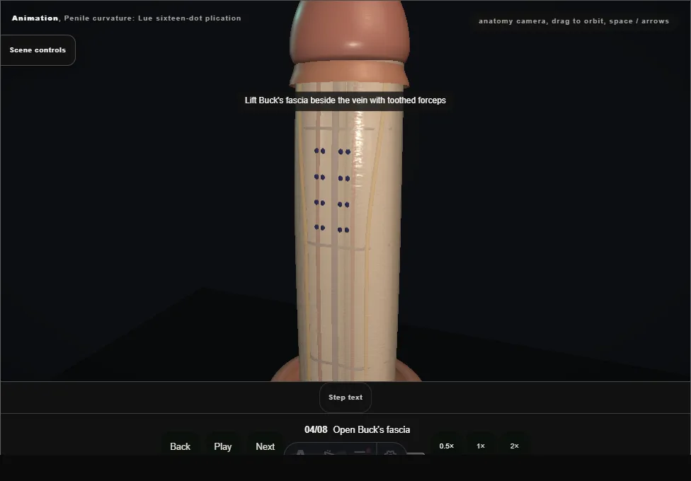
- Toothed forceps lift Buck's fascia beside the vein and scissors open two paramedian windows; white tunica shows through.
- The dorsal nerves stay lateral and untouched; the vein is retracted, not divided.
- 05
Place the bites
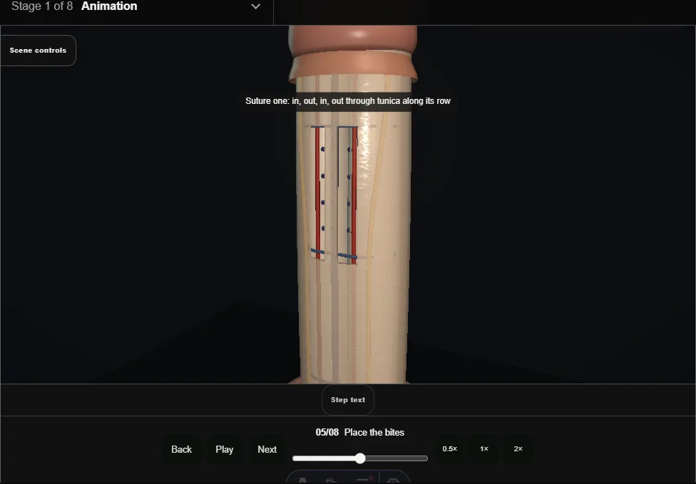
- Each suture passes in, out, in, out through tunica along its row: two bites that take the tunica and never the cavernosal tissue beneath.
- The inset shows one bite in section.
- Four sutures lie loose before any is tied.
- 06
Tie and bury
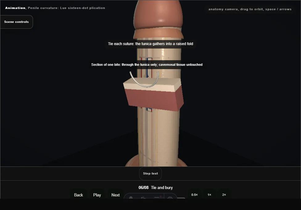
- The knots tighten one by one and the tunica between the dots gathers into a raised fold; the convex face shortens and the bend reduces.
- Each knot is turned under its fold.
- Over-tension tears tunica; a knot left proud is palpable.
- 07
Reassess
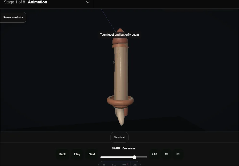
- Tourniquet and butterfly again.
- The erect shaft is straight against the same guide; the urethra and the dorsal bundle are inspected.
- A residual bend means another parallel pair, not more tension on the pair already tied.
- 08
Close and counsel
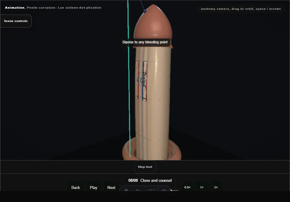
- Bipolar to any bleeding point.
- Buck's fascia closes over the folds, the sleeve returns and the subcoronal line is sutured; a light dressing.
- Counsel: shortening and palpable knots are expected, and sensation may change for a time.
Derived from PenilePlicationScene.tsx at build time: every step title and caption above is the authored
ProcedureDef text, reproduced verbatim, and this page shares its readable-points renderer with the animation
itself. 8 of 8 steps have a captured frame.
Text fingerprint 90f73f1d38a4e825. A caption edit without a re-capture fails the production
check, so the sheet and the animation cannot drift apart silently.