UrOops3D, danger-zone atlas

Schematic model.

Operative animation, Posterior urethral reconstruction

Posterior urethral reconstruction: perineal anastomotic repair after pelvic fracture

The delayed repair of a pelvic fracture urethral injury.

Through a perineal incision the bulbar urethra is freed and cut across at the scar, the scar of the distraction defect is excised onto a sound passed through the suprapubic tract, and healthy urethra is joined to healthy urethra without tension.

The gap is bridged step by step, using only the manoeuvres it needs. The lesson is the order of those manoeuvres, and what lies around the scar: the rectum behind, the cavernous nerves beside it and a continence mechanism that now rests on the bladder neck.

Advanced tier FRCS urological trauma, reconstructive urology Interactive 3D

Operative visualisation

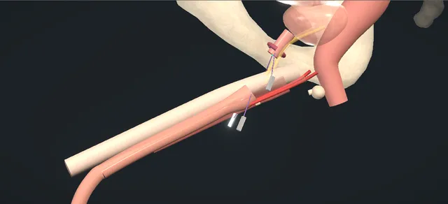
Schematic scene preview: Posterior urethral reconstruction
Preparing the 3D anatomy…

UrOops3D · UroRef · © Nity G

How to use this module. Play through the nine steps, or use Back and Next. Watch the scar excised onto the sound, the crura parted so the urethra takes a shorter course, and the sutures placed clockwise from twelve. The triggers show the rectum, a narrowed repair, the cavernous nerves and the bladder neck.

Click me next Section 2 of 8 Anatomy The structures this operation works between, and the one it must not cross.

The structures in the scene

Posterior urethral reconstruction: how the structures relateThe perineum from the foot of the table, down onto the distraction defect. Scar of the distraction defect: its distal end; the urethra is cut across here Bulbar urethra: freed and cut across. Scar of the distraction defect: its proximal end; opened onto the sound Prostatic apex: found with a sound. Scar of the distraction defect: lies behind and below; crura parted for length Pubis and the crura, above it. Scar of the distraction defect: lies on it; the deep excision Rectum, behind its deepest part (danger). Scar of the distraction defect: run beside it to the crura Cavernous nerves, beside the apex (danger). Scar of the distraction defect: above it; the sphincter is lost in the scar Bladder neck: continence rests on it (danger).Scar of the distractiondefectBulbar urethra:freed and cut acrossProstatic apex:found with a soundPubis and the crura,above it!Rectum, behind itsdeepest part!Cavernous nerves,beside the apex!Bladder neck:continence rests onitits distal end; theurethra is cutacross hereits proximal end;opened onto thesoundlies behind andbelow; crura partedfor lengthlies on it; thedeep excisionrun beside it tothe cruraabove it; thesphincter is lostin the scarTargetDangerLandmarkContext
The perineum from the foot of the table, down onto the distraction defect. Schematic, not to scale; relationships only.
  • From the foot of the table in lithotomy, the bulbar urethra runs under the midline of the perineum and ends in pale scar.
  • The scar runs deep, behind and below the pubic symphysis, up to the prostatic apex, which the fracture has lifted out of line.
  • Looking into the wound, the surgeon looks straight along the defect.
  • The rectum lies behind its deepest part and a cavernous nerve passes on each side of the apex toward the crura.
  • Dorsally the crura part toward the pubis when they are separated, and the freed urethra drops between them on a shorter course to the apex.
  • ›The distraction defect - scar between the end of the bulbar urethra and the prostatic apex, behind and below the pubic symphysis; excised whole.
  • ›Bulbar urethra and corpus spongiosum - freed from the corpora, cut across at the scar and mobilised to the penoscrotal junction.
  • ›Prostatic apex and the proximal stump - displaced upward by the injury; found with a sound passed through the suprapubic tract.
  • ›Bulbar arteries - enter the bulb from behind; preserved during the proximal mobilisation.
  • ›Corpora cavernosa and the crura - separated in the midline for a longer defect, giving the urethra a shorter course.
  • ›Dorsal vein in the intracrural space - met under the pubis as the crura are separated; ligated and divided.
  • ›Inferior pubic margin - removed by an inferior pubectomy when the crural separation does not give enough length.
  • ›Rectum - immediately behind the deepest part of the scar.
  • ›Cavernous nerves - run posterolateral to the prostate and past the apex to the crura; often injured by the fracture itself.
  • ›External sphincter remnant and bladder neck - the sphincter is disrupted by the injury; continence after repair rests on the bladder neck.
Click me next Section 3 of 8 Steps The operation in order, with the landmark that guards each move.

Operative steps

  1. The defect The repair is delayed until the haematoma has resolved, with the bladder drained by a suprapubic catheter in the meantime. Contrast is run up the urethra from the meatus and down from the bladder through the suprapubic tract. The two columns show the distal and proximal ends; the gap between them is the length of the defect, and its relation to the pubis predicts how much exposure will be needed.
    where it bites: a bladder neck that is open on the cystogram, or injured, is noted before surgery: after the repair continence depends on it.
  2. Lithotomy and exposure Intravenous antibiotics and general or regional anaesthesia, then high dorsal lithotomy in well-padded stirrups, a bolster under the buttocks, the images in the room. Under loupe magnification a vertical midline perineal incision runs from the base of the scrotum to just above the anus. Colles fascia is opened, bulbospongiosus is divided in the midline and dissected off the corpus spongiosum, and a self-retaining retractor is set.
    where it bites: the tunica of the corpus spongiosum is thin over the bulb and is perforated by careless dissection of the muscle.
  3. Free and transect A catheter passed from the meatus stops at the distal end of the scar. The mid-bulbar urethra is dissected all the way round and freed dorsally from the corporal bodies. It is cut across at the level of the scar and mobilised distally to the penoscrotal junction, off the tunica of the corpora. Proximally it is freed from its posterior midline attachments, the bulbar arteries are preserved and haemostasis is secured.
    where it bites: the bulbar arteries enter the bulb from behind; they are kept, because the mobilised urethra depends on its blood supply.
  4. Find the proximal urethra A curved metal sound is passed through the cystostomy tract, gently into the bladder neck and down to the proximal end of the obstruction. The scar between the bulb and the prostatic apex is excised from below until the sound can be felt through it. The obliterated proximal urethra is then incised directly onto the sound.
    where it bites: the excision follows the sound; without it the dissection can wander behind the prostate toward the rectum.
  5. Excise to healthy urethra The remaining fibrosis is excised entirely, down to healthy tissue all the way round the proximal end. The sound is withdrawn and cystourethroscopy confirms that there is no false passage and no bladder pathology. Both healthy urethral ends are tagged with stay sutures and calibrated progressively with bougies à boule.
    where it bites: the anterior rectal wall lies immediately behind the deepest part of the scar.
  6. Bridge the gap The ends are brought together. For a longer defect the space between the crura is developed sharply in the midline down to the inferior surface of the pubis, without entering the tunica of the corpora, and the dorsal vein met there is ligated and divided. The anterior urethra then takes a shorter course to the proximal stump. If more length is needed, an inferior pubectomy follows; rerouting round the left corporal body is the further option.
    where it bites: the corporal tunica during the crural separation, and the cavernous nerves beside the apex: only the manoeuvres the gap needs are used.
  7. Spatulate With a tension-free approximation confirmed, the distal end is spatulated along its dorsal wall and the proximal end along its ventral wall, so that the two openings overlap and the join is wider than either lumen.
    where it bites: any tension left at this point is carried into the anastomosis; the gap is bridged first.
  8. The anastomosis Full-thickness interrupted sutures are placed outside-in through the proximal end, clockwise from twelve o'clock, then inside-out through the distal end, taking spongiosum and mucosa. Each is held on a numbered clamp to keep its order. After half the distal sutures are placed, a urethral catheter is passed easily into the bladder. When all are in, they are tied one by one, clockwise from twelve.
    where it bites: the deep anterior sutures lie against the back of the pubis and the posterior sutures toward the rectum; each bite is placed under vision.
  9. Layered closure With a tension-free anastomosis and a dry field, the retractor is removed and the wound irrigated. Bulbospongiosus is closed with a running suture, then Colles fascia and the subcutaneous tissue separately to obliterate dead space, then the skin with interrupted sutures. The cystostomy is left to close by secondary intention; the urethral catheter goes to a drainage bag, taped to the abdominal wall.
    where it bites: dead space in the perineal wound collects blood and urine; the deep layers are closed separately to obliterate it.
Click me next Section 4 of 8 Danger zones Where this operation bites, and why it bites there.
Critical relationship

Danger zones

The spatial relationships the animation is built to make automatic. Before you read each one, ask yourself: at this point in the operation, what is about to go wrong, and what is my first move? The save for each sits in the Complications tab.

  • RiskThe rectum behind the scar - the deepest excision lies on the anterior rectal wall; the sound guides the dissection.
  • RiskExternal sphincter and bladder neck - the sphincter is disrupted by the injury; a bladder neck that is open or injured predicts incontinence after repair.
  • RiskCavernous nerves and the crura - beside the apex and between the crura, where the dissection and the crural separation run.
  • RiskThe bulbar arteries - kept during the proximal mobilisation so that the urethra stays perfused.
  • RiskThe ureters and the bladder neck in an abdominal approach - at risk when the repair needs abdominoperineal exposure.
  • RiskThe lithotomy position - neuropraxia after a long operation in high lithotomy; the stirrups are well padded.
Practise it Take me into the scene Opens the animation at a danger moment and asks you to predict the mechanism and your first move before it reveals.
Click me next Section 5 of 8 Complications The save for each one: the first move, the escalation trigger, the op-note line.

Complications and immediate saves

Trigger each of these live in the Animation tab, then read the save: the first move, the escalation trigger, and the line that goes in the op note.

Rectal injury in the dissection

  • the anterior rectal wall is opened while the deep scar is excised, most often in a complex or previously manipulated case.
The save
  1. Recognise it at once: a finger in the rectum shows the opening and its relation to the urethral stump. first move
  2. Close the rectal wall in layers, clear of the line of the anastomosis.
  3. Place healthy, well-vascularised tissue such as bulbospongiosus or a gracilis flap between the rectal repair and the anastomosis.
  4. A large or contaminated injury, or doubt about the repair, is discussed with a colorectal surgeon for faecal diversion. escalation trigger
  5. Record the site and size of the injury, the layers closed, the tissue interposed and the plan for diet and follow-up imaging. document

Recurrent stricture

  • the anastomosis narrows again and the stream slows, usually within the first year.
The save
  1. Confirm it objectively: a flow rate, then a urethrogram or cystoscopy to show the site and length of the narrowing. first move
  2. A short recurrence that is not obliterative may be treated with a urethrotomy or dilatation, no more than twice if lasting patency is the aim.
  3. Repeated endoscopic treatment that keeps failing only delays the definitive repair.
  4. A failed repair is referred to a high-volume reconstructive centre for redo urethroplasty. escalation trigger
  5. Record the original manoeuvres used, the site and length of the recurrence and the flow rate before and after each intervention. document

Erectile dysfunction

  • erections are weaker or absent; the fracture is usually the cause, and the repair adds a smaller risk.
The save
  1. Ask about erectile function before the repair and record it, so that the effect of the injury and of the operation can be told apart. first move
  2. Keep the dissection close to the scar at the apex and use the crural separation only when the gap needs it.
  3. Erectile function can recover for some time after the injury, so assessment is repeated at follow-up.
  4. Persisting dysfunction is referred to andrology for assessment and treatment. escalation trigger
  5. Record the preoperative function, the manoeuvres used and the function reported at each review. document

Urinary incontinence

  • leakage after repair, usually because the bladder neck no longer holds; the external sphincter was lost with the injury.
The save
  1. Assess the bladder neck before surgery on the cystogram and by cystoscopy through the suprapubic tract, and consent accordingly. first move
  2. Leave the bladder neck undisturbed during the perineal repair.
  3. After the repair, separate stress leakage from urgency with a history, a bladder diary and urodynamics if needed.
  4. Persisting stress incontinence is referred for continence surgery once the urethra is stable. escalation trigger
  5. Record the bladder neck findings before surgery and the type and severity of any leakage after it. document
Click me next Section 6 of 8 Troubleshooting When the case turns: the problem, then the next move.

Intra-operative troubleshooting

When the case turns: the problem, then the next move.

  • ActionThe ends do not meet without tension after mobilisation - develop the space between the crura in the midline down to the pubis and divide the dorsal vein; the urethra takes a shorter course between them.
  • ActionStill too short after the crural separation - an inferior pubectomy removes a wedge of the lower pubis with an osteotome and rongeur, bone wax on the raw surface; then bring the urethra up again.
  • ActionStill too short after the pubectomy - reroute the anterior urethra round the left corporal body to shorten its course further.
  • ActionThe sound cannot be felt from the perineum - the proximal urethra is not reached safely from below; an abdominoperineal approach may be required.
  • ActionA false passage is seen on cystourethroscopy - identify the true lumen with the scope and the sound before any suture is placed; the anastomosis is made only to healthy urethra.
  • ActionBleeding from the crura or the dorsal vein - ligate the dorsal vein before dividing it, stay out of the corporal tunica during the separation, and use bone wax on a raw pubic surface.
Click me next Section 7 of 8 FRCS viva Say it out loud, then mark yourself against the model answer.

FRCS viva questions

The examiner stems this scene prepares you for. Try to answer out loud first, then reveal, and learn the discriminator, the detail that turns a pass into a strong pass.

QA man has a suprapubic catheter after a pelvic fracture with a complete urethral disruption. When and how would you plan his repair?reveal answer
  • I would wait.
  • The European trauma guideline recommends suprapubic diversion and deferred urethroplasty after at least three months for a complete disruption, by which time the haematoma has nearly always resolved.
  • He must also be stable and able to lie in lithotomy.
  • I would image both ends at once: a retrograde urethrogram with a voiding or antegrade cystourethrogram through the suprapubic tract, which the strictures guideline recommends for these injuries.
  • That shows the length of the gap, its relation to the symphysis and whether the bladder neck is open.
  • I would add cystoscopy through the tract, and MRI if the anatomy is unclear, because MRI measures a posterior defect more accurately.
  • I would not offer endoscopic treatment for an obliterated segment: the guideline advises against it, and it risks a false passage.
Strong pass

Say all three

  • Naming the three-month delay
  • Imaging from above and below at the same time
  • Assessing the bladder neck before surgery
Examiner note
Naming the three-month delay, imaging from above and below at the same time, and assessing the bladder neck before surgery.
QTalk me through the operation.reveal answer
  • High lithotomy in padded stirrups, antibiotics given, the images in the room.
  • I make a vertical midline perineal incision from the base of the scrotum to just above the anus, open Colles fascia and divide bulbospongiosus in the midline.
  • I pass a catheter to the distal end of the scar, free the bulbar urethra all the way round and cut it across at the scar.
  • I mobilise it distally to the penoscrotal junction and proximally off its posterior attachments, keeping the bulbar arteries.
  • I pass a curved sound through the cystostomy into the bladder neck and down to the obstruction, and excise the scar until I can feel it.
  • I open the proximal urethra onto the sound and excise all the remaining fibrosis.
  • Cystourethroscopy excludes a false passage.
  • I tag and calibrate both ends, then separate the crura or remove the lower pubis only if the gap needs it.
  • I spatulate the distal end dorsally and the proximal end ventrally.
  • I place interrupted sutures outside-in proximally, clockwise from twelve, then inside-out distally, each on a numbered clamp.
  • I pass the catheter after half the distal bites and tie them clockwise.
  • I close in layers and let the cystostomy close by itself.
Strong pass

Say both

  • Using the sound through the suprapubic tract to find the proximal urethra
  • Excising the scar completely before any manoeuvre to gain length
Examiner note
Using the sound through the suprapubic tract to find the proximal urethra, and excising the scar completely before any manoeuvre to gain length.
QThe ends will not meet. What do you do?reveal answer
  • I would follow the stepwise perineal approach and stop at the first step that gives a tension-free join.
  • After distal mobilisation the next step is separating the corporal bodies in the midline, then an inferior pubectomy, then supracrural rerouting.
  • Webster and Ramon described this sequence: 66 of their 74 men needed added manoeuvres, and they reported excellent results in 96%.
  • In the multicentre series of Kizer and colleagues, which includes the author of the operative text, 67% of 142 repairs needed only excision and mobilisation.
  • Corporal splitting was added in 17%, inferior pubectomy in 10% and rerouting in only 3%, three of those four recurring.
  • An abdominoperineal approach was needed in 4%.
  • The European guideline keeps that approach for rectourethral fistula, prostatic or bladder neck injury, and advises against total pubectomy.
Strong pass

Say both

  • Giving the order of the manoeuvres
  • Knowing that most repairs need only excision and mobilisation
Examiner note
Giving the order of the manoeuvres and knowing that most repairs need only excision and mobilisation.
QWhat outcomes do you quote him?reveal answer
  • The European strictures guideline gives an overall patency of 85.7% after deferred anastomotic repair, and says complete excision of the scar strongly predicts success.
  • In series, Kizer and colleagues reported 92% success and Cooperberg and colleagues a recurrence rate of 14%, with 93% success if one urethrotomy is allowed.
  • Erectile dysfunction is common after the injury itself: about 34% in the meta-analysis of Blaschko and colleagues, with delayed urethroplasty adding about 3%.
  • The guideline gives incontinence after repair as 6.8 to 8.5%, usually from an incompetent bladder neck, and rectal injury as 0 to 4.9%, more often in complicated cases.
  • I would follow him with flow rates and imaging, and treat a short recurrence endoscopically no more than twice.
Strong pass

Say both

  • Separating erectile dysfunction caused by the injury from that caused by the repair
  • Linking incontinence to the bladder neck
Examiner note
Separating erectile dysfunction caused by the injury from that caused by the repair, and linking incontinence to the bladder neck.
QHow does this differ from a bulbar anastomotic urethroplasty?reveal answer
  • Both excise scar and join healthy, spatulated ends without tension, but the problems differ.
  • A bulbar stricture is spongiofibrosis in the anterior urethra; here the urethra has been torn apart and the gap is filled with scar behind the pubis.
  • The proximal end lies at the prostatic apex, so I need a sound through the suprapubic tract to find it.
  • The gap may need corporal separation, pubectomy or rerouting, never needed in a bulbar repair.
  • The rectum lies behind the dissection.
  • The external sphincter has been lost, so continence depends on the bladder neck.
  • Erectile dysfunction is usually caused by the fracture.
  • The imaging differs too: I need contrast from above as well as below.
Strong pass

Say both

  • Explaining that continence rests on the bladder neck because the sphincter is disrupted
  • That the proximal end is found from above
Examiner note
Explaining that continence rests on the bladder neck because the sphincter is disrupted, and that the proximal end is found from above.
Click me next Section 8 of 8 Evidence The guidance each of these answers is built on.

Evidence and guidance

Operative Dictations in Urologic Surgery
Armenakas NA, Fracchia JA, Golan R. Posterior urethral reconstruction. Chapter 64. Wiley, 2019
The source of the sequence drawn and of every step, danger and troubleshooting line: the ten essential steps, the template dictation, the three variations and the complication list. Book sizes kept off screen: the catheter and sound sizes, calibration to 28 or 30 Fr, 8 to 12 sutures, 5 cm of crural separation and a 2 cm pubic wedge. The chapter leaves the cystostomy to close by secondary intention and drains with a urethral catheter, so no suprapubic catheter is drawn at the end; many UK units also leave one, which is general practice and not shown. The chapter gives no catheter duration; the scene places eight sutures. Not in the chapter: the timing, the imaging, the outcome figures and the save steps for each complication, which come from the sources below or are general practice.
Open guideline
EAU Guidelines
Urethral Strictures: Disease Management in Males, section 6.3.5.b, post-traumatic posterior stenosis, read 18 October 2026
Deferred management at earliest three months after the trauma. Recommendations: progressive perineal excision and primary anastomosis for obliterative stenosis (strong); midline perineal incision (strong); no endoscopic treatment for obliterative stenosis (strong); no more than two urethrotomies or dilatations for a short nonobliterative recurrence (weak); abdominoperineal repair only for rectourethral fistula, prostatic or bladder neck injury (weak); no total pubectomy (strong); open repair in high-volume centres (weak). Overall patency 85.7%. Incontinence 6.8 to 8.5%, usually from bladder neck incompetence. Rectal injury 0 to 4.9%. Read through a summarising fetch tool.
Open guideline
EAU Guidelines
Urethral Strictures: Diagnostic Evaluation, sections 5.2.1.d, e and g, read 18 October 2026
Combine retrograde urethrography with voiding cystourethrography for pelvic fracture urethral injuries (strong). Combined retrograde urethroscopy and antegrade cystoscopy through the suprapubic tract as an adjunct (weak). Consider MRI urethrography in posterior stenosis (strong); MRI measures the length of a posterior stenosis more accurately than urethrography.
Open guideline
EAU Guidelines
Urological Trauma: Urogenital trauma, section 4.4, urethral trauma, read 18 October 2026
Erectile dysfunction occurs in about 34% of men after a pelvic fracture urethral injury, unrelated to urethroplasty. For a complete disruption: suprapubic diversion and deferred urethroplasty after at least three months (strong); no immediate urethroplasty within 48 hours (strong); no repeated endoscopic treatment after failed realignment (strong).
Open guideline
The Journal of Urology
Kizer WS, Armenakas NA, Brandes SB, Cavalcanti AG, Santucci RA, Morey AF. Simplified reconstruction of posterior urethral disruption defects: limited role of supracrural rerouting. J Urol 2007;177:1378-81
Abstract read through PubMed, 18 October 2026. Retrospective, five centres, 142 patients with more than a year of follow-up. Excision and mobilisation alone in 67%, corporal splitting in 17%, inferior pubectomy in 10%, supracrural rerouting in 3% (three of four recurred), abdominoperineal approach in 4%. Overall success 92%.
Open guideline
The Journal of Urology
Cooperberg MR, McAninch JW, Alsikafi NF, Elliott SP. Urethral reconstruction for traumatic posterior urethral disruption: outcomes of a 25-year experience. J Urol 2007;178:2006-10
Abstract read through PubMed, 18 October 2026. 134 men, single surgeon. Partial pubectomy in 22%, abdominoperineal approach in 4%; pubectomy needed in 34% with a closed bladder neck on imaging against 7% with an open one. Recurrence 14%; success 93% allowing one urethrotomy.
Open guideline
Arab Journal of Urology
Blaschko SD, Sanford MT, Schlomer BJ, et al. The incidence of erectile dysfunction after pelvic fracture urethral injury: a systematic review and meta-analysis. Arab J Urol 2015;13:68-74
Abstract read through PubMed, 18 October 2026. 24 studies. Erectile dysfunction after the injury 34%; delayed urethroplasty added 3%.
Open guideline
The Journal of Urology
Webster GD, Ramon J. Repair of pelvic fracture posterior urethral defects using an elaborated perineal approach: experience with 74 cases. J Urol 1991;145:744-8
Abstract read through PubMed, 18 October 2026. 74 patients, one-stage perineal anastomosis in all; 66 needed added manoeuvres, used in sequence as needed: distal mobilisation, corporal separation, inferior pubectomy, supracrural rerouting. Excellent results in 96%.
Open guideline
BJU International
Fu Q, Zhang J, Sa YL, Jin SB, Xu YM. A retrospective series of 573 transperineal bulboprostatic anastomoses after pelvic fracture. BJU Int 2013;112:E358-63
Abstract read through PubMed, 18 October 2026 (the title here describes the paper). Success 88%; an intraoperative rectal injury was repaired primarily in 28. Cited for primary repair of a rectal injury found at operation. Interposing tissue and the threshold for diversion are general practice, supported by the EAU recommendation of a local flap after a concomitant rectourethral fistula is corrected (weak).
Open guideline

Curriculum. Supports learning relevant to the ISCP Urology Curriculum syllabus topics Genitourinary trauma and Urethral stricture disease. Not a workplace-based assessment and not evidence of competence.

Two minutes

Close the lap: recall it before you leave.

Say it aloud, then reveal. Reading is not remembering.

  1. 01 The relationship Say the critical spatial relationship out loud, then reveal. reveal
    Memory frame

    From the foot of the table in lithotomy, the bulbar urethra runs under the midline of the perineum and ends in pale scar.

    Why?
    The scar runs deep, behind and below the pubic symphysis, up to the prostatic apex, which the fracture has lifted out of line. Looking into the wound, the surgeon looks straight along the defect. The rectum lies behind its deepest part and a cavernous nerve passes on each side of the apex toward the crura. Dorsally the crura part toward the pubis when they are separated, and the freed urethra drops between them on a shorter course to the apex.
  2. 02 The first move Rectal injury in the dissection. Your first move? reveal
    First move

    Recognise it at once: a finger in the rectum shows the opening and its relation to the urethral stump.

  3. 03 The escalation trigger Rectal injury in the dissection. What makes you call for help? reveal
    Escalate when

    A large or contaminated injury, or doubt about the repair, is discussed with a colorectal surgeon for faecal diversion.

  4. 04 The discriminator What turns a pass into a strong pass? reveal
    Strong pass

    Say all three

    • Naming the three-month delay
    • Imaging from above and below at the same time
    • Assessing the bladder neck before surgery
    Examiner note
    Naming the three-month delay, imaging from above and below at the same time, and assessing the bladder neck before surgery.
  5. 05 The pitfall Where does this operation bite first? reveal
    Pitfall

    The rectum behind the scar

    Why?
    the deepest excision lies on the anterior rectal wall; the sound guides the dissection.

Revision practice with self-marked answers, not an assessment of clinical competence. Kept in this browser only; it does not sync between devices.