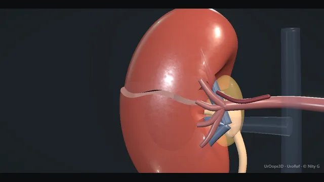
A blunt injury to the right kidney walked up the AAST grades: cortex, collecting system, pedicle. The scene teaches the thresholds where management changes: embolise the stable blush, explore the failing resuscitation, and never lose track of a leak.
One kidney from capsule to hilum, one axis of severity. Schematic, not to scale; relationships only.
One kidney, four zones, one axis of severity.
The grade climbs as the injury deepens: capsule, cortex, calyx, pedicle.
Two imaging findings override the grade in practice: the blush, which sends the stable patient to angiography, and extravasation, which commits to follow-up until it resolves.
›Cortex + capsule - grade I to III territory; a subcapsular haematoma is contained by the capsule and can compromise the kidney if it expands.
›Medulla + collecting system - the grade IV threshold: a laceration into a calyx means urine extravasation and a leak to follow.
›Hilar pedicle - artery, vein and segmental branches; injury here is grade IV to V and the mechanism of the non-enhancing kidney.
›Perinephric space - where the haematoma and the urinoma collect; contained here they are a finding, not an indication.
The kidney in situThe kidney is read in layers from outside in: capsule and cortex, medulla, collecting system, then the hilar pedicle where artery, vein and segmental branches sit. The AAST grade climbs with depth, driven by two axes: how deep the laceration runs, and whether the collecting system or the pedicle is involved. The habit this step teaches is to name the injury in those anatomical terms before any decision is made, because the grade is an anatomical statement while the decisions come later from physiology. A grade quoted without the finding behind it cannot be handed over safely at shift change.
Grade I-II: contusion / subcapsular haematomaThese injuries are confined to the cortex and capsule: contusion, or a subcapsular or perinephric haematoma without any laceration, with the collection held by the fascia around it. Management is non-operative by default: rest until the urine clears, serial examination of abdomen and flank, and serial haemoglobin at the intervals the local protocol sets. Observation is a treatment with its own observation chart, not a decision to do nothing. The specific failure of this grade is the subcapsular collection that quietly expands and strangles the kidney, which is why the patient who is observed must actually be observed. where it bites: a subcapsular haematoma can grow and strangle the kidney: observe, but actually observe.
Grade III: deep cortical lacerationA grade III injury is a laceration that runs deep into the cortex without entering the collecting system and without segmental-vessel injury, and the perinephric haematoma stays contained around it. The stable patient remains on the non-operative pathway, with the same discipline of serial examination and haemoglobin as the lower grades. The CT is read as a whole study rather than at its most reassuring slice, because a laceration that looks purely cortical on one cut may reach a calyx on the next, and that finding moves the patient to grade IV and changes the follow-up. A deep laceration with a contained haematoma is also the injury that can declare itself again days later, which the discharge plan should anticipate. where it bites: the laceration that looks cortical on one slice may reach a calyx on the next: read the whole study, not the worst single image.
Grade IV: collecting system / segmental vesselGrade IV covers two injuries: a laceration into the collecting system, seen as contrast extravasation on the delayed phase, or a segmental-vessel injury with a contained haematoma or an active blush. The stable patient with a blush goes to angiography for selective embolisation, which treats the bleeding while preserving the parenchyma that surgery in this setting would take. The stable patient with extravasation is observed with planned early re-imaging, and a persistent or symptomatic leak is decompressed with a stent or nephrostomy and followed until imaging shows resolution. The failure to design against is the leak that is never re-imaged: new fever or ileus after extravasation is an untreated leak until proven otherwise. where it bites: extravasation plus fever or ileus is a missed leak until proven otherwise.
Grade V: shattered kidney / hilar avulsionGrade V is parenchyma shattered into devitalised, non-perfused fragments, or an avulsed hilum, and the signature on CT is a kidney that does not enhance. This is the grade that kills and the grade that explores: the management question is the resuscitation, not the kidney, and the patient who fails to respond belongs in theatre rather than the angiography suite. If exploration happens, proximal control of the pedicle at its origin is obtained before the haematoma is opened, because entering Gerota's fascia over an uncontrolled perinephric haematoma converts a contained bleed into exsanguination. In the shocked patient that exploration usually ends in nephrectomy, and the team should say so plainly before it starts. where it bites: exploration of a grade V in shock usually ends in nephrectomy; proximal control is obtained before the haematoma is opened.
Management: embolise or exploreThe decision fork is physiological, not anatomical. The stable patient with a contrast blush goes to angiography and selective embolisation, because that stops the bleeding and preserves parenchyma in one act, with serial haemoglobin and examination as the endpoints afterwards. The failing resuscitation goes to a midline laparotomy with proximal vascular control of the pedicle before the haematoma is touched, accepting that renal exploration in shock usually means nephrectomy, and that this is the correct trade. Between the poles sit the stable patients with a leak or devitalised fragments, who are followed on their leak and re-imaged to resolution rather than operated on. Whatever the fork, the grade, the findings and the timings are documented, because the patient who deteriorates overnight is managed by the record, not the memory.
The spatial relationships the animation is built to make automatic. Before you read each one, ask yourself: at this point in the operation, what is about to go wrong, and what is my first move? The save for each sits in the Complications tab.
RiskThe blush on CT - contrast extravasation in a stable patient is an angioembolisation referral, not an observation.
RiskThe failing resuscitation - the only absolute indication to explore; renal exploration in shock usually becomes nephrectomy.
RiskThe missed urine leak - extravasation that is never re-imaged becomes urinoma, ileus and sepsis.
When the case turns: the problem, then the next move.
ActionBlush on CT, patient stable - angiography and selective embolisation; preserve parenchyma and re-image afterwards.
ActionGrade IV leak on follow-up imaging, patient well - stent or nephrostomy to decompress, then image to resolution; most leaks close on drainage alone.
ActionNon-responder in the emergency department - theatre, not more fluid or more CT: proximal control first and a damage-control mindset.
ActionTrauma in a solitary kidney - lower every threshold: image regardless of haematuria, embolise rather than explore, and reconstruct where at all possible.
The examiner stems this scene prepares you for. Try to answer out loud first, then reveal, and learn the discriminator, the detail that turns a pass into a strong pass.
QClassify blunt renal injuries and explain what actually changes management.reveal answer
The AAST scale runs from grade I, subcapsular haematoma or contusion, through grade II, perinephric haematoma or a laceration under a centimetre, grade III, a laceration deeper than a centimetre without collecting-system involvement, grade IV, a laceration into the collecting system with extravasation or a segmental-vessel injury with contained haemorrhage, to grade V, a shattered kidney or hilar avulsion.
The grade describes anatomy, but management is driven by physiology: haemodynamic stability decides between imaging and theatre, and it keeps deciding, hourly, on the ward.
Within the grades, three features change management independently: contrast blush, which points to angiography and embolisation;
extravasation, which commits to follow-up imaging and, usually, drainage of the collecting system;
and devitalised fragments or absent enhancement, which raises the prospect of intervention for the kidney itself.
The grade therefore frames prognosis, while stability, blush and leak drive the actual decisions.
Strong pass
Say both
Separating anatomical grading from physiological decision-making
Naming blush and extravasation as the two imaging findings that act
Examiner note
Separating anatomical grading from physiological decision-making, and naming blush and extravasation as the two imaging findings that act.
QWhen do you embolise a renal injury?reveal answer
Embolisation is for the patient with radiological evidence of arterial bleeding and a physiology that tolerates the angiography suite.
The typical settings are a contrast blush or pseudoaneurysm on CT in a stable patient, a segmental injury with contained haematoma, and delayed haemorrhage or pseudoaneurysm days after injury or surgery.
It is chosen precisely because it stops bleeding while preserving parenchyma, which surgery in this setting often does not.
Embolisation is not the answer for the patient failing resuscitation, who belongs in theatre with proximal control, nor for venous extravasation, which is not an angiographic target.
Afterwards, the patient is observed with serial haemoglobin and examination as the endpoints, and re-imaged if bleeding declares itself again;
in a stable patient a failed first embolisation is usually followed by a second attempt before surgery is considered.
Strong pass
Say both
Blush plus stability as the entry condition
Naming the failed-resuscitation patient as the one who must not be sent to the angiography suite
Examiner note
Blush plus stability as the entry condition, and naming the failed-resuscitation patient as the one who must not be sent to the angiography suite.
QWhen do you explore a blunt renal injury, and what does exploration usually mean?reveal answer
The absolute indication is haemodynamic non-response: a patient who does not respond to resuscitation, or who destabilises again, with a known or suspected renal source.
The other classical triggers are an expanding or pulsatile perinephric haematoma found at laparotomy done for other injuries, and renal vascular injury in a patient already undergoing surgery.
Exploration is by midline laparotomy with proximal control: the renal pedicle is controlled at its origin before the haematoma is entered, because opening Gerota's fascia on an uncontrolled haematoma converts a contained bleed into an exsanguination.
What exploration usually means is nephrectomy: in the shocked patient a shattered kidney or an avulsed pedicle is rarely reconstructible, and the decision is life over nephrons.
Reconstruction is attempted in the stable patient with a salvageable kidney, with vascular control, consideration of cooling, and collaboration where available.
Consent and counselling before and after must be honest about that trajectory.
Strong pass
Say both
Proximal pedicle control before the haematoma is opened
Stating plainly that renal exploration in shock usually ends in nephrectomy
Examiner note
Proximal pedicle control before the haematoma is opened, and stating plainly that renal exploration in shock usually ends in nephrectomy.
QA grade IV injury with extravasation: how is the urine leak managed?reveal answer
The first question is whether the patient is well: fever, ileus or rising inflammatory markers convert a radiological finding into a clinical problem.
Minor extravasation in a well patient is observed with early repeat imaging, and much of it resolves spontaneously.
A persistent or symptomatic leak is decompressed: a retrograde JJ stent is usually first-line, with a nephrostomy reserved for the failed retrograde attempt, the septic patient or a disrupted collecting system.
A large urinoma is drained percutaneously.
Antibiotics are used for infection rather than routinely for every leak, following local policy.
The endpoint is documented resolution on imaging, because the missed leak is the classic cause of the delayed septic readmission.
If leakage persists despite drainage, or a devitalised fragment keeps the system open, delayed endoscopic or surgical intervention addresses it once the patient is stable.
Strong pass
Say all three
Observation for the well patient
Drainage for the symptomatic one
Documented radiological resolution as the discharge criterion
Examiner note
Observation for the well patient, drainage for the symptomatic one, and documented radiological resolution as the discharge criterion.
QDoes the management change with a solitary kidney?reveal answer
Yes, in the direction of nephron preservation at every fork.
Imaging thresholds drop: a solitary kidney is imaged whatever the haematuria, because the penalty for a missed injury is dialysis rather than compensation by the other side.
Intervention thresholds move toward embolisation and reconstruction and away from nephrectomy: a blush is embolised selectively, a leaking collecting system is stented or drained early and definitively, and a shattered but partially perfused solitary kidney is reconstructed by a surgeon who does that work, with vascular control and a realistic staged plan.
Nephrectomy in a solitary kidney is dialysis by another route and is a last resort accepted only for uncontrollable haemorrhage in a dying patient.
Follow-up is stricter, with imaging to confirm perfusion and drainage and close monitoring of renal function, because a silently obstructed or ischaemic solitary kidney presents late and badly.
Strong pass
Say both
Imaging regardless of haematuria
Nephrectomy named explicitly as dialysis by another route in this population
Examiner note
Imaging regardless of haematuria, and nephrectomy named explicitly as dialysis by another route in this population.
Say it aloud, then reveal. Reading is not remembering.
01The relationshipSay the critical spatial relationship out loud, then reveal.reveal
Memory frame
One kidney, four zones, one axis of severity.
Why?
The grade climbs as the injury deepens: capsule, cortex, calyx, pedicle. Two imaging findings override the grade in practice: the blush, which sends the stable patient to angiography, and extravasation, which commits to follow-up until it resolves.
02The first moveDelay to embolisation. Your first move?reveal
First move
Reassess now: pulse, pressure, abdomen and haemoglobin; a rising pulse or falling haemoglobin reclassifies the patient.
03The escalation triggerDelay to embolisation. What makes you call for help?reveal
Escalate when
Physiological decline, or embolisation unavailable → theatre for exploration with proximal control.
04The discriminatorWhat turns a pass into a strong pass?reveal
Strong pass
Say both
Separating anatomical grading from physiological decision-making
Naming blush and extravasation as the two imaging findings that act
Examiner note
Separating anatomical grading from physiological decision-making, and naming blush and extravasation as the two imaging findings that act.
05The pitfallWhere does this operation bite first?reveal
Pitfall
The blush on CT
Why?
contrast extravasation in a stable patient is an angioembolisation referral, not an observation.
Revision practice with self-marked answers, not an assessment of clinical competence. Kept in
this browser only; it does not sync between devices.