UrOops3D, danger-zone atlas

Schematic model.

Operative animation, Retropubic sling

Retropubic mid-urethral sling

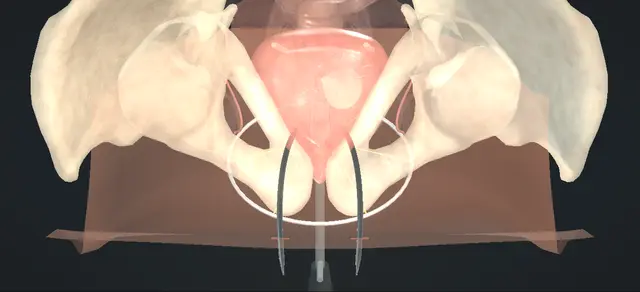
A synthetic tape placed under the mid-urethra through a short anterior vaginal incision, each arm carried up behind the pubis to the suprapubic skin on a curved trocar.

The trocars are passed from the vagina upward on the back of the bone, with the bladder drained and its neck held away from the side of the pass. The bladder is inspected with both trocars still in place, before any tape is drawn through.

The tape is then set flat and loose over a spacer, its sheaths are taken off and its ends are cut at the skin.

Core tier FRCS continence, female urology Interactive 3D

Operative visualisation

Schematic scene preview: Retropubic sling
Preparing the 3D anatomy…

UrOops3D · UroRef · © Nity G

How to use this module. Play through the eight steps, or use Back and Next. Watch the bladder fall back as it drains, each trocar climb the back of the pubis, the cystoscope look with both trocars in, and the tape settle loose over its spacer. The triggers show a perforation, retropubic bleeding and a tight tape.

Click me next Section 2 of 8 Anatomy The structures this operation works between, and the one it must not cross.

The structures in the scene

Retropubic sling: how the structures relateThe path of each trocar, from the vaginal wound to the skin above the pubis. Path of the trocar, then of the tape: kept on its posterior surface all the way up Back of the pubis: the guide. Path of the trocar, then of the tape: lies immediately behind and medial Bladder: drained and carried away (danger). Path of the trocar, then of the tape: begins beside it, in the para-urethral tunnel Mid-urethra: the tape lies under it. Path of the trocar, then of the tape: pierced at the lower border of the pubis Endopelvic fascia. Path of the trocar, then of the tape: bleeding here is out of sight Retropubic space (danger). Path of the trocar, then of the tape: ends at a stab incision above the pubis Suprapubic skin, each side of the midline.Path of the trocar,then of the tapeBack of the pubis:the guide!Bladder: drained andcarried awayMid-urethra: thetape lies under itEndopelvic fascia!Retropubic spaceSuprapubic skin,each side of themidlinekept on itsposterior surfaceall the way uplies immediatelybehind and medialbegins beside it,in thepara-urethral…pierced at thelower border of thepubisbleeding here isout of sightends at a stabincision above thepubisTargetDangerLandmarkContext
The path of each trocar, from the vaginal wound to the skin above the pubis. Schematic, not to scale; relationships only.
  • From the foot of the table in lithotomy, the urethra runs up and back from the meatus to the bladder neck in front of the anterior vaginal wall, and the pubis lies above and in front of it.
  • Each trocar leaves the vaginal wound beside the mid-urethra, passes under the lower border of the pubis through the endopelvic fascia and climbs the back of the bone to the skin just above it.
  • The bladder fills the space behind that path, and a full bladder lies against the bone.
  • ›Mid-urethra - the level the tape supports; well short of the bladder neck.
  • ›Anterior vaginal wall - opened in the midline under the mid-urethra; its flaps are lifted off the fascia and closed at the end.
  • ›Periurethral fascia - the layer the vaginal flap is dissected off; the tape lies on it, against the ventral urethra.
  • ›Endopelvic fascia - met under the pubis on each side; pierced there to enter the retropubic space.
  • ›Pubic symphysis - its posterior surface guides the tip of each trocar from the fascia to the skin.
  • ›Retropubic space - between the pubis in front and the bladder behind; the corridor of the pass.
  • ›Bladder - drained for the passes, filled for the cystoscopy, drained again before the tape is brought up.
  • ›Bladder neck - carried away from the side of each pass by the guide in the catheter.
  • ›Ureteric orifices - efflux from both is seen at the cystoscopy.
  • ›Suprapubic skin - the two exit points lie directly over the pubic symphysis, one each side of the midline.
Click me next Section 3 of 8 Steps The operation in order, with the landmark that guards each move.

Operative steps

  1. Landmarks A woman with stress incontinence who has chosen a retropubic mesh sling after counselling that it is a permanent implant. Antibiotics, then dorsal lithotomy on padded stirrups; the genitalia, the perineum and the lower abdomen are prepared. A urethral catheter drains the bladder, which is kept empty throughout the passes. A weighted speculum is placed on the posterior vaginal wall and a self-retaining retractor is set. The mid-urethra is identified on the anterior vaginal wall, and an exit point is marked on the skin directly over the pubic symphysis on each side of the midline.
    where it bites: a full bladder lies against the pubis, in the line of the pass.
  2. Incision and tunnels The anterior vaginal wall is infiltrated with local anaesthetic and adrenaline, which lifts the plane and limits bleeding. With the bladder empty, a midline incision is made in the anterior vaginal wall under the mid-urethra. By sharp and blunt dissection the vaginal flap on each side is lifted off the periurethral fascia and the dissection is carried laterally toward the ischiopubic ramus. This leaves a short tunnel on each side of the urethra that ends at the endopelvic fascia, at the lower border of the pubis.
    where it bites: the tunnels stay lateral to the urethra and on the vaginal side of its fascia; a tunnel that strays medially opens the urethra.
  3. Right trocar A small stab incision is made at each skin mark. A rigid guide is passed down the catheter and its handle is moved toward the side of the pass, which carries the bladder neck and urethra to the other side. The trocar is introduced into the right tunnel, pierces the endopelvic fascia at the lower border of the pubis and enters the retropubic space. It is advanced upward with its tip kept on the posterior surface of the pubic symphysis until it emerges through the right skin incision. It is left in place.
    where it bites: the bladder lies immediately behind and medial; a trocar that leaves the bone is heading for the bladder.
  4. Left trocar The handle of the guide is moved to the left, so that the bladder neck now lies to the right. The left trocar is passed in the same way: into its tunnel, through the endopelvic fascia under the pubis, and up the back of the bone to the left skin incision. The two trocars now lie in their tracks and neither has been pulled up.
    where it bites: the guide is moved before the second trocar is; the second pass carries the same risk as the first.
  5. Cystoscopy with the trocars in The guide and the urethral catheter are removed and cystourethroscopy is performed with the bladder filled, using 30-degree and 70-degree lenses. Both lateral walls and the dome are inspected for a trocar, the proximal urethra is inspected, and clear efflux is seen from both ureteric orifices. If a trocar is seen in the bladder it is withdrawn and passed again, and the inspection is repeated. The cystoscope is removed, the catheter is replaced and the bladder is drained.
    where it bites: a perforation that is not looked for now is found later, as tape inside the bladder.
  6. Draw the tape through Only when the bladder and urethra have been seen to be intact is each trocar drawn up and out through its skin incision. The tape, covered by its plastic sheath, follows each trocar through the tunnel and the retropubic space. Its middle is brought to lie flat against the ventral urethral wall at the level of the mid-urethra.
    where it bites: a tape drawn through before the bladder has been inspected may already lie inside it.
  7. Set it loose Scissors or a similar instrument are held between the tape and the urethra as a spacer. The tape is adjusted until it lies against the spacer without tension, and each plastic sheath is then pulled off through its skin incision while the spacer holds the gap. With the spacer removed, the tape lies flat and loose under the mid-urethra.
    where it bites: a tape set under tension obstructs the urethra; urinary retention is a listed complication.
  8. Trim and close Each end of the tape is cut flush with the skin, so that it retracts beneath it, and the skin incisions are closed. The wounds are irrigated and haemostasis is checked. The anterior vaginal wall is approximated with interrupted absorbable sutures, and the self-retaining retractor and the weighted speculum are removed. The urethral catheter is connected to a drainage bag.
    where it bites: tape left proud at the skin, or caught in the vaginal suture line, is tape that is exposed later.
Click me next Section 4 of 8 Danger zones Where this operation bites, and why it bites there.
Critical relationship

Danger zones

The spatial relationships the animation is built to make automatic. Before you read each one, ask yourself: at this point in the operation, what is about to go wrong, and what is my first move? The save for each sits in the Complications tab.

  • RiskThe back of the pubis - the guide for the tip all the way up; a trocar that leaves the bone is heading for the bladder.
  • RiskBladder - drained before the passes and carried away from the passing side; inspected while the trocars are still in.
  • RiskLevel of the tape - under the mid-urethra, short of the bladder neck.
  • RiskTension - the tape is set over a spacer and left loose; a tape that indents the urethra obstructs it.
  • RiskRetropubic space - bleeding here is out of sight; it shows as blood down the tunnel or a swelling above the pubis.
  • RiskUrethra - the tunnels stay lateral to it, and it is inspected at the cystoscopy.
Practise it Take me into the scene Opens the animation at a danger moment and asks you to predict the mechanism and your first move before it reveals.
Click me next Section 5 of 8 Complications The save for each one: the first move, the escalation trigger, the op-note line.

Complications and immediate saves

Trigger each of these live in the Animation tab, then read the save: the first move, the escalation trigger, and the line that goes in the op note.

Bladder perforation

  • the tip of a trocar leaves the bone and enters the bladder.
  • It is found at the cystoscopy, which is done before any tape is drawn, when it can still be put right by passing the trocar again.
The save
  1. Draw no tape through. Withdraw the trocar that is in the bladder. first move
  2. Drain the bladder, move the guide so the bladder neck lies away from that side, and pass the trocar again with its tip on bone.
  3. Fill the bladder and inspect it again, both sides, before anything is brought up.
  4. A second entry on the same side, a large hole or a hole close to a ureteric orifice needs a senior colleague before the operation goes on. escalation trigger
  5. A catheter is left to drain the bladder afterwards, for as long as local practice asks.
  6. Op note: the side, what was seen at each cystoscopy, the second pass and the catheter plan. document

Retropubic bleeding

  • bleeding from the space behind the pubis during a pass: blood running down the tunnel beside the trocar, or a swelling above the pubis.
The save
  1. Pressure: a finger in the vagina pressed up against the back of the pubis and a hand on the abdomen above it, and held. first move
  2. Tell the anaesthetist and have the loss measured.
  3. If it settles, the pass is completed on the bone and the bladder is inspected as usual; the vagina may be packed with gauze at the end to help haemostasis.
  4. Bleeding that continues under pressure, an enlarging suprapubic swelling or a falling blood pressure needs senior and anaesthetic help at once. escalation trigger
  5. Op note: when it happened, the measured loss, what controlled it and the plan for observation. document

Urinary retention

  • she cannot void, or leaves a large residual, after a tape that was set without a gap and indents the urethra.
The save
  1. Measure the residual and drain the bladder with a catheter. first move
  2. Look for what else may be stopping her: pain, constipation, a pack still in the vagina.
  3. A trial without the catheter follows once those are dealt with, with the residual measured again.
  4. Retention that does not settle goes back early to the surgeon who placed the tape, who can loosen or divide it. escalation trigger
  5. Notes: the residual volumes, the catheter plan and when she is to be seen again. document
Click me next Section 6 of 8 Troubleshooting When the case turns: the problem, then the next move.

Intra-operative troubleshooting

When the case turns: the problem, then the next move.

  • ActionThe tip of the trocar loses the bone on the way up - stop advancing. The tip is brought back until bone is felt again and the pass continues on it. A tip that has been off the bone is assumed to have been near the bladder until the cystoscopy shows otherwise.
  • ActionThe tip does not come out at the skin incision - it is not forced along a new line. The trocar is brought back below the pubis and passed again toward the incision of its own side.
  • ActionEfflux is hard to see from a ureteric orifice - indigo carmine or methylene blue given intravenously before the cystoscopy makes the efflux easier to see. An orifice that stays dry is not left unexplained: ureteric obstruction and laceration are listed complications.
  • ActionThe tape looks tight once the sheaths are off - it is loosened before the wounds are closed and while its ends are still long, by drawing the middle of the tape down over the spacer.
  • ActionA midline incision gives poor exposure - an inverted-U incision in the anterior vaginal wall is the described alternative.
  • ActionVoiding after the operation is in doubt - a suprapubic catheter may be placed as well as the urethral catheter, to help with the trial of voiding.
Click me next Section 7 of 8 FRCS viva Say it out loud, then mark yourself against the model answer.

FRCS viva questions

The examiner stems this scene prepares you for. Try to answer out loud first, then reveal, and learn the discriminator, the detail that turns a pass into a strong pass.

QA woman with stress incontinence has not improved with pelvic floor training and asks about surgery. What do you offer?reveal answer
  • I would use the NICE patient decision aid and go through every operation NICE recommends, whether or not we offer it locally: colposuspension, open or laparoscopic, an autologous rectus fascial sling, and a retropubic mid-urethral mesh sling, with a bulking agent for a woman for whom those are unsuitable or unacceptable.
  • I would say plainly that there is evidence of benefit for all of them and limited evidence about long-term adverse effects, particularly where mesh is implanted.
  • If she is considering the retropubic sling I would tell her that it is a permanent implant and that complete removal might not be possible, and if she has it I would give her written details of the implant.
  • I would also be clear that in England vaginally inserted mesh for stress incontinence has been under a high vigilance restriction since July 2018.
  • I would use a device made of type 1 macroporous polypropylene mesh.
  • I would not offer a transobturator approach unless there was a specific reason to keep out of the retropubic space, such as previous pelvic procedures, and I would not use a top-down retropubic approach or a single-incision sling outside a clinical trial.
  • On results, the European guideline asks me to tell her that long-term outcomes are better with the retropubic route than with the transobturator route.
  • Her operation would be recorded in a national registry, and I would see her within 6 months and examine her for exposure of the tape.
Strong pass

Say all four

  • Naming all three first-line operations
  • The NICE limits on mesh slings: a retropubic sling by default
  • The bottom-up pass only
  • A permanent implant that may not be fully removable
Examiner note
Naming all three first-line operations, and the NICE limits on mesh slings: a retropubic sling by default, the bottom-up pass only, and a permanent implant that may not be fully removable.
QTalk me through a retropubic mid-urethral sling.reveal answer
  • Antibiotics and lithotomy.
  • I drain the bladder with a catheter and keep it empty.
  • With a weighted speculum and a self-retaining retractor in place I infiltrate the anterior vaginal wall and make a midline incision under the mid-urethra.
  • I lift the vaginal flap on each side off the periurethral fascia and carry the dissection laterally toward the ischiopubic ramus, which gives me a short tunnel on each side of the urethra.
  • I make a stab incision over the pubic symphysis on each side of the midline.
  • I pass a rigid guide down the catheter and use it to move the bladder neck away from the side I am about to pass.
  • I then pass each trocar from the vaginal wound upward: along the tunnel, through the endopelvic fascia under the pubis and up the back of the bone to its skin incision, with the tip on bone all the way.
  • I leave both trocars in, remove the catheter, fill the bladder and inspect it with 30-degree and 70-degree lenses: both lateral walls, the dome, the urethra, and efflux from both orifices.
  • Only then do I draw each trocar through with its tape.
  • I set the tape under the mid-urethra over a spacer so that it lies flat and loose, pull off the sheaths, cut the ends flush with the skin and close the vaginal wall with interrupted absorbable sutures.
Strong pass

The order at the centre of the operation: trocars, then cystoscopy, then tape; and a tape left without tension.

QHow do the retropubic and transobturator routes compare?reveal answer
  • I would quote the Cochrane review by Ford and colleagues, which included 81 trials and 12,113 women.
  • Subjective cure up to one year was similar by the two routes.
  • The retropubic route had the higher operative morbidity, although the overall rate of adverse events was low: the bladder was perforated in 4.5 per cent against 0.6 per cent by the transobturator route, voiding dysfunction afterwards was more frequent, and major vascular or visceral injury was more frequent.
  • Groin pain ran the other way, 6.4 per cent after the transobturator route against 1.3 per cent.
  • Tape erosion, exposure or extrusion was uncommon after either, about 24 and 21 per 1,000.
  • On limited data, repeat incontinence surgery in the long term was more likely after the transobturator route.
  • The same review found the bottom-to-top retropubic pass more effective than the top-to-bottom pass, with less voiding dysfunction and fewer bladder perforations and tape erosions.
  • The European guideline reads the evidence the same way: it grades the higher risk of perforation and voiding dysfunction behind the pubis as level 1a, and it makes a strong recommendation to tell women that long-term outcomes are better with that route.
  • NICE reserves the transobturator approach for women in whom the retropubic approach should be avoided.
Strong pass

Say all four

  • Knowing which way each difference runs: more bladder perforation
  • Voiding dysfunction behind the pubis
  • More groin pain through the obturator foramen
  • Better long-term results with the retropubic sling
Examiner note
Knowing which way each difference runs: more bladder perforation and voiding dysfunction behind the pubis, more groin pain through the obturator foramen, and better long-term results with the retropubic sling.
QAt cystoscopy you see a trocar inside the bladder. What do you do?reveal answer
  • This is the reason I look before I draw anything through.
  • The European guideline makes cystoscopy for bladder perforation a strong recommendation whenever a synthetic mid-urethral sling is inserted, and I do it while the trocars are still in and no tape has been drawn, because a trocar can be withdrawn and passed again and a tape drawn through the bladder cannot be left there.
  • I would withdraw that trocar, drain the bladder, check that the guide is carrying the bladder neck away from that side and pass the trocar again, keeping its tip on the bone.
  • I would then fill the bladder and inspect it again, both sides, before going on.
  • If the second pass also entered the bladder, or the hole was large or close to a ureteric orifice, I would stop and ask a senior colleague.
  • I would leave a catheter afterwards, record the side and what I saw at each inspection, and tell the patient what had happened.
Strong pass

Say both

  • The timing: with the trocars still in and no tape through
  • A perforation is put right by one clean second pass and a second look
Examiner note
The timing: with the trocars still in and no tape through, a perforation is put right by one clean second pass and a second look.
QShe is in retention the morning after a retropubic sling. Why, and what do you do?reveal answer
  • The cause I would think of first is a tape that is too tight.
  • Urinary retention is on the list of complications in the operative text, and voiding dysfunction is more frequent after a retropubic sling: in the Cochrane review by Ford and colleagues the risk after the transobturator route was about half that after the retropubic.
  • I would examine her, measure the residual and drain the bladder with a catheter, and I would look for the simpler contributors: pain, constipation, a pack left in the vagina.
  • I would not leave persistent retention to settle on its own; I would discuss her early with the surgeon who placed the tape, because an obstructing tape can be loosened or divided.
  • The prevention is in theatre: I set the tape with an instrument between it and the urethra, I take the sheaths off with that spacer still in, and I leave the tape loose.
Strong pass

Say both

  • Linking retention to tension
  • Treating an obstructing tape as a problem for early loosening or division by the surgeon who placed it
Examiner note
Linking retention to tension, and treating an obstructing tape as a problem for early loosening or division by the surgeon who placed it.
Click me next Section 8 of 8 Evidence The guidance each of these answers is built on.

Evidence and guidance

Operative Dictations in Urologic Surgery
Armenakas NA, Fracchia JA, Golan R. Midurethral retropubic sling. Chapter 94. Wiley, 2019
Followed here: the catheter and the empty bladder, the weighted speculum and self-retaining retractor, the infiltration, the midline anterior vaginal incision, the flap dissected off the periurethral fascia and laterally to the ischiopubic rami to the endopelvic fascia, the rule of keeping on the posterior surface of the pubic symphysis to avoid the bladder, the paired skin incisions over the symphysis, the cystoscopy with the catheter out (30-degree and 70-degree lenses, bladder and proximal urethra intact, efflux from both sides), the closure, the variations (an inverted-U incision, an intravenous dye before cystoscopy, a suprapubic catheter, a vaginal pack) and the complication list, from which the three error states are taken: bladder perforation, bleeding, urinary retention. Where this module differs from the chapter's dictation: the dictation passes a needle from the suprapubic incisions down to the vagina, draws the sling's sutures up, performs cystoscopy before those sutures are tied, and positions the sling at the bladder neck and proximal urethra. Here the trocars are passed from the vagina upward, the tape lies under the mid-urethra and is left loose over a spacer, and the bladder is inspected with the trocars in place before the tape is drawn through; the next three entries give the reasons. The chapter does not describe a catheter guide, a spacer, plastic sheaths or how far from the midline the skin incisions lie; those are drawn generically. It lists complications without their management: the first, escalate and document steps, most of the danger lines and the troubleshooting entries on a lost tip, a missed exit and a tight tape are general surgical practice. The scene is schematic: the bones, bladder and rectum are those of the atlas, the female soft tissue is drawn on them, the cystoscopy is shown as the scope in a translucent bladder with the trocars seen outside its wall, and no figure in the scene is a measurement.
Open guideline
NICE
Urinary incontinence and pelvic organ prolapse in women: management. NICE guideline NG123, recommendations 1.5.1 to 1.5.16, read 13 October 2026
Use the patient decision aid and discuss all recommended procedures (1.5.1); offer the choice of colposuspension, an autologous rectus fascial sling or a retropubic mid-urethral mesh sling (1.5.2), with bulking agents where those are unsuitable or unacceptable (1.5.3); record procedures in a national registry (1.5.6); advise that the sling is a permanent implant and complete removal might not be possible (1.5.7); give written information about the implant (1.5.8); use a type 1 macroporous polypropylene mesh device (1.5.9); do not offer a transobturator approach unless the retropubic approach should be avoided (1.5.10); do not use the top-down retropubic approach or a single-incision sling except in a clinical trial (1.5.11); follow-up within 6 months, with a vaginal examination for exposure or extrusion after a retropubic sling (1.5.15, 1.5.16). Recommendation 1.5.11 is the reason the pass is drawn from the vagina upward.
Open guideline
NICE
Urinary incontinence and pelvic organ prolapse in women: management. NICE guideline NG123, overview page (last reviewed 3 July 2026), read 14 October 2026
NICE notes that in July 2018 the Government announced a period of high vigilance restriction in England on the use of a group of procedures to treat stress urinary incontinence and pelvic organ prolapse, and that the restriction period was extended in March 2019. The page gives no later change.
Open guideline
EAU Guidelines
Non-neurogenic Female LUTS, 2026: Disease management, section 4.2.4.c.2.e Mid-urethral slings and its summary of evidence and recommendations, read 13 October 2026
Summary of evidence: the retropubic route carries a higher intraoperative risk of bladder perforation and a higher rate of voiding dysfunction than the transobturator route (level 1a); the top-to-bottom direction in the retropubic approach carries a higher risk of postoperative voiding dysfunction (level 1b); patient-reported cure in the longer term is higher with the retropubic route (level 1b). Recommendations, all strong: offer a mid-urethral sling after a thorough discussion of risks and benefits; perform cystoscopy to check for intraoperative bladder perforation when inserting a synthetic mid-urethral sling; inform women that long-term outcomes are better with the retropubic route; inform women of the complications and discuss the alternatives. The guideline asks for the cystoscopy but does not say at which moment; doing it with both trocars in place, before the tape is drawn through, is this module's fixed choice.
Open guideline
Cochrane Database of Systematic Reviews
Ford AA, Rogerson L, Cody JD, Aluko P, Ogah JA. Mid-urethral sling operations for stress urinary incontinence in women. Cochrane Database Syst Rev 2017;7:CD006375
Abstract read 13 October 2026. Eighty-one trials, 12,113 women. Transobturator against retropubic route: similar subjective cure up to one year; bladder perforation 0.6% against 4.5% (40 trials, 6,372 women); postoperative voiding dysfunction less frequent after the transobturator route (relative risk 0.53); major vascular or visceral injury lower with the transobturator route; groin pain 6.4% against 1.3%; tape erosion, exposure or extrusion 24 against 21 per 1,000; repeat incontinence surgery in the long term more likely after the transobturator route (4 trials, low-quality evidence). A retropubic bottom-to-top route was more effective for subjective cure than a top-to-bottom route (3 trials, 477 women), with less voiding dysfunction and fewer bladder perforations and vaginal tape erosions. The source of every figure in the viva.
Open guideline
International Urogynecology Journal
Ulmsten U, Henriksson L, Johnson P, Varhos G. An ambulatory surgical procedure under local anesthesia for treatment of female urinary incontinence. Int Urogynecol J Pelvic Floor Dysfunct 1996;7:81-85
Abstract read 13 October 2026. The first report of the tension-free mid-urethral tape, as a modified intravaginal slingplasty in 75 women followed for two years. The abstract does not describe the pass, so it supports the origin of the operation only and no statement of technique on this page.
Open guideline

Curriculum. Supports learning relevant to the ISCP Urology Curriculum syllabus topic Stress urinary incontinence in men and women. Not a workplace-based assessment and not evidence of competence.

Two minutes

Close the lap: recall it before you leave.

Say it aloud, then reveal. Reading is not remembering.

  1. 01 The relationship Say the critical spatial relationship out loud, then reveal. reveal
    Memory frame

    From the foot of the table in lithotomy, the urethra runs up and back from the meatus to the bladder neck in front of the anterior vaginal wall, and the pubis lies above and in front of it.

    Why?
    Each trocar leaves the vaginal wound beside the mid-urethra, passes under the lower border of the pubis through the endopelvic fascia and climbs the back of the bone to the skin just above it. The bladder fills the space behind that path, and a full bladder lies against the bone.
  2. 02 The first move Bladder perforation. Your first move? reveal
    First move

    Draw no tape through.

    More detail
    Withdraw the trocar that is in the bladder.
  3. 03 The escalation trigger Bladder perforation. What makes you call for help? reveal
    Escalate when

    A second entry on the same side, a large hole or a hole close to a ureteric orifice needs a senior colleague before the operation goes on.

  4. 04 The discriminator What turns a pass into a strong pass? reveal
    Strong pass

    Say all four

    • Naming all three first-line operations
    • The NICE limits on mesh slings: a retropubic sling by default
    • The bottom-up pass only
    • A permanent implant that may not be fully removable
    Examiner note
    Naming all three first-line operations, and the NICE limits on mesh slings: a retropubic sling by default, the bottom-up pass only, and a permanent implant that may not be fully removable.
  5. 05 The pitfall Where does this operation bite first? reveal
    Pitfall

    The back of the pubis

    Why?
    the guide for the tip all the way up; a trocar that leaves the bone is heading for the bladder.

Revision practice with self-marked answers, not an assessment of clinical competence. Kept in this browser only; it does not sync between devices.