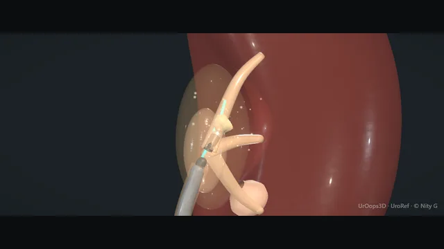
Left kidney atlas, the same sequence on the right. Schematic bladder and distal ureter. Owner-supplied values.
Operative animation, RIRS
Flexible ureterorenoscopy and intrarenal lithotripsy
A flexible scope passed through a suction access sheath stopped at the renal pelvis, into the pelvis and every calyx. The case is won by the map of the collecting system, by deflection discipline in the lower pole, and by keeping intrarenal pressure low.
Core tierFRCS endourology, stone diseaseInteractive 3D
Up the ureter through the access sheath into the collecting system. Schematic, not to scale; relationships only.
Everything happens inside a translucent shell: the pelvis is the crossroads, the lower-pole infundibulum is the narrow angulated branch where the fibre meets mucosa, and the access sheath is what keeps the pressure that drives irrigant back into the veins low.
›Renal pelvis - the crossroads every calyx drains to; the place you re-orient when lost.
›Lower-pole infundibulum - narrow, angulated, and the thinnest lining in the system - the classic site of perforation.
›Suction access sheath - the pressure relief, the outflow and the clearance route; tip at the renal pelvis, never forced.
›Ureter - three narrowings on the way up; the sheath must respect each one.
Wire up under screeningCystoscopy finds the left orifice at the corner of the trigone, with the image intensifier already over the flank. A 0.035 inch guidewire goes up the orifice soft tip first and its course is read on the screen against each narrowing of the ureter until it reaches the kidney and curls in the collecting system. The wire is the rail for everything that follows, so it is placed under screening, not by feel alone. where it bites: a wire forced against resistance dissects or perforates the ureter.
Stop thereNothing follows the wire until its curl is seen in the renal pelvis on the screen. The pause is the check that the rail lies in the collecting system rather than submucosally or through a perforation. That first wire stays as the safety wire for the whole case: the rail for a stent if the case has to end early. In the technique this module teaches, the second (working) wire is passed under direct vision through the semirigid ureteroscope into the renal pelvis; the scope comes out over it, and the working wire guides the access sheath. Other routes for the second wire are also in use.
Access sheath under screeningThe suction access sheath, inner dilator in place, is railroaded over the working wire under screening. Length is chosen by sex: about 35 cm for a woman and about 45 to 46 cm for a man (owner-supplied typical values; sizes vary by maker). The tip is watched on the image intensifier the whole way up. It should slide to its mark without force; a sheath that stops is downsized or abandoned, and a ureter that will not accept one is stented and the case rebooked. where it bites: forcing a sheath past a narrow, unstented ureter tears the wall or avulses a segment.
Suction connected, tip stopped at the renal pelvisThe suction tubing is connected to the side port of the sheath, but suction stays off until the tip is in the kidney. On the image intensifier the sheath tip is brought up to the renal pelvis and stopped there, at the PUJ, not pushed through it. A tip seated at the pelvis gives the scope a straight run into the kidney and lets the sheath carry irrigant and fragments out for the rest of the case. where it bites: a sheath driven through the PUJ tears the last narrowing.
Dilator out, scope inThe inner dilator is withdrawn with the working wire, so the working wire is out before lasering, leaving the sheath as the working channel and the safety wire outside it, and the flexible ureteroscope is passed up inside the sheath to the renal pelvis. The scope never meets the ureteric wall: it travels inside the sheath, so every exchange later in the case is low trauma.
Map the entire kidneyBefore the stone is touched, every calyx is inspected in a fixed order: upper, middle, lower. The map is what the case navigates by when the view clouds or a fragment migrates. If the map is complete and every calyx is clear, the next move is straight to the stone and the laser.
Contrast, find the stone, move itIf the map is not certain, dilute contrast injected through the scope outlines the renal pelvis and all the calyces on the screen, and the stone is found against that picture. A stone in the lower pole is basketed and carried up to the renal pelvis or the upper pole, where the scope is straight, the fibre passes freely and the irrigation works. It is lasered there rather than in the flexed lower pole. where it bites: lasering in the fully flexed lower pole strains the scope and the infundibulum.
The fibre that will not come outWith the tip fully flexed into the lower pole the fibre will not advance out of the channel. It is never forced. The flexion is released, the scope comes back to the renal pelvis, the fibre is advanced out of the channel in the straight scope, and only then is the tip flexed again with the fibre already out. where it bites: forcing a fibre round a fully flexed tip breaks the channel or the fibre.
Dusting and the settingsWith the stone where it can be worked, it is dusted at low energy and high frequency, the fibre tip seen on the stone and off the mucosa before every activation, the face painted until the stone is dust the irrigation carries into the sheath. Typical Ho:YAG ranges (owner-supplied):
Technique
Energy
Frequency
Typical power
Aim
Dusting
0.2 to 0.5 J
20 to 40 Hz
about 4 to 20 W
Fine particulate, minimal retropulsion
Fragmentation
0.8 to 1.2 J
6 to 12 Hz
about 5 to 14 W
Discrete basketable fragments
Pop-dusting
0.3 to 0.5 J
30 to 50 Hz
about 10 to 25 W
Finish fragments in a calyx
where it bites: firing on mucosa rather than stone perforates a thin infundibulum within seconds.
Popcorn the tiny fragmentsThe last small fragments are pop-dusted: the fibre is held still just above them in a confined calyx and fired, so the fragments dance in the beam and are reduced without being chased or basketed one by one.
Suction clearance to the markerThumb on the suction port, the access sheath is brought over the scope until the fragments are in view, and the scope is withdrawn slowly all the way down to the marker with the suction running, so the fragments are drawn down the sheath and out. A last look under vision on the way out; a stent only for a marked ureter, oedema or residual dust.
The spatial relationships the animation is built to make automatic. Before you read each one, ask yourself: at this point in the operation, what is about to go wrong, and what is my first move? The save for each sits in the Complications tab.
RiskLower-pole infundibulum - thin lining plus acute angle: perforation and bleeding territory.
RiskIntrarenal pressure - unrelieved irrigation pressurises the system and drives pyelovenous backflow - the sepsis route.
RiskPUJ on sheath placement - the last narrowing the sheath crosses; resistance here means stent and stage, not push.
When the case turns: the problem, then the next move.
ActionSheath will not pass the ureter - never force; leave a stent and return in one to two weeks to a passively dilated ureter.
ActionPoor deflection to the lower pole - empty the working channel, use a 200 µm-class fibre, or relocate the stone to the upper calyx or pelvis.
ActionFibre will not advance with the tip fully flexed - release the flexion, return to the pelvis, advance the fibre in the straight scope, then flex again.
ActionVision lost in dust - pause lasing, let the sheath clear the field, then resume at lower energy.
ActionStone pushed into an upper calyx - follow with deflection rather than chasing with irrigation bursts; irrigation only moves stones you cannot see.
The examiner stems this scene prepares you for. Try to answer out loud first, then reveal, and learn the discriminator, the detail that turns a pass into a strong pass.
QHow does intrarenal pressure cause sepsis during RIRS, and how do you prevent it?reveal answer
The collecting system is a closed basin once a scope occludes the infundibulum or pelvis.
Irrigation that has nowhere to escape raises intrarenal pressure, and above a threshold it drives fluid through fornices and pyelovenous channels directly into the venous system.
If the stone or urine is infected, that is bacteraemia by design.
Prevention is mechanical: a correctly sized ureteral access sheath to guarantee outflow, the lowest irrigation pressure that maintains vision, limiting case duration, and treating infection before elective stone therapy - a positive culture gets treated, and an obstructed infected system gets drained first.
If the patient shows physiological instability, the answer is to stop, not to irrigate harder for a cleaner view.
Strong pass
Say both
Naming the access sheath as a pressure-relief device
Decompression-before-stone-therapy in the infected obstructed kidney
Examiner note
Naming the access sheath as a pressure-relief device, and decompression-before-stone-therapy in the infected obstructed kidney.
QA 1.5 cm lower-pole stone: RIRS or PCNL, and why?reveal answer
A 1.5 cm lower-pole stone sits at the boundary where lower-pole anatomy decides the modality.
For RIRS the issues are the infundibulopelvic angle, infundibular length and width: an acute angle and a long narrow infundibulum make dust clearance from the lower pole unreliable, so a stone that fragments well may still not pass.
PCNL through a lower-pole puncture clears that same stone in one sitting with a higher clearance but at the cost of an invasive tract and its bleeding risk.
I would take the anatomy from the CT: a favourable angle supports RIRS; an unfavourable lower pole, a hard stone or a solitary kidney tips me to PCNL or to a staged approach.
I would also discuss ESWL, which is weaker exactly where RIRS struggles, since lower-pole fragments clear poorly.
Strong pass
Say all three
Using infundibulopelvic anatomy as the deciding variable
Not stone size alone
Naming fragment clearance (not fragmentation) as the RIRS weakness in the lower pole
Examiner note
Using infundibulopelvic anatomy as the deciding variable, not stone size alone, and naming fragment clearance (not fragmentation) as the RIRS weakness in the lower pole.
QYou lose vision in a blood-filled calyx mid-dusting. What next?reveal answer
Stop lasing immediately - a fibre that cannot see its target is a perforation device.
I would stop irrigation briefly and let the field settle, because most intrarenal bleeding is low-pressure ooze that clots when the system is still.
I would withdraw the scope to the pelvis, confirm my orientation, and re-advance only when vision returns.
If the field will not clear, the case ends: place a stent, image the kidney post-operatively to exclude a subcapsular or collecting-system haematoma, and either stage the procedure or reconsider PCNL.
Persisting against a blood-filled field is how perforation, pressure injury and lost stone fragments compound.
Strong pass
Say both
Stopping as the first move
Imaging post-operatively rather than assuming a benign outcome
Examiner note
Stopping as the first move, and imaging post-operatively rather than assuming a benign outcome.
QWhat is the role of the ureteral access sheath, and when would you leave it out?reveal answer
The sheath gives three things: repeated low-trauma exchange of the scope, continuous irrigant outflow that keeps intrarenal pressure low, and better vision through flow.
For anything beyond a small single stone it is standard.
I would leave it out when the ureter will not accept it without force - a tight, unstented ureter is avulsed by sheaths, not by scopes - and in that case I would either work without a sheath using low irrigation, or stent and return to a dilated ureter.
I would also avoid rough sheath placement in a solitary kidney or a known stricture.
The decision is always the same trade: pressure control and access versus the mechanical risk to the ureteric wall.
Strong pass
Naming avulsion as the sheath-specific risk and the staged stent-and-return option for the tight ureter.
Say it aloud, then reveal. Reading is not remembering.
01The relationshipSay the critical spatial relationship out loud, then reveal.reveal
Memory frame
Everything happens inside a translucent shell
Why?
Everything happens inside a translucent shell: the pelvis is the crossroads, the lower-pole infundibulum is the narrow angulated branch where the fibre meets mucosa, and the access sheath is what keeps the pressure that drives irrigant back into the veins low.
02The first moveCalyceal perforation. Your first move?reveal
First move
Stop lasing and stop advancing the instant the mucosa disappears or fat is seen.
03The escalation triggerCalyceal perforation. What makes you call for help?reveal
Escalate when
Free extravasation, fever or a lost tract to the calyx → terminate, stent or nephrostomy drainage, senior review.
04The discriminatorWhat turns a pass into a strong pass?reveal
Strong pass
Say both
Naming the access sheath as a pressure-relief device
Decompression-before-stone-therapy in the infected obstructed kidney
Examiner note
Naming the access sheath as a pressure-relief device, and decompression-before-stone-therapy in the infected obstructed kidney.
05The pitfallWhere does this operation bite first?reveal
Pitfall
Lower-pole infundibulum
Why?
thin lining plus acute angle: perforation and bleeding territory.
Revision practice with self-marked answers, not an assessment of clinical competence. Kept in
this browser only; it does not sync between devices.